Jump to content
In this section we have spot diagnoses posted on a daily basis since June 2010, now over 4000! You can review the archived cases and read the suggested diagnoses by users and the final comment by the contributors.
Case are uploaded each week day by 10 am UK time with the correct diagnosis will generally be posted at 8 pm UK time. Why not view the most recent spot diagnosis and proffer a diagnosis?

Case Number : Case 909 - 12th December Posted By: Guest

Please read the clinical history and view the images by clicking on them before you proffer your diagnosis.
Submitted Date :
   (0 reviews)

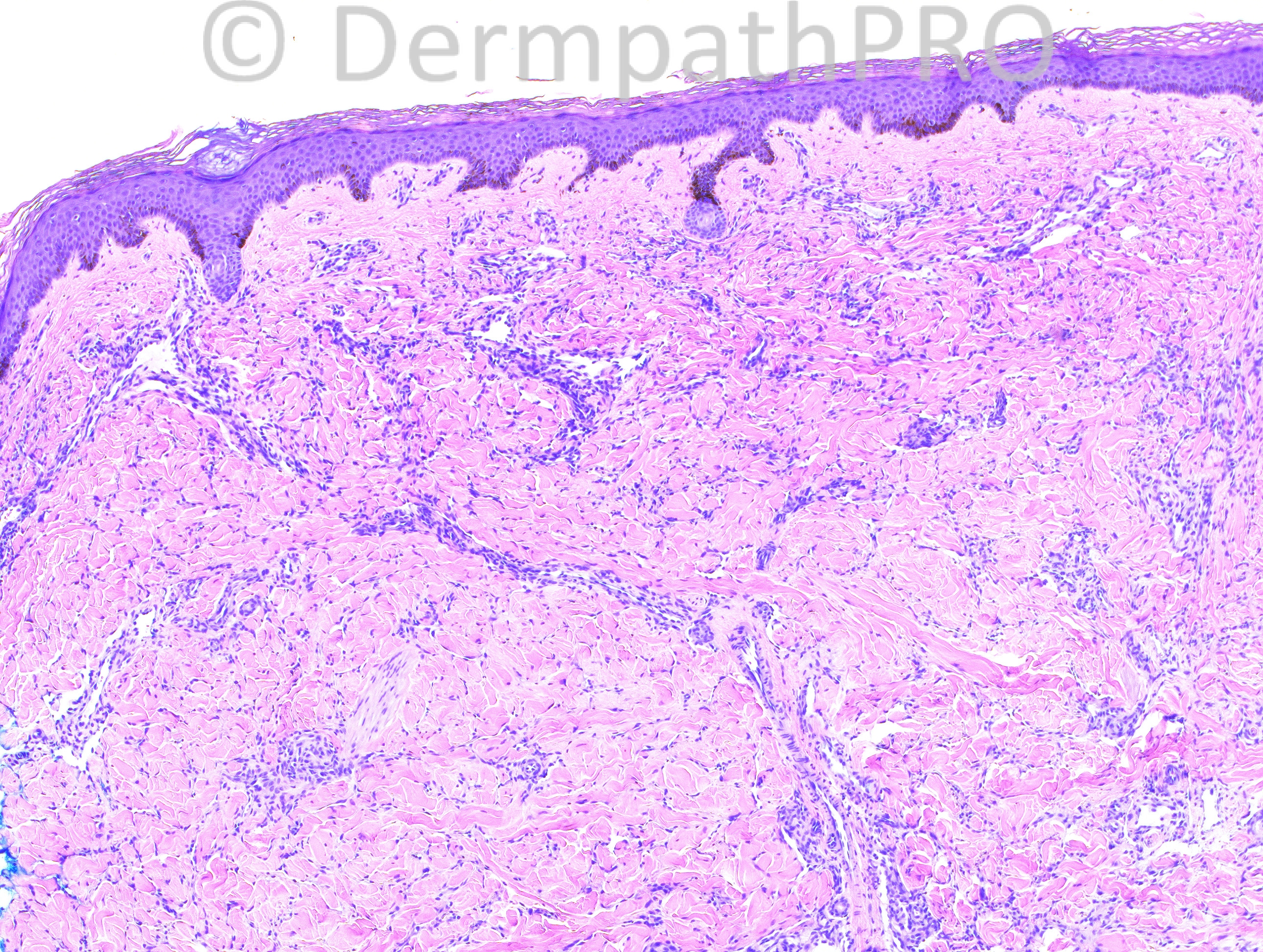
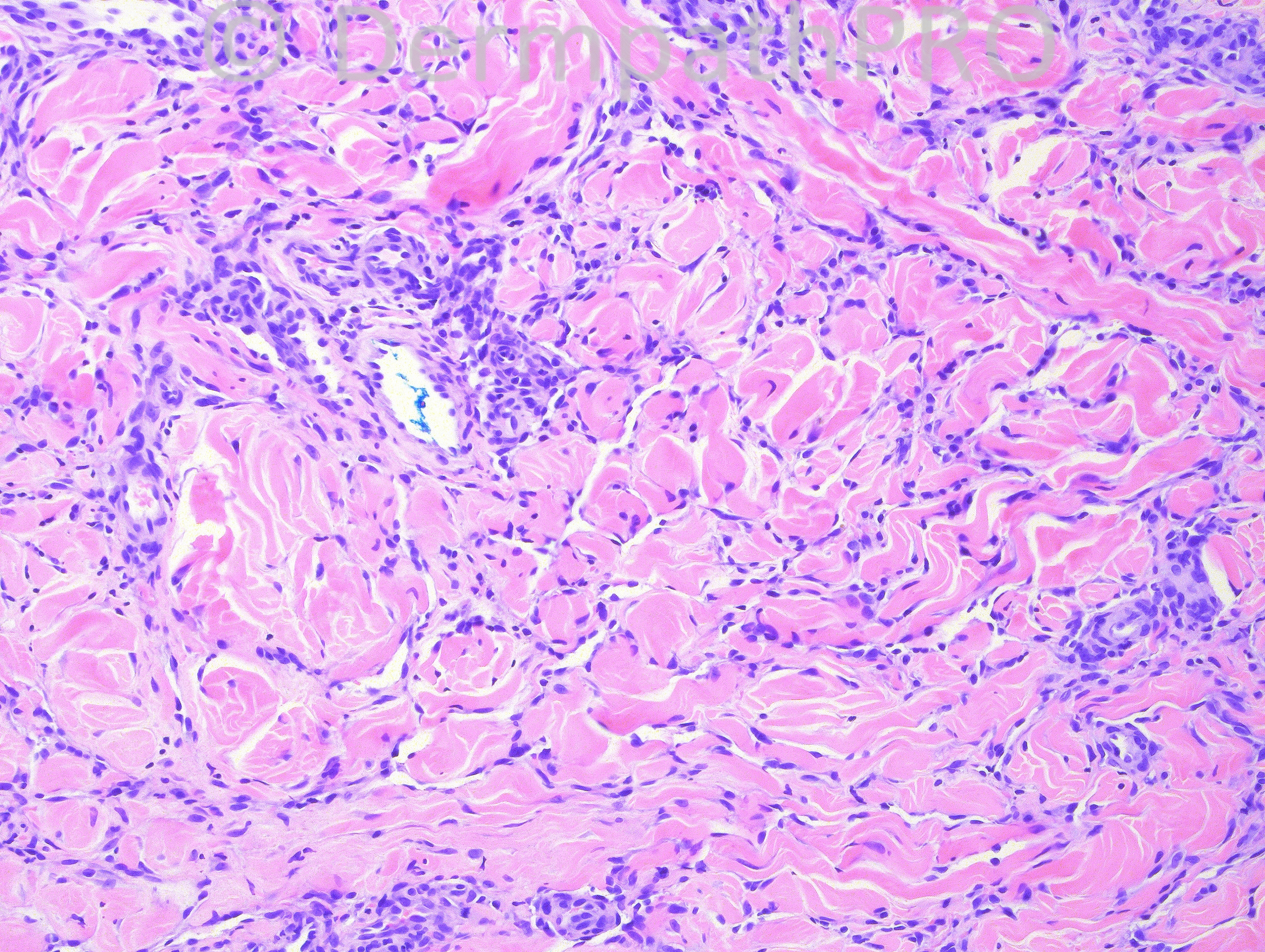
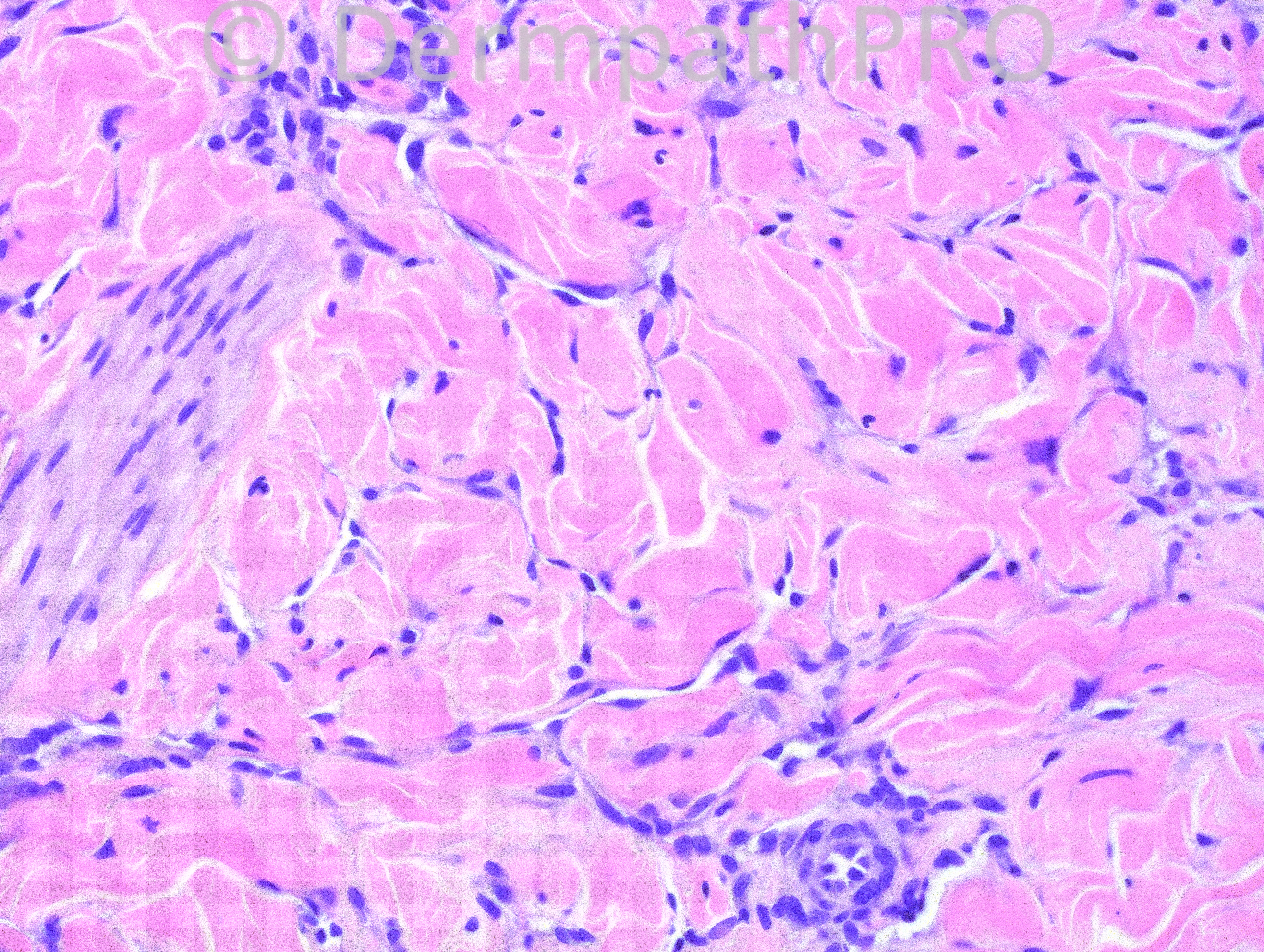
24 year-old male with right arm lesions.

Case posted by Dr. Hafeez Diwan.


  Report Record

User Feedback


Guest Romualdo

Posted

Despite the general resemblance to a dermatofibroma the perivascular concentration of spinde cells and the muliplicity of lesions ar more in keeping with Kaposi's sarcoma.

Share this comment


Link to comment
share_externally

I agree, KS.
The most difficult feature of this case, in my opinion, is the lack of red blood cells and plasma cells, I can not see them, but there´s no other vascular differential diagnosis for this case (neither angiosarcoma nor the different kinds of hemangioendotheliomas).

Share this comment


Link to comment
share_externally

Robledo F. Rocha

Posted

A very early patch stage of Kaposi sarcoma. Without extravasated erythrocytes and fascicles of spindle cells, it´s hard to rule out definitely an inflammatory skin disease, like morphea. Nevertheless, jagged vascular spaces dissecting collagen bundles and a putative promontory sign on picture #2 make me favor Kaposi sarcoma.

Share this comment


Link to comment
share_externally

Guest Jim Davie MD

Posted

Interesting case. Agree that Kaposi's patch stage is most likely diagnosis, vs. hobnail hemangioma, based on these images.

Despite the bland cytology, there is sparse mitotic activity [lowest left image], increased cellularity, and promontory sign with indisputable single endothelial layers (lacking supporting pericytes) closely applied to surfaces of collagen bundles, and connection to pre-existent vessels. There may be plasma cells in lowest left image. A confirmatory positive stain for HHV8 (and/or clinical correlation) would be of utility.
Differential diagnosis would include:
1. microvenular hemangioma (which had been my first-glance consideration, given classic clinical presentation on arm of a young adult male). However, these lesions are usually solitary, should have easily visible pericytes associated with the dissecting vascular spaces, and show absence of mitotic activity. They often infiltrate arrector pilae, which appears to not be the case in these images. SmActin stain would help highlight the pericytes for equivocal cases.
2. Hobnail hemangioma. (another classic clinical presentation on arm of a young adult male). These are rarely multiple. They can have dissecting architecture with bland endothelial cells deeper in the dermis, and more classic papillary telangiectatic vessels superficially, which may be argued as present in the top two images. Also may have sparse inflammatory infiltrate with plasma cells.

Share this comment


Link to comment
share_externally


Join the conversation

You can post now and register later. If you have an account, sign in now to post with your account.

Guest
Add a comment...

×   Pasted as rich text.   Paste as plain text instead

  Only 75 emoji are allowed.

×   Your link has been automatically embedded.   Display as a link instead

×   Your previous content has been restored.   Clear editor

×   You cannot paste images directly. Upload or insert images from URL.

Loading...

×
×
  • Create New...