Jump to content
In this section we have spot diagnoses posted on a daily basis since June 2010, now over 4000! You can review the archived cases and read the suggested diagnoses by users and the final comment by the contributors.
Case are uploaded each week day by 10 am UK time with the correct diagnosis will generally be posted at 8 pm UK time. Why not view the most recent spot diagnosis and proffer a diagnosis?

Case Number : Case 912 - 17th December Posted By: Guest

Please read the clinical history and view the images by clicking on them before you proffer your diagnosis.
Submitted Date :
   (0 reviews)

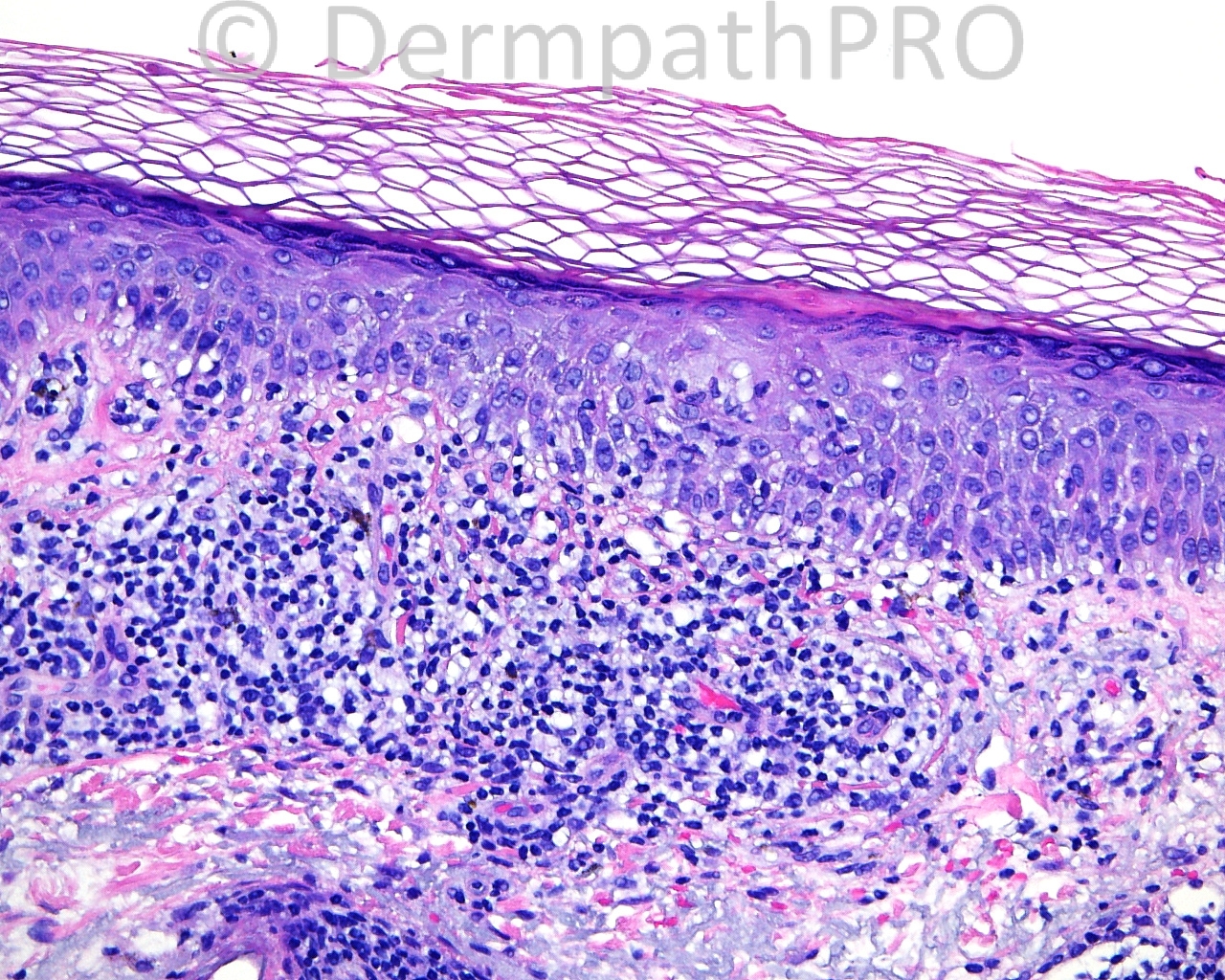
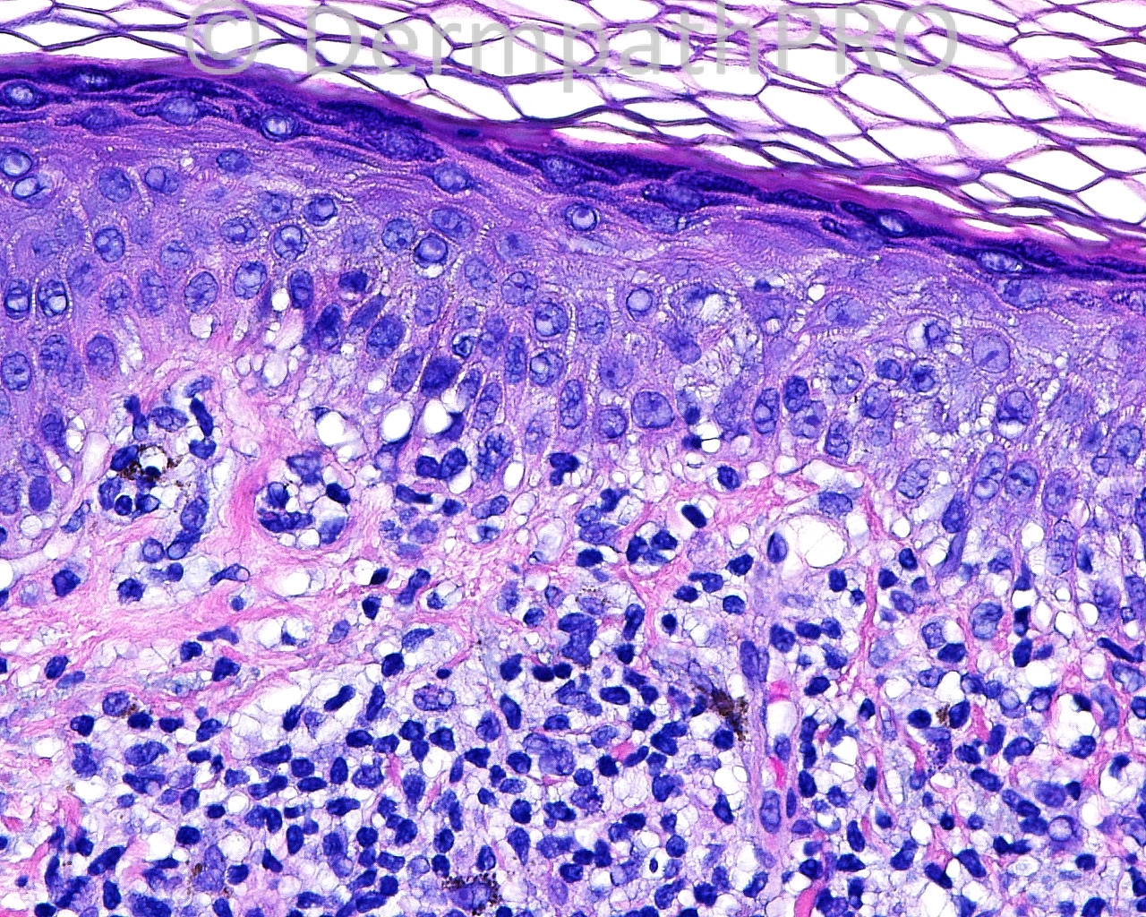
The patient is a 72 year old white woman with a shave biopsy of a pink papule taken from the left distal forearm.

Case posted by Dr. Mark Hurt


  Report Record

User Feedback


Guest Maria George

Posted

Extravasated RBC, siderophages points to Pigmented purpura , most likely lichen aurus.
Lupus, perniosis among others in the differential diagnoses.

Share this comment


Link to comment
share_externally

Eman El-Nabarawy

Posted

DD of lichenoid tissue reaction. Basket weave stratum corneum points to erythema multiforme.

Share this comment


Link to comment
share_externally

A shave biopsy of a pink papule points towards a clinical suspicion of a benign skin lesion (tumor). The histopathological figures points to a Lichen planus-like keratosis (Lichenoid keratosis).

Share this comment


Link to comment
share_externally

Sasi Attili

Posted

I think the DD of this LTR is wide. But if the clinical is correct (single lesion- pink papule), then we would have to call this a lichenoid keratosis.

Share this comment


Link to comment
share_externally

Guest Romualdo

Posted

Agree with Igor: lichen planus-like keratosis.

Share this comment


Link to comment
share_externally

Dr. Mona Abdel-Halim

Posted

If the pink papule is all what the patient has, then this is lichen planus like keratosis. If it is part of a widespread papular eruption, could be acute LE or EM. More clinical details are needed...

Share this comment


Link to comment
share_externally

Guest Juan Carlos Garcés, Ecuador

Posted

I would add lichen nitidus to the DD

Share this comment


Link to comment
share_externally

Robledo F. Rocha

Posted

I favor erythema multiforme. There is an interface vacuolar lymphocytic infiltrate rather than a band-like one. Also, incipient subepidermal blister with sprinkling of lymphocytes, extravased erythrocytes in papillary dermis, and basket-woven stratum corneum. The very subtle ballooning of epidermis and the lack of noticeable necrotic keratinocytes are findings of an early lesion.
I'd include early Mucha-Habermann disease to the differential diagnosis, albeit this only rarely occurs in eldery.

Share this comment


Link to comment
share_externally

Guest Engin Sezer

Posted

Fixed drug eruption based on conspicious melanophages and interphase changes. By the way I am missing Dr McKee's comments..

Share this comment


Link to comment
share_externally

Guest Jim Davie MD

Posted

Favor early lichenoid keratosis in context of a solitary pink papule. Agree with the above excellent differentials for interface vacuolar dermatitis. I don't see convincing evidence of a junctional melanocytic proliferation hiding within the lichenoid inflammatory process.

Share this comment


Link to comment
share_externally

I think this is a lichen planus like keratosis with pseudonests.

Share this comment


Link to comment
share_externally

Dr. Richard Carr

Posted

I like the suggesions of lichen plans-like reaction and inflammatory pseudonests (this assumes a solitary lesion of course). I routinely examine mulitple levels if I think a melanocytic lesion might be hiding but can't see it here.

Share this comment


Link to comment
share_externally

Guest Jim Davie MD

Posted

The melanocytes show low-grade, dendritic cytology and low lentiginous density; this seems within normal range in the context of inflammation and chronic actinic exposure as evidenced by solar elastosis. Of interest, however, is the presence of sparse melanocytes spreading down the acrosyringium in the center, which raises the differential of an atypical lentiginous junctional proliferation... I would have to compare to the non-inflamed adjacent epidermis to see the 'background' melanocytic density to properly assess the significance of this finding.
Regardless, the inflammatory process does not seem to be targeting the melanocytic component, so I will stick with lichenoid keratosis, based on the above images.

Share this comment


Link to comment
share_externally


Join the conversation

You can post now and register later. If you have an account, sign in now to post with your account.

Guest
Add a comment...

×   Pasted as rich text.   Paste as plain text instead

  Only 75 emoji are allowed.

×   Your link has been automatically embedded.   Display as a link instead

×   Your previous content has been restored.   Clear editor

×   You cannot paste images directly. Upload or insert images from URL.

Loading...

×
×
  • Create New...