Jump to content
In this section we have spot diagnoses posted on a daily basis since June 2010, now over 4000! You can review the archived cases and read the suggested diagnoses by users and the final comment by the contributors.
Case are uploaded each week day by 10 am UK time with the correct diagnosis will generally be posted at 8 pm UK time. Why not view the most recent spot diagnosis and proffer a diagnosis?

Case Number : Case 915 - 20th December Posted By: Guest

Please read the clinical history and view the images by clicking on them before you proffer your diagnosis.
Submitted Date :
   (0 reviews)

No history

Case posted by Dr. Richard Carr.

  Report Record

User Feedback


Guest Tiberiu Tebeica

Posted

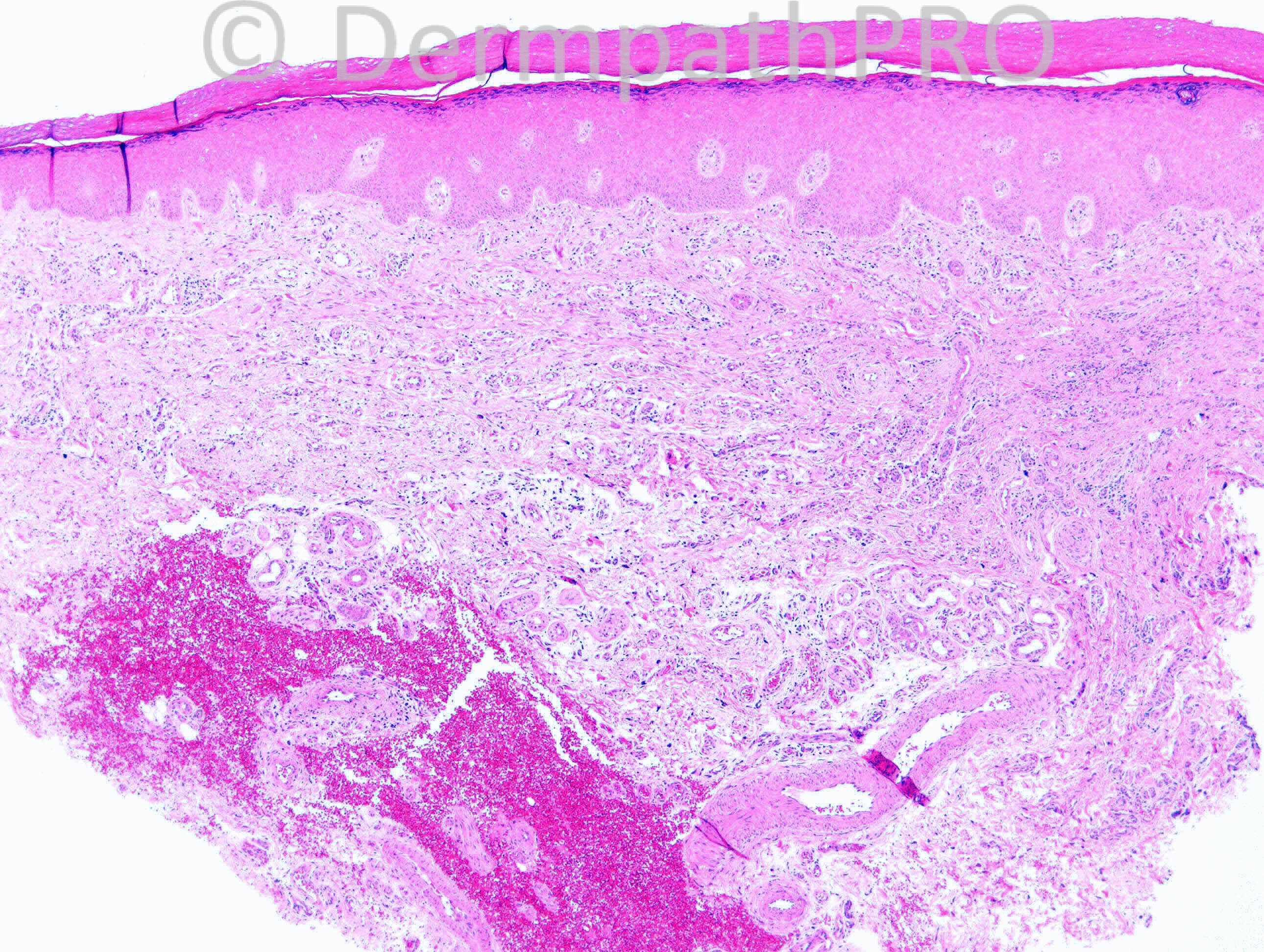
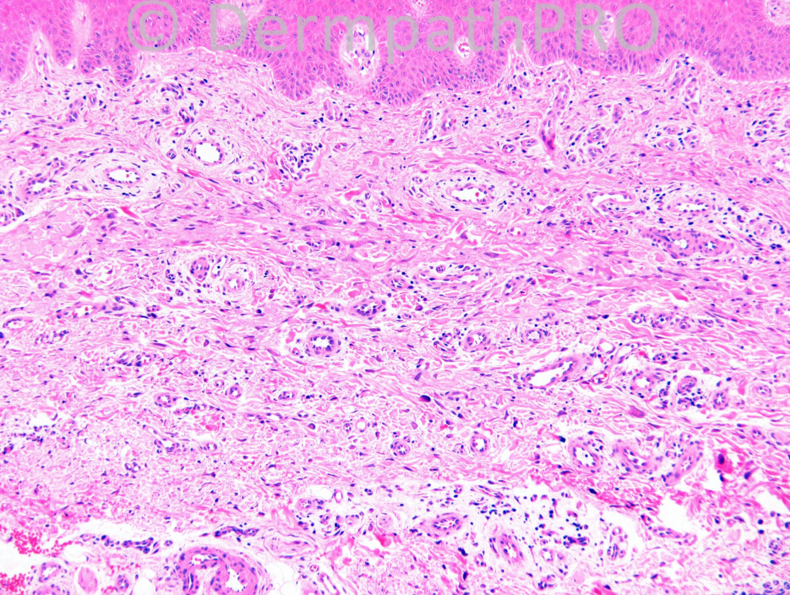
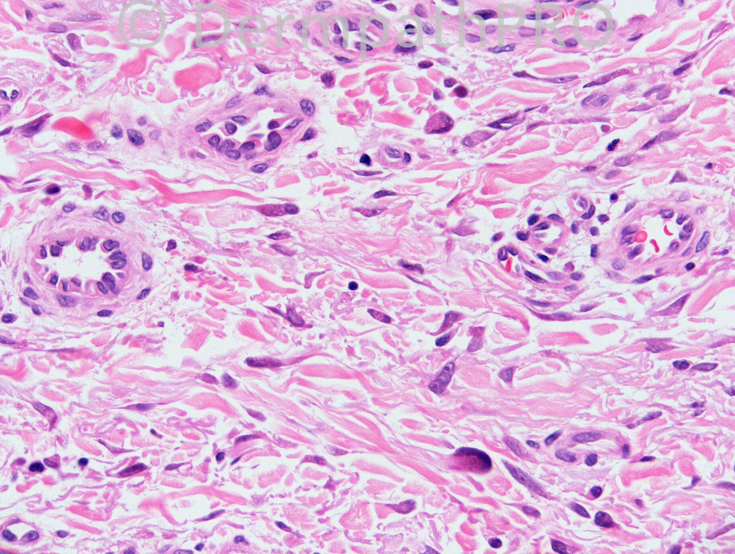
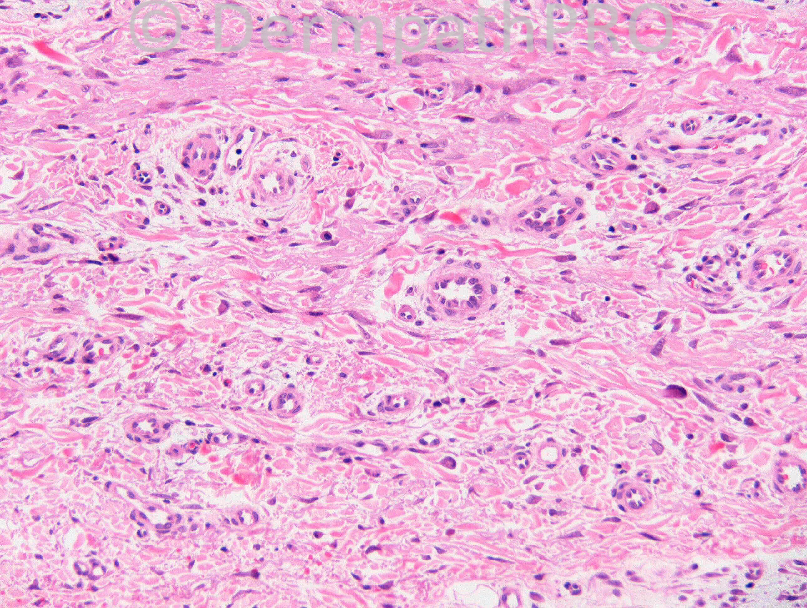
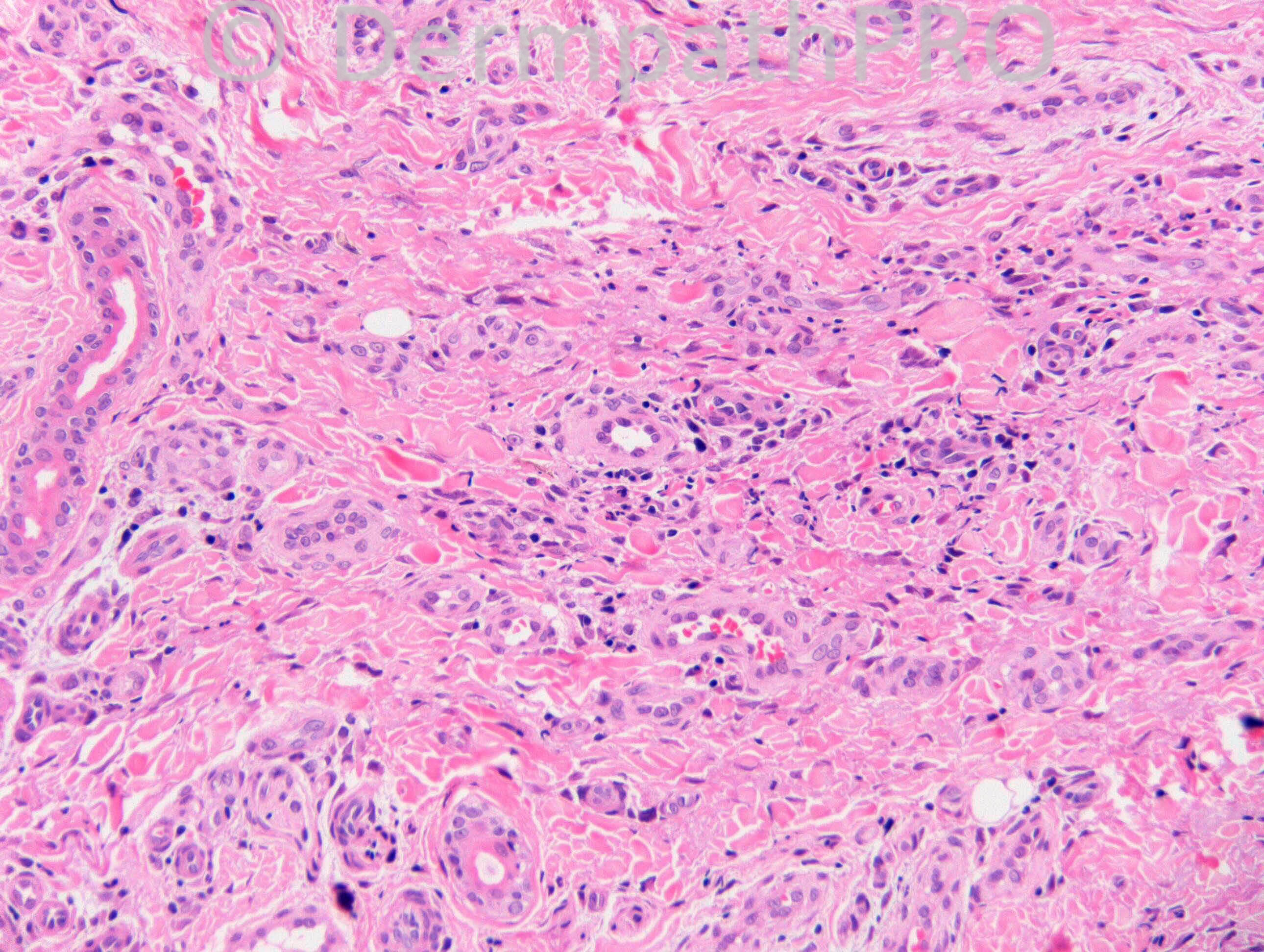
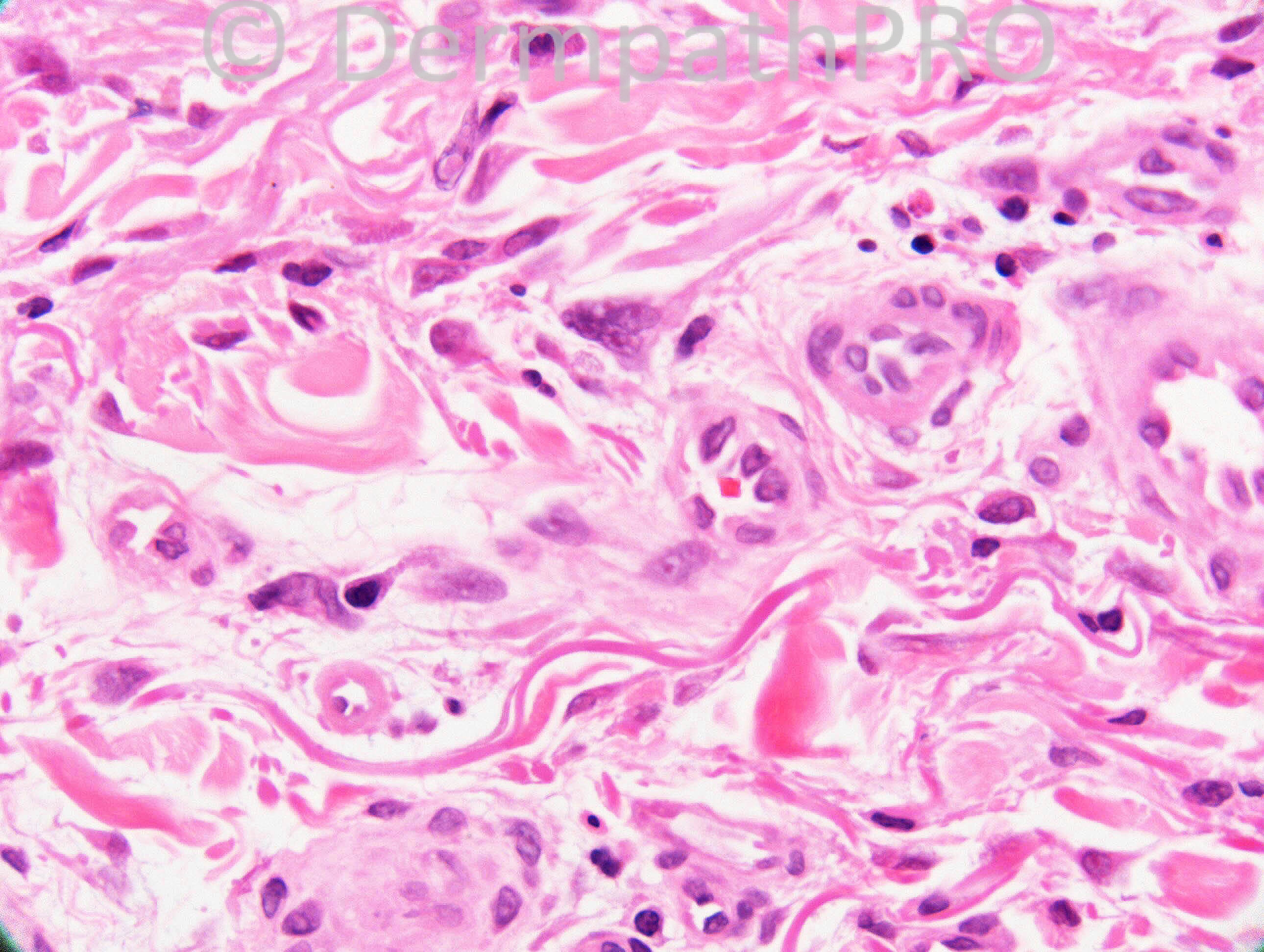
This biopsy was taken from a female patient with age over 40, presenting with red papules on her legs and/or hands. My histologic diagnosis is multinucleate cell angiohistiocytoma.

Share this comment


Link to comment
share_externally

Dr. Mona Abdel-Halim

Posted

First impression: multinucleate cell angiohistiocytoma,,, lovely :-)

Share this comment


Link to comment
share_externally

Guest Maria George

Posted

Epidermal hyperplasia overlying a thick wall capillary proliferations which show hobnailing .
I agree with Tiberiu and Mona.Capilary nevus is 2ed the dfferential.
By the way.The site needs Maintainance.I write the comments with geat difficulity.
Did other members feel the same?

Share this comment


Link to comment
share_externally

Guest Romualdo

Posted

Agree with Tiberiu and Mona: multinucleate cell angiohistiocytoma.

Share this comment


Link to comment
share_externally

Guest Romualdo

Posted

About yesterday's case, it would very instructive if we could see some photographs of the elastic stains!

Share this comment


Link to comment
share_externally

Guest Pedro Aleixo

Posted

I favor multinucleate cell angiohistiocytoma. Mast cells are not prominent in this case.

Share this comment


Link to comment
share_externally

Robledo F. Rocha

Posted

Multinucleate cell angiohistiocytoma. Geometric-shaped multinucleated cells and dilated vascular spaces are characteristic.

Share this comment


Link to comment
share_externally

Sasi Attili

Posted

Seems like there is a consensus. Would be stupid for me to disagree on this spotter.

Share this comment


Link to comment
share_externally

Dr. Richard Carr

Posted

Yes no tricks from me today. I called the multinucleate cell angiohistiocytoma and I did not think you needed the history which proved correct but for the record:
M75. Dorsum L hand. ?Dermatofibroma ?scar tissue. 6 mm punch excision.
Enjoy your weekends and the festive season!!!!

Share this comment


Link to comment
share_externally


Join the conversation

You can post now and register later. If you have an account, sign in now to post with your account.

Guest
Add a comment...

×   Pasted as rich text.   Paste as plain text instead

  Only 75 emoji are allowed.

×   Your link has been automatically embedded.   Display as a link instead

×   Your previous content has been restored.   Clear editor

×   You cannot paste images directly. Upload or insert images from URL.

Loading...

×
×
  • Create New...