Jump to content
In this section we have spot diagnoses posted on a daily basis since June 2010, now over 4000! You can review the archived cases and read the suggested diagnoses by users and the final comment by the contributors.
Case are uploaded each week day by 10 am UK time with the correct diagnosis will generally be posted at 8 pm UK time. Why not view the most recent spot diagnosis and proffer a diagnosis?

Case Number : Case 918 - 27th December Posted By: Guest

Please read the clinical history and view the images by clicking on them before you proffer your diagnosis.
Submitted Date :
   (0 reviews)

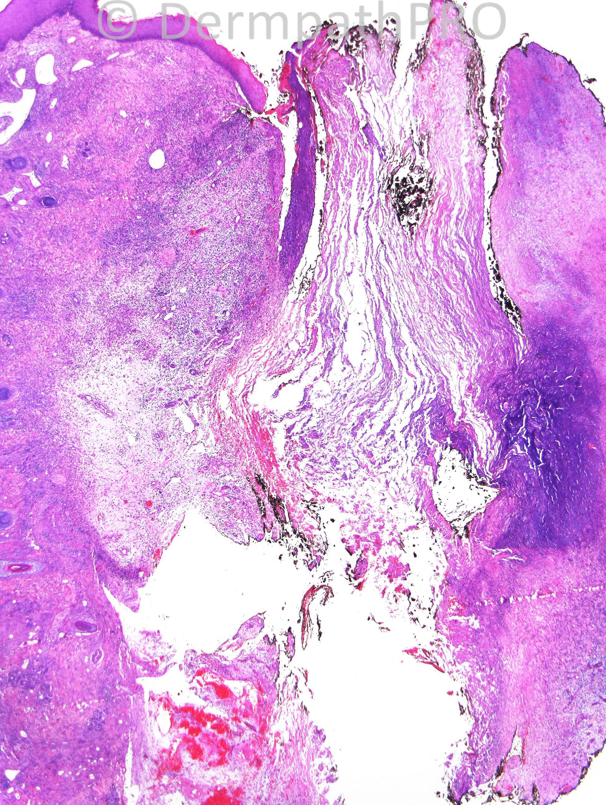
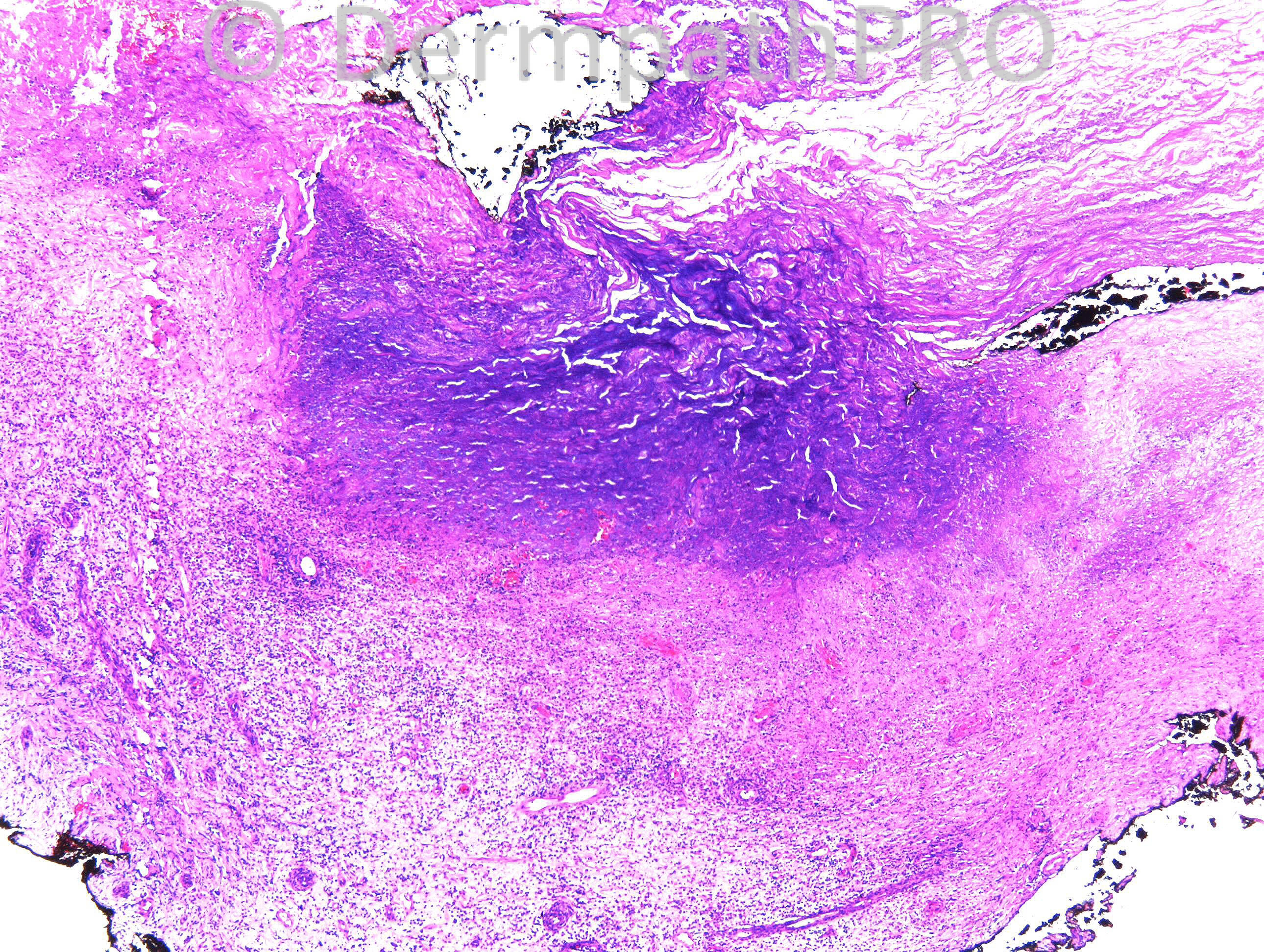
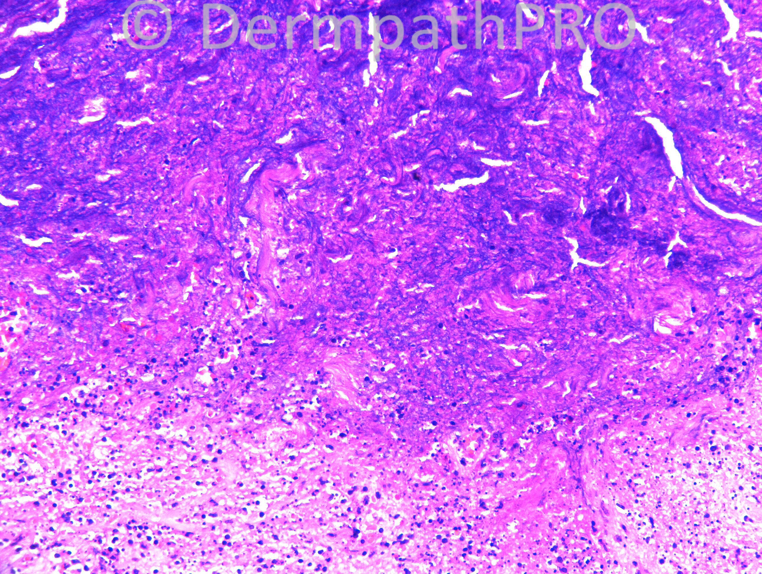
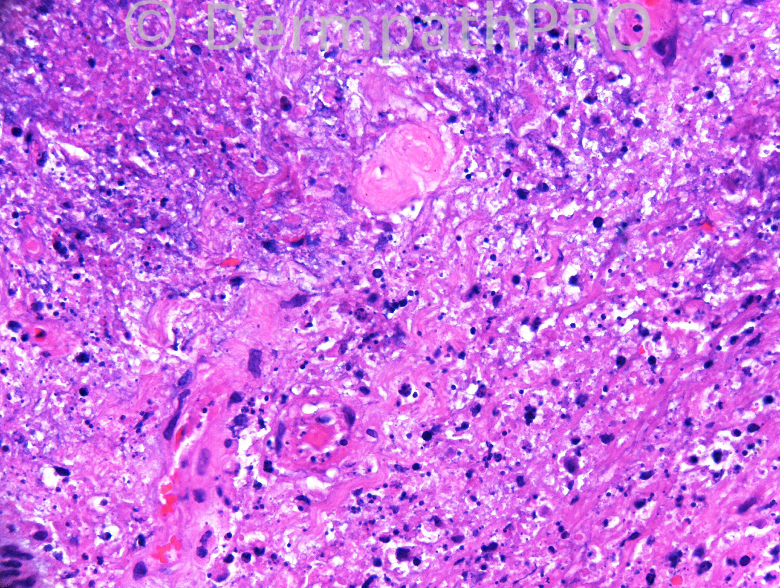
F69. Has CLL. Scalp vesicles Rx’d by GP as Zoster but developed large unilateral area of black ulcerated scalp. Swabs growing pseudomonas.

Case posted by Dr. Richard Carr.

  Report Record

User Feedback


Dr. Richard Carr

Posted

Hope I am not the only one working today!!!

Share this comment


Link to comment
share_externally

Guest Juan Carlos Garcés, Ecuador

Posted

You are not, Dr Carr, happy holidays

Ectima?

Share this comment


Link to comment
share_externally

Eman El-Nabarawy

Posted

Necrotizing fasciitis in HZ lesions in immunocompromized patient, severe HZ infection in immunocompromized patient,or leukemic infiltrate in HZ lesions with secondary vasculitis.

Share this comment


Link to comment
share_externally

Dr. Mona Abdel-Halim

Posted

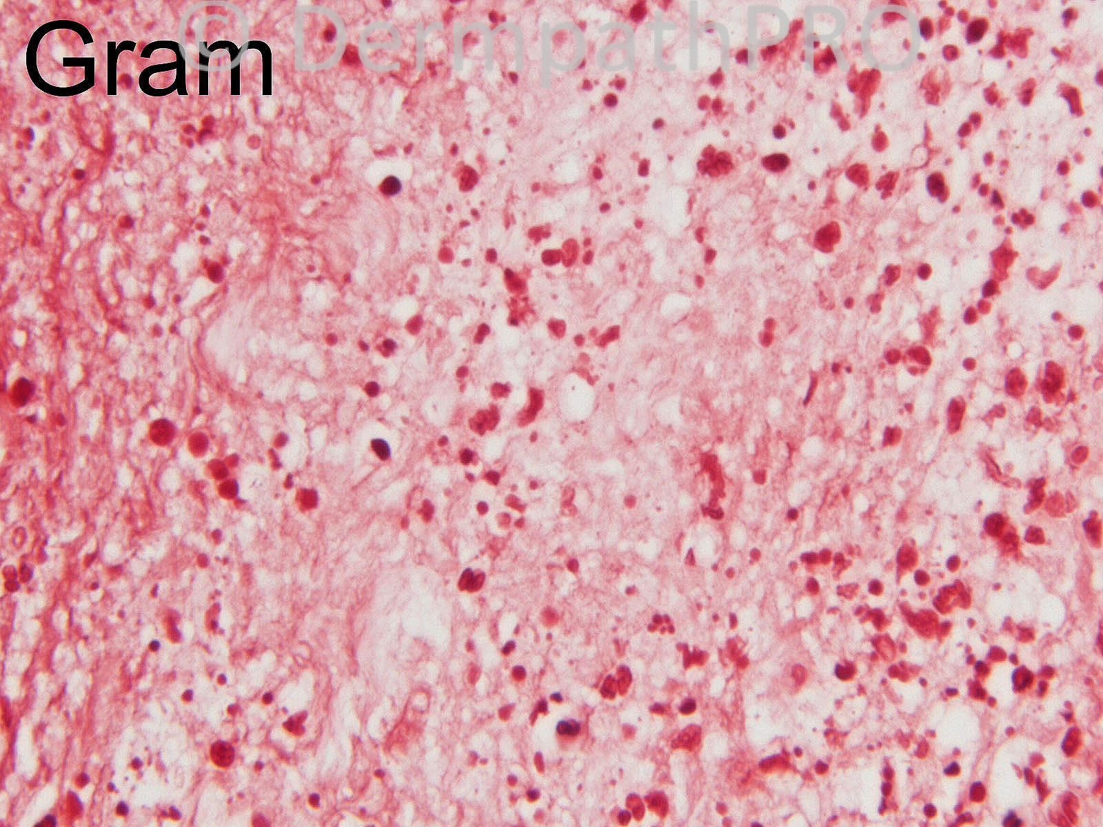
Echthyma gangrenosum. Pseudomonus is on the top list of causative organisms but candida can also cause EG like picture. Pseudomonas is gram negative and does not look like the organism appearing in this gram stain. I believe what I am seeing in the gram stain is budding yeasts and few hyphae, indicating candida which can be stained by gram. Aspergillus, and Fusarium species, and Zygomycetes can also cause EG like picture but they do not stain with gram. The other yeasts that can stain with gram are the cryptococci, but they produce different clinical and pathological picture.

Share this comment


Link to comment
share_externally

Robledo F. Rocha

Posted

I’m working too, Dr. Carr!
I think this is solitary ecthyma gangrenosum.

Share this comment


Link to comment
share_externally

Dr. Richard Carr

Posted

This was a dramatic clinicopathological case (to end the year).There were 3 separate biopsies over a 6 week period (about half of one side of the scalp was involved by an extensive black ulcer). Pseudomonas grew repeatedly and finally (much later) some aspergillus (I assumed a red herring). I did not see any organisms on any of the stains on any of the biopsies (although you might imagine some rods on the Gram). Thromboembolic was considered but no other clin-path evidence for this. I looked for giant cell arteritis and could find no evidence for that. I did consider lymphoma but did not persue that route as I felt this was an infection. We sent a block to Dr Kutzner (sincere thanks) for PCR which was negative for HZV and positive for pseudomonas (not aspergillus). She started to improve steadily and dramatically after being given intra-venous immunoglobulins. Final diagnosis: Pseudomonas ecthyma gangrenosum (uncertain if preceded by Zoster). Thank you for your excellent comments today and my very best wishes to all for the new year!!

Share this comment


Link to comment
share_externally


Join the conversation

You can post now and register later. If you have an account, sign in now to post with your account.

Guest
Add a comment...

×   Pasted as rich text.   Paste as plain text instead

  Only 75 emoji are allowed.

×   Your link has been automatically embedded.   Display as a link instead

×   Your previous content has been restored.   Clear editor

×   You cannot paste images directly. Upload or insert images from URL.

Loading...

×
×
  • Create New...