Jump to content
In this section we have spot diagnoses posted on a daily basis since June 2010, now over 4000! You can review the archived cases and read the suggested diagnoses by users and the final comment by the contributors.
Case are uploaded each week day by 10 am UK time with the correct diagnosis will generally be posted at 8 pm UK time. Why not view the most recent spot diagnosis and proffer a diagnosis?

Case Number : Case 687 - 1 Feb Posted By: Guest

Please read the clinical history and view the images by clicking on them before you proffer your diagnosis.
Submitted Date :
   (0 reviews)

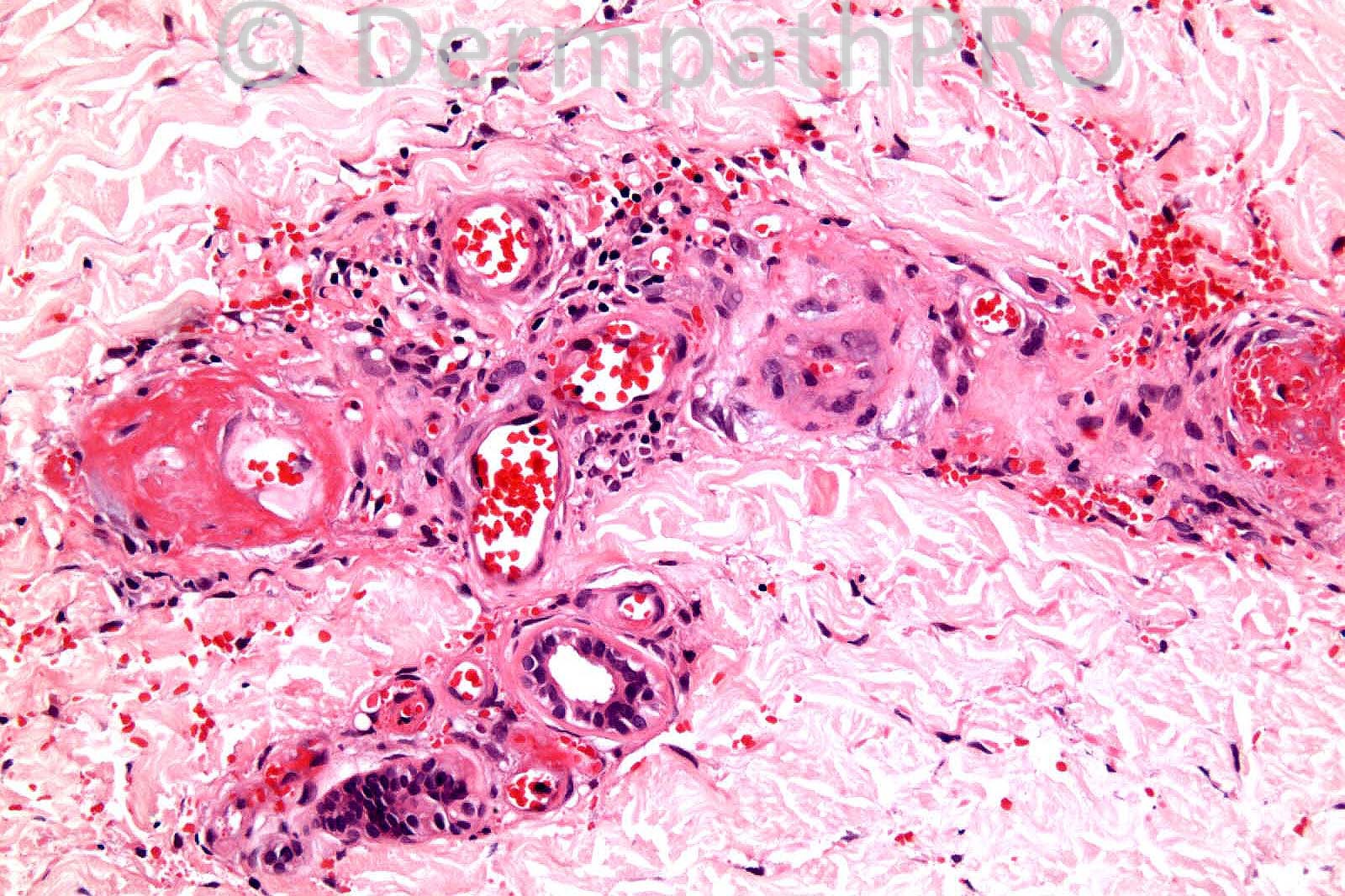
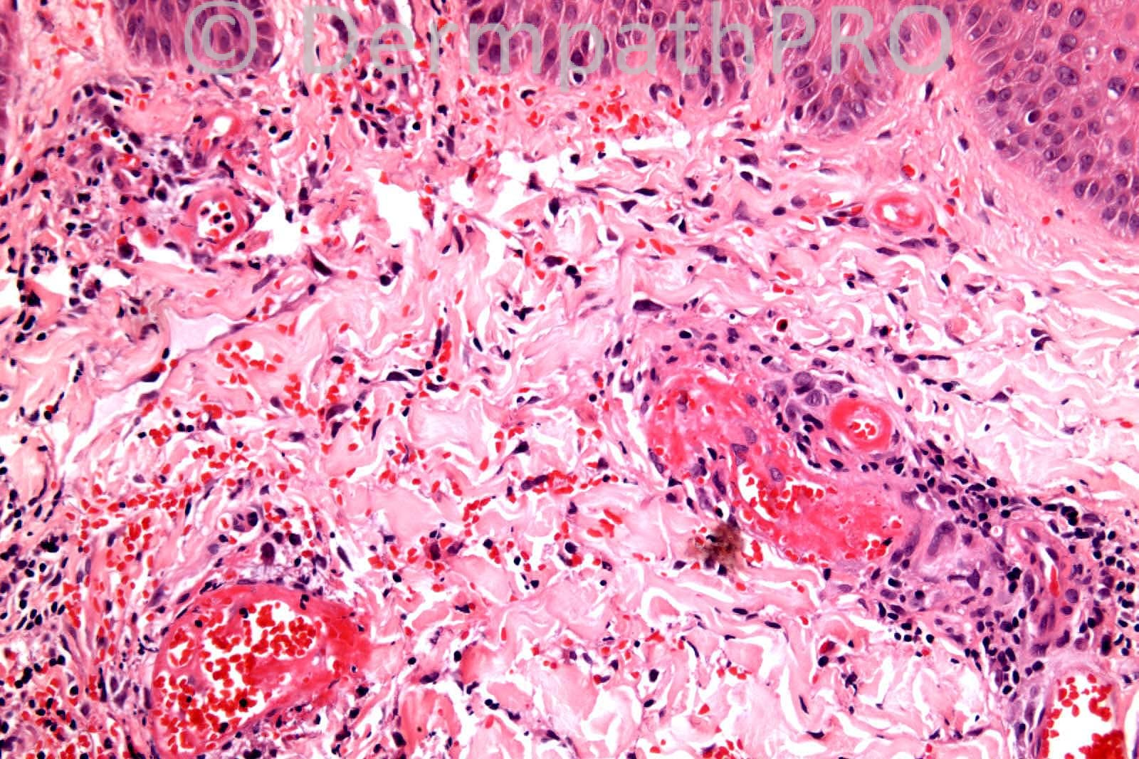
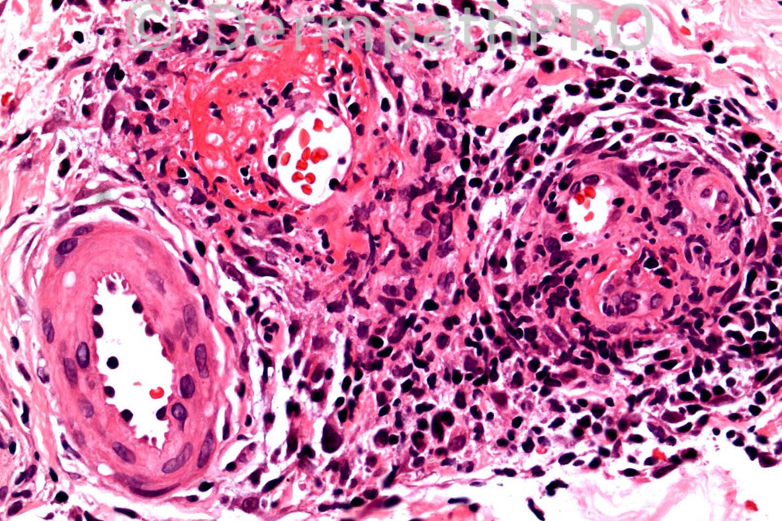
26 years-old female. Necrotic and purpuric lesions on lower legs.

Case posted by Dr. Richard Carr.


  Report Record

User Feedback


nick turnbull

Posted

LCV, but is it quite deep? and lots of extravasd rbcs especially in papillary dermis? LCV related to CTD? cryoglobulins? Parvo? HSP?

Share this comment


Link to comment
share_externally

Guest Dr Engin Sezer

Posted

Livedoid vasculitis

Share this comment


Link to comment
share_externally

Robledo F. Rocha

Posted

Fully developed leukocytoclastic vasculitis of both superficial and deep plexuses.

Share this comment


Link to comment
share_externally

Dr. Richard Carr

Posted

I did not diagnose LCV in this case. Neutrophils and sparse eosinophils
were present but the infiltrate is mainly lymphocytic. I deliberately
photographed the most "inflammatory" areas. She had a long history of recurrent
crops of lesions that also involved the dorsa of the feet.

Share this comment


Link to comment
share_externally

Dr. Mona Abdel-Halim

Posted

As the infiltrate was mainly lymphocytic, and these represent most inflammatory areas which means that some areas are non or less inflammatory and with clinical correlation, I will revise my diagnosis to atrophie blanche, lividoid vasculitis.

Share this comment


Link to comment
share_externally

nick turnbull

Posted

PLEVA? febrile ulceronecrotic MH disease? or thrombocclusive? maybe APLS?

Share this comment


Link to comment
share_externally

Eman El-Nabarawy

Posted

Lividoid vasculitis.

Share this comment


Link to comment
share_externally

Robledo F. Rocha

Posted

[font="Palatino Linotype, serif"][size="3"]Further information gave me a chance for revising my opinion. Gender, affected regions, and a history of chronic course with spontaneous remissions and exacerbations favor livedoid vasculitis, as well as the microscopic features of mild lymphocytic vasculitis with fibrin deposition in the vessel wall. Nevertheless, this disese is expected in older patients and there's no notice about white atrophic plaques of healed lesions.[/size][/font]
[font="Palatino Linotype, serif"][size="3"]Collagen tissue disease vasculitis may be an alternative hypothesis since some neutrophils and nuclear dust can be found.[/size][/font]

Share this comment


Link to comment
share_externally

I would add levamisole-induced necrosis to the differential.

Share this comment


Link to comment
share_externally

Dr. Richard Carr

Posted

Many thanks all. In fact we did think there might be an assocition with the
oral contraceptive pill in this case. We diagnosed livedoid vasculopathy based
on clinicopathological correlation. There were very nice PAS positive fibrin
rings. It just shows you that as in the previous case (thrombrophlebitis) you
can get prominent inflammatory infiltrates in the response / healing phase
of coagulopathies and thrombotic disorders.

Share this comment


Link to comment
share_externally


×
×
  • Create New...