Jump to content
In this section we have spot diagnoses posted on a daily basis since June 2010, now over 4000! You can review the archived cases and read the suggested diagnoses by users and the final comment by the contributors.
Case are uploaded each week day by 10 am UK time with the correct diagnosis will generally be posted at 8 pm UK time. Why not view the most recent spot diagnosis and proffer a diagnosis?

Case Number : Case 690 - 6 Feb Posted By: Guest

Please read the clinical history and view the images by clicking on them before you proffer your diagnosis.
Submitted Date :
   (0 reviews)

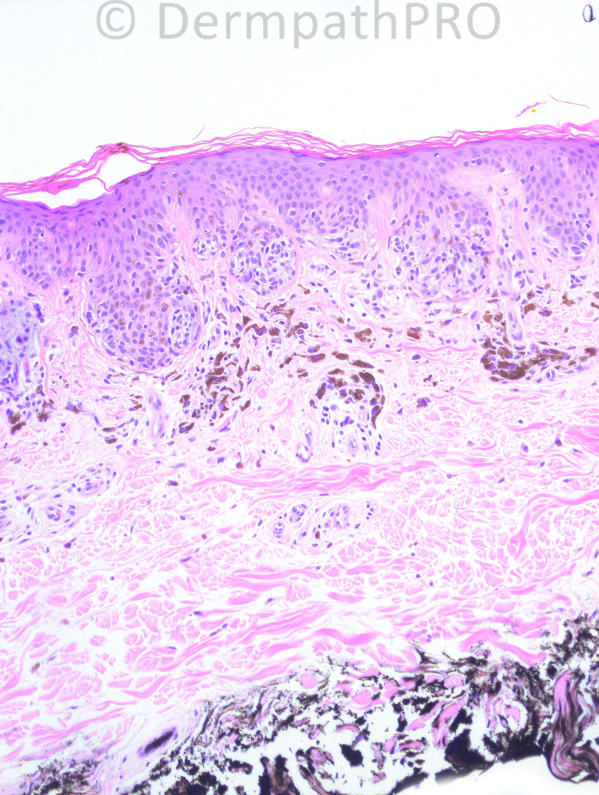
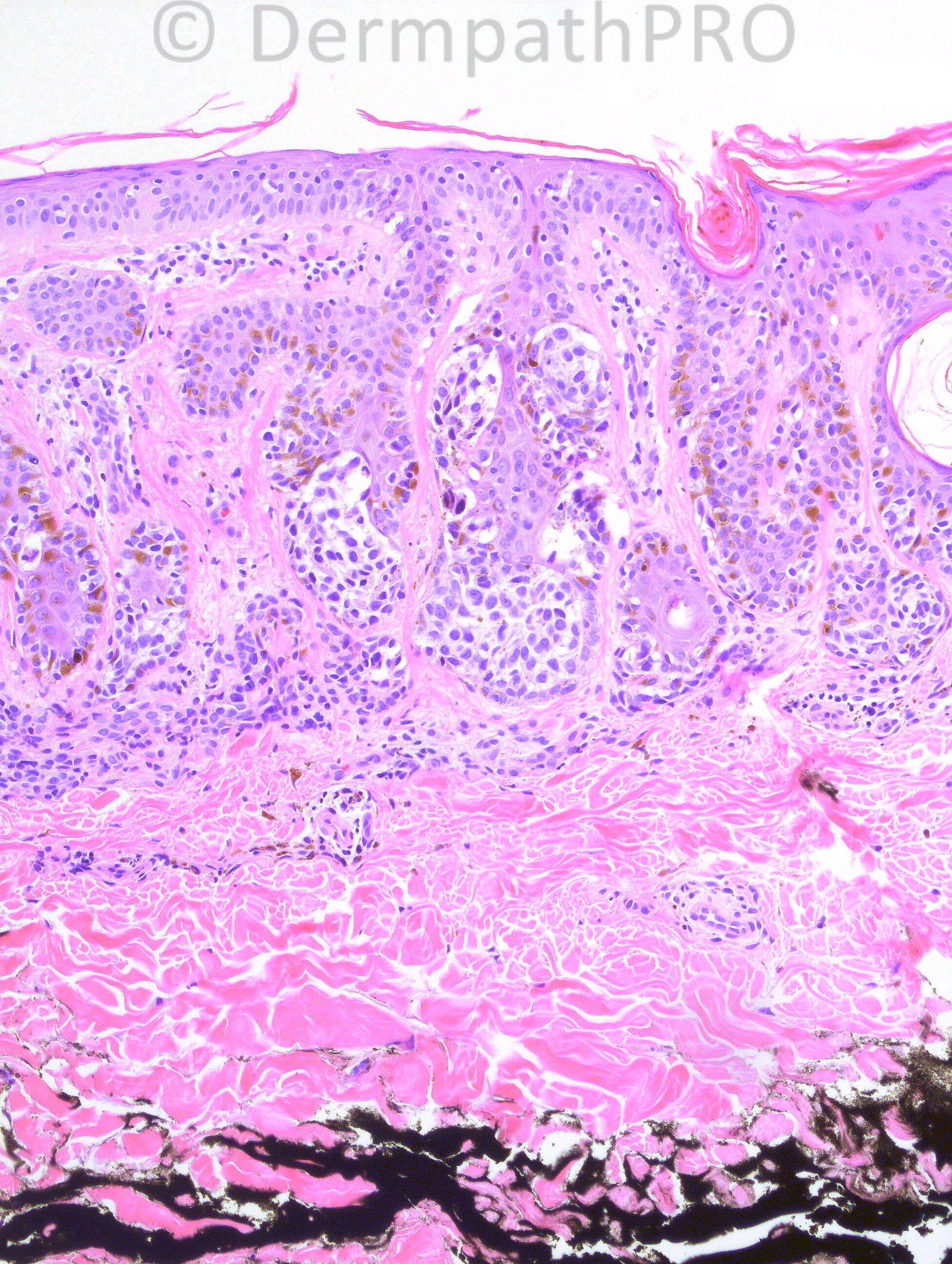
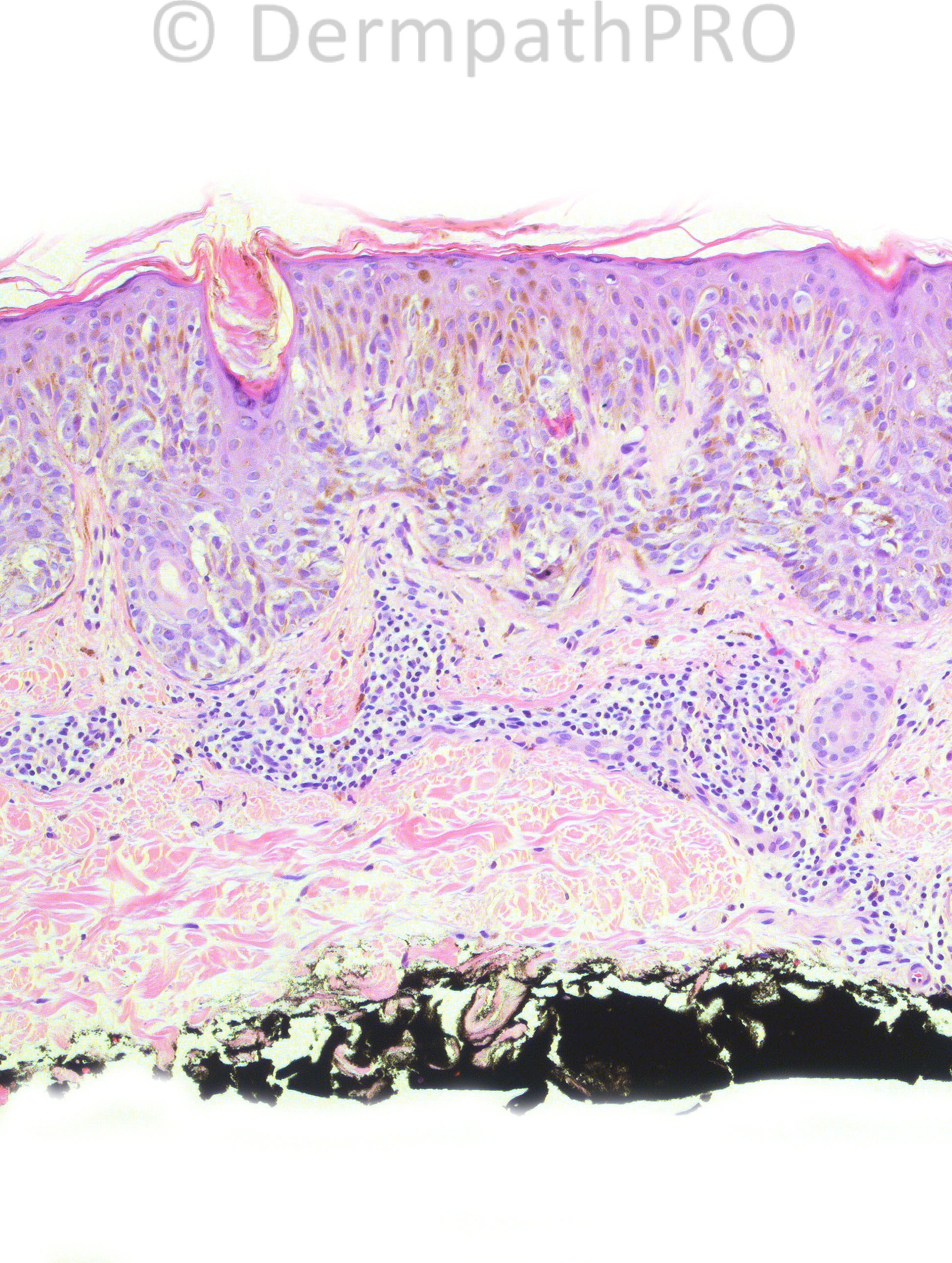
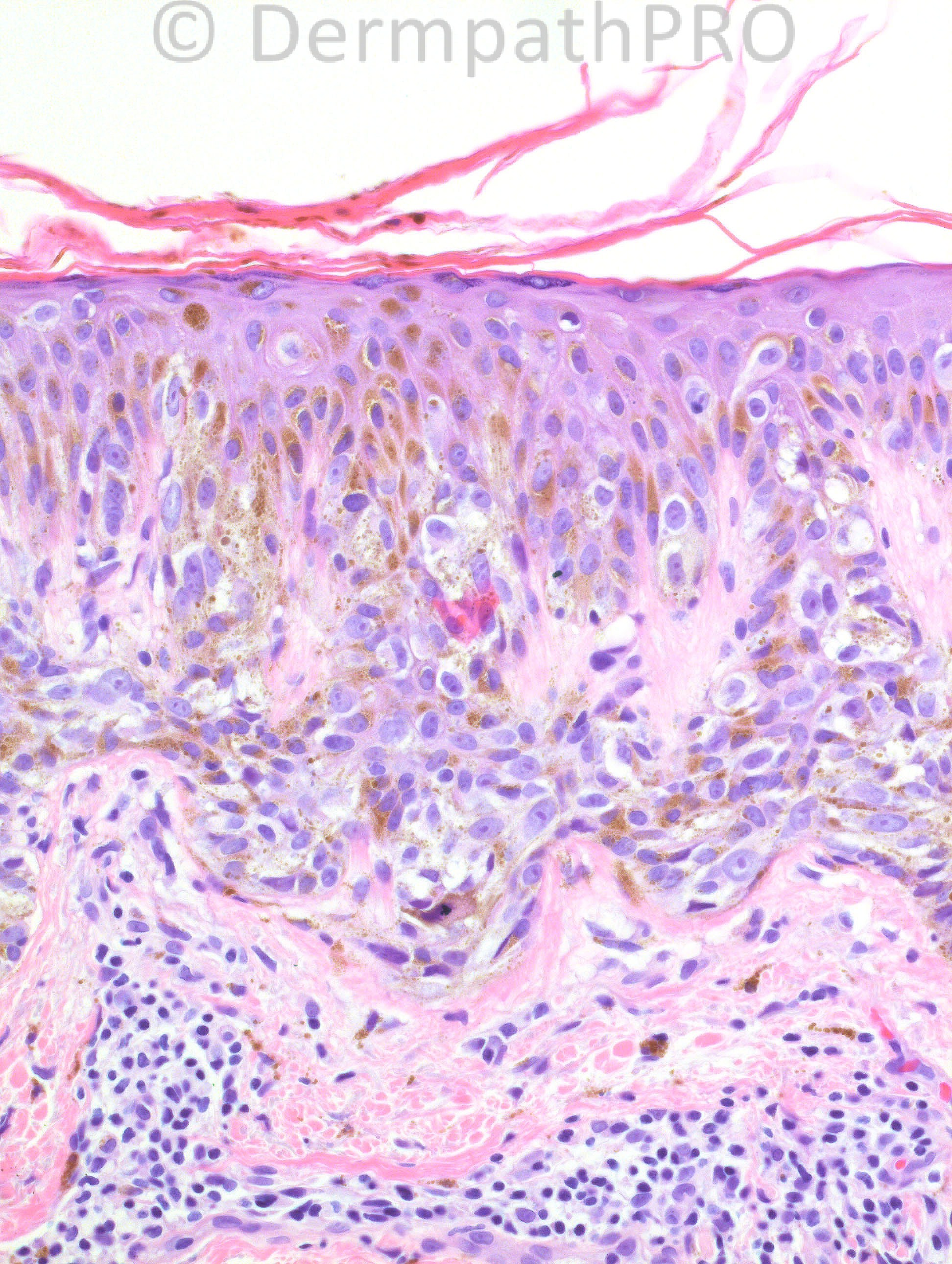
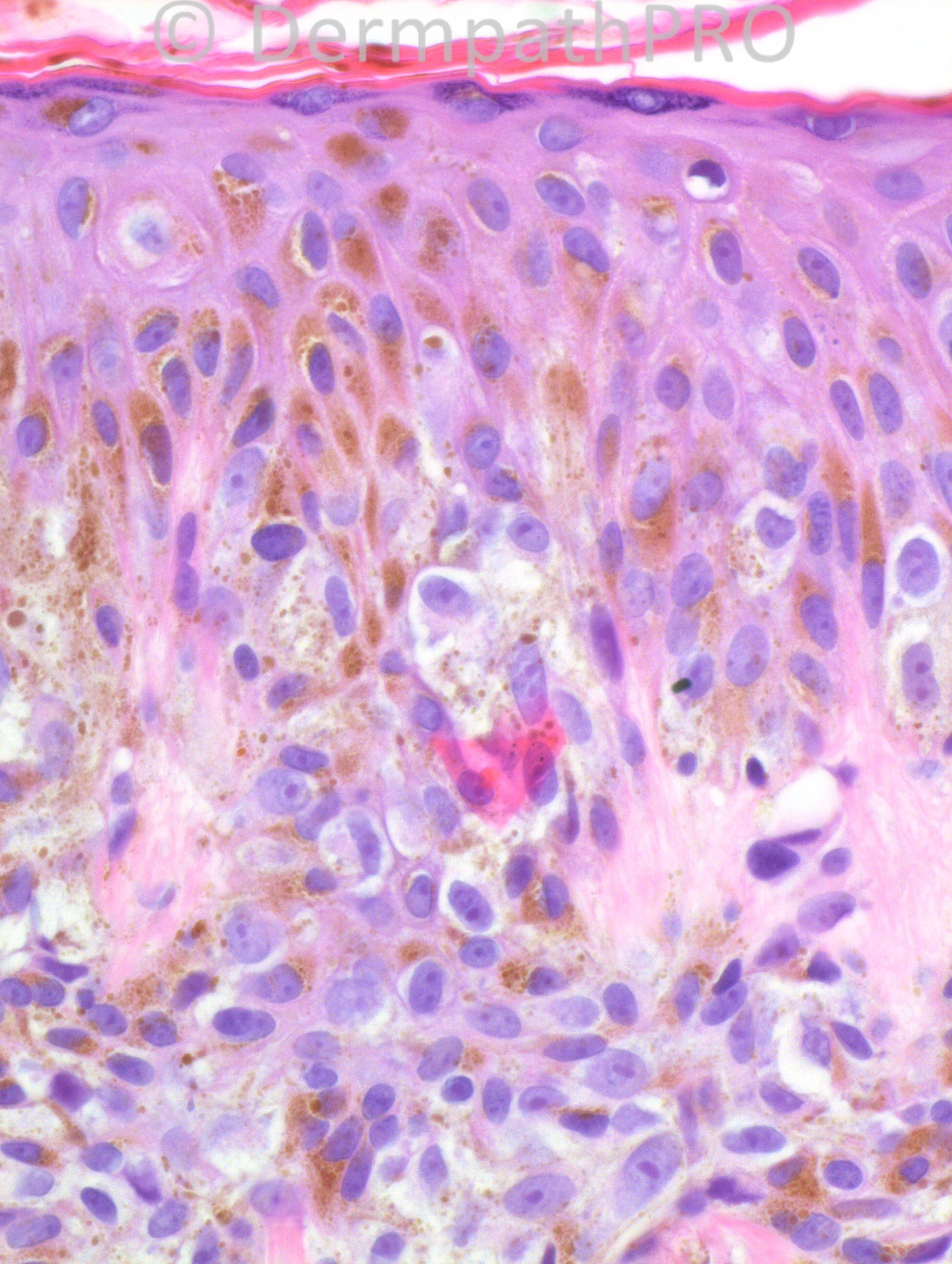
61-years-old white male with a pigmented lesion on his back.

Case posted by Dr. Hafeez Diwan


  Report Record

User Feedback


I think melanoma on top of atypical junctional nevus ???

Share this comment


Link to comment
share_externally

DD: dysplastic naevus, in situ melanoma. When does a dysplastic naevus definitively become in situ melanoma ? Other than pagetoid spread, are there any other parameters?

Share this comment


Link to comment
share_externally

Dr. Mona Abdel-Halim

Posted

Severly dysplastic nevus evolving into melanoma in situ.

Share this comment


Link to comment
share_externally

Guest Dr. Francisco Vílchez

Posted

Clark nevus

Share this comment


Link to comment
share_externally

Eman El-Nabarawy

Posted

Severely dysplastic nevus.

Share this comment


Link to comment
share_externally

Sasi Attili

Posted

severely dysplastic nevus/ MIS

Share this comment


Link to comment
share_externally

Robledo F. Rocha

Posted

Severe dysplastic nevus. Pagetoid spread indicates coexisting in-situ melanoma.

Share this comment


Link to comment
share_externally

Dr. Richard Carr

Posted

Mild uniform cytological atypia (always a concern with this clincal). I could
not be sure this is a melanoma in these images. Like to see the rest of the
lesion! Some dysplastic features. Recommend complete excision!

Share this comment


Link to comment
share_externally

Dysplastic naevus. While I would go upto severe based on these limited views of the architecture, I'm not convinced there is pagetoid spread.

Share this comment


Link to comment
share_externally

Dr. Hafeez Diwan

Posted

I called this melanoma in situ arising in association with dysplastic nevus. The top two images have a different morphology as compared to the bottom three (the latter shows upward scatter as well).

Share this comment


Link to comment
share_externally

Guest Dr Ian McColl

Posted

I think there is enough upward spread of atypical melanocytes to make this melanoma in situ.

Share this comment


Link to comment
share_externally


×
×
  • Create New...