Jump to content
In this section we have spot diagnoses posted on a daily basis since June 2010, now over 4000! You can review the archived cases and read the suggested diagnoses by users and the final comment by the contributors.
Case are uploaded each week day by 10 am UK time with the correct diagnosis will generally be posted at 8 pm UK time. Why not view the most recent spot diagnosis and proffer a diagnosis?

Case Number : Case 695 - 13 Feb Posted By: Guest

Please read the clinical history and view the images by clicking on them before you proffer your diagnosis.
Submitted Date :
   (0 reviews)

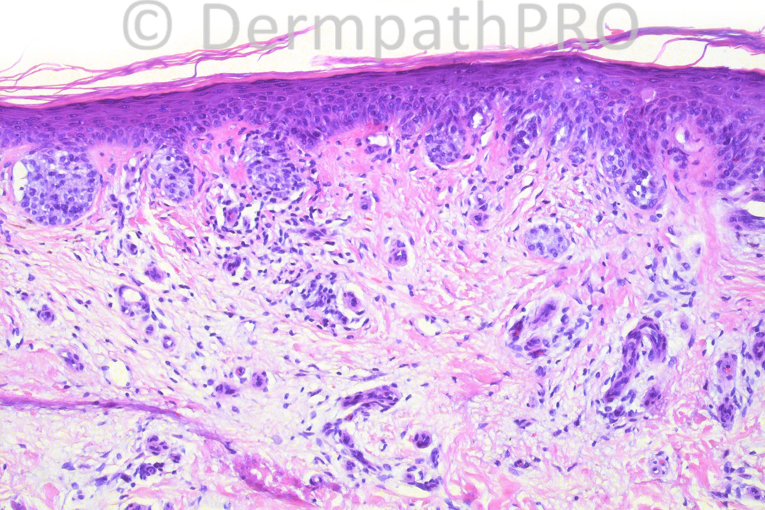
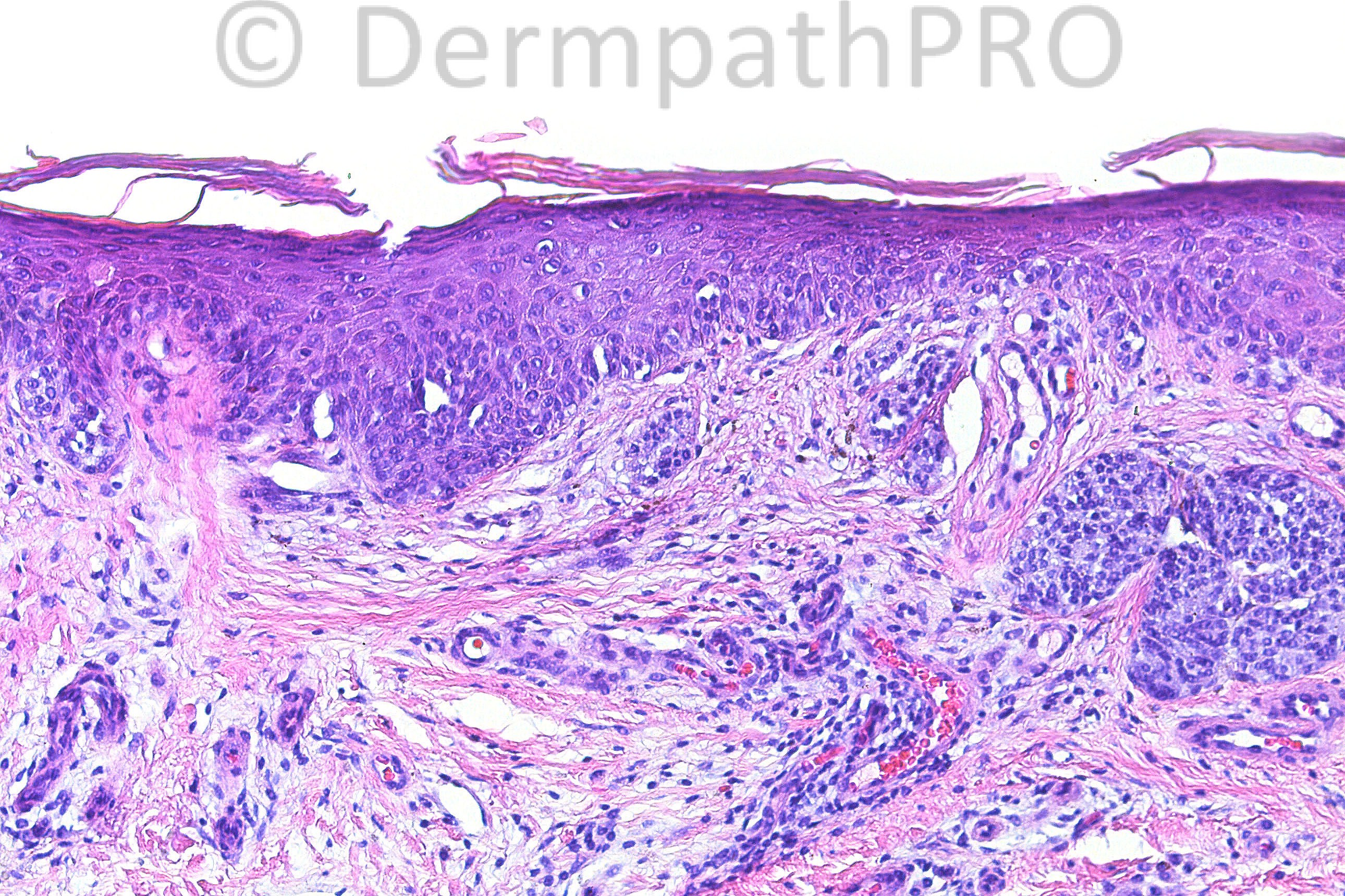
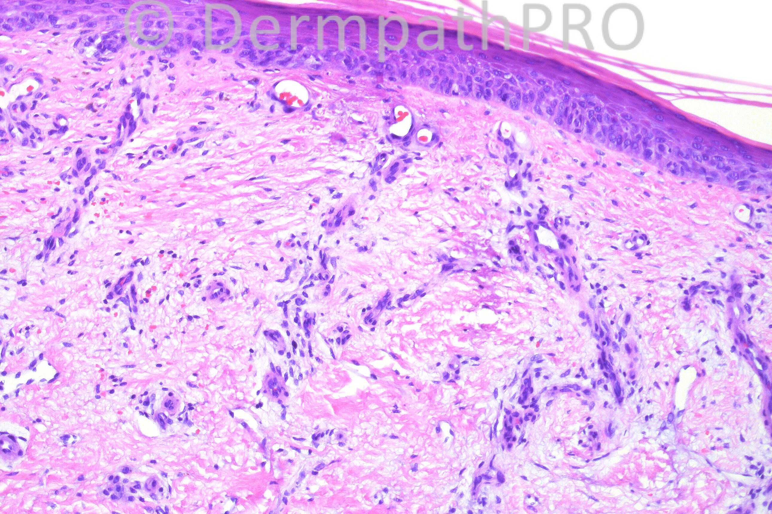
61-year-old white male with traumatized pigmented lesion on left calf. Clinical is dysplastic nevus vs. pigmented BCC vs. SK vs. other.

Case posted by Dr. Hafeez Diwan


  Report Record

User Feedback


Small cell melanoma with regression vs recurrent nevus. Very difficult, would love to see a panoramic view.

Share this comment


Link to comment
share_externally

Hanan Vaknine MD

Posted

I do not see any signs of obvious malignancy, I would diagnosed it as [b]benign nevus[/b] with areas of scar formation, most likely due to trauma

Share this comment


Link to comment
share_externally

I think this is a melanoma ?naevoid ? small cell. I too wish to see a scanning / low power view.

Share this comment


Link to comment
share_externally

Guest nick turnbull

Posted

benign, with scar related psudomelanoma changes?

Share this comment


Link to comment
share_externally

Sasi Attili

Posted

[quote name='Dr. Marcela Saeb' timestamp='1360750748']
Small cell melanoma with regression vs recurrent nevus. Very difficult, would love to see a panoramic view.
[/quote]
Agree. This is certainly not a 'spot' diagnosis.

Share this comment


Link to comment
share_externally

Guest Dr Engin Sezer

Posted

Recurrent naevus (secondary to trauma) with congenital features. The atypical nest appears to be restricted to scar..

Share this comment


Link to comment
share_externally

Robledo F. Rocha

Posted

This melanocytic compound lesion has small junctional nests in an atrophic epidermis (Picture #1) but expansile dermal ones in a scar dermis (Picture #2). Obvious maturation with depth is shown in Picture #3. There’s no cytological atypia nor mitotic activity. So, I think this lesion is a traumatized banal melanocytic nevus.
If the expansile dermal nest in Picture #5 represents focal epithelioid type A cell component that is deeper than lymphocyte-like type B cells, this lesion should be a traumatized inverted nevus.

Share this comment


Link to comment
share_externally

Recurrent nevus. I'd like HMB45, Ki-67, but given the banal nevus w/ scar and the reported history of trauma I really don't feel this is melanoma. There's no significant atypia or mitotic activity, no pagetoid spread that I can make out, no confluent growth.

Share this comment


Link to comment
share_externally

Dr. Mona Abdel-Halim

Posted

I do not see enough criteria to consider this malignant. I believe the fibrosis, vascular proliferation and inflammatory cell infiltrate are due to the trauma. It is just a traumatized nevus.

http://www.ncbi.nlm.nih.gov/pubmed/17414433

Am J Dermatopathol. 2007 Apr;29(2):134-6.
Melanocytic nevi with nonsurgical trauma: a histopathologic study.
Selim MA, Vollmer RT, Herman CM, Pham TT, Turner JW.
Source
Department of Pathology, Duke University Medical Center, Durham, North Carolina 27710, USA. selim001@mc.duke.edu
Abstract
There is a belief among dermatopathologists that benign melanocytic nevi (BMN) may display atypical histologic characteristics when traumatized. However, to our knowledge, a systematic study of nonsurgically traumatized melanocytic nevi (TMN) has not been published. We studied a series of 92 TMN. Cases were analyzed for histologic evidence of architectural and cytologic criteria associated with atypia. Of the patients, 54 were female and 37 were male. The mean age was 38 years old (range 8-74 years old). Nevi were present, in order of frequency, on the extremities, trunk, and head/neck, but there were no acral sites. Histologic findings of trauma were as follows: parakeratosis (92%), dermal telangiectasias (61%), ulceration (51%), dermal inflammation (49%), melanin within stratum corneum (24%), and dermal fibrosis (25%). Pagetoid spread of melanocytes was limited to the site of trauma in 20% of cases and was identified away from areas of trauma in 8% of cases. Melanocytic atypia was seen in three cases. Dermal mitoses were rare (one mitotic figure in three cases). Pagetoid spread under a traumatized epidermis was relatively frequent and, in isolation, is compatible with a benign TMN. Any traumatized melanocytic lesion that displays cytologic atypia, pagetoid spread outside of the area of the traumatized epidermis, or dermal mitoses should be treated with caution because these findings were rarely seen in TMN.

Share this comment


Link to comment
share_externally

Iam agree Traumatized or Recurrent nevus
Which shows mild atypica with scar

Share this comment


Link to comment
share_externally

Thanks Mona for the reference. Recurrent / traumatised naevus it most likely is then.

Share this comment


Link to comment
share_externally

Eman El-Nabarawy

Posted

Recurrent / traumatised nevus.

Share this comment


Link to comment
share_externally

Dr. Hafeez Diwan

Posted

I agree this is a very challenging case. Given the fibrosis, I called this a traumatized dysplastic nevus. The lesion was re-excised.

Thanks to Dr. Mona for attaching the abstract.

Share this comment


Link to comment
share_externally


×
×
  • Create New...