Jump to content
In this section we have spot diagnoses posted on a daily basis since June 2010, now over 4000! You can review the archived cases and read the suggested diagnoses by users and the final comment by the contributors.
Case are uploaded each week day by 10 am UK time with the correct diagnosis will generally be posted at 8 pm UK time. Why not view the most recent spot diagnosis and proffer a diagnosis?

Case Number : Case 696 - 14 Feb Posted By: Guest

Please read the clinical history and view the images by clicking on them before you proffer your diagnosis.
Submitted Date :
   (0 reviews)

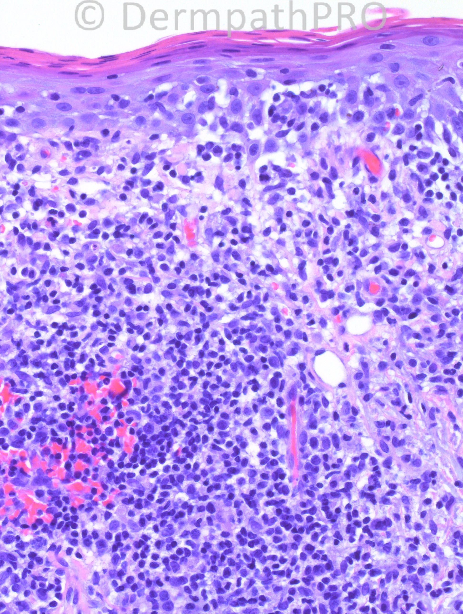
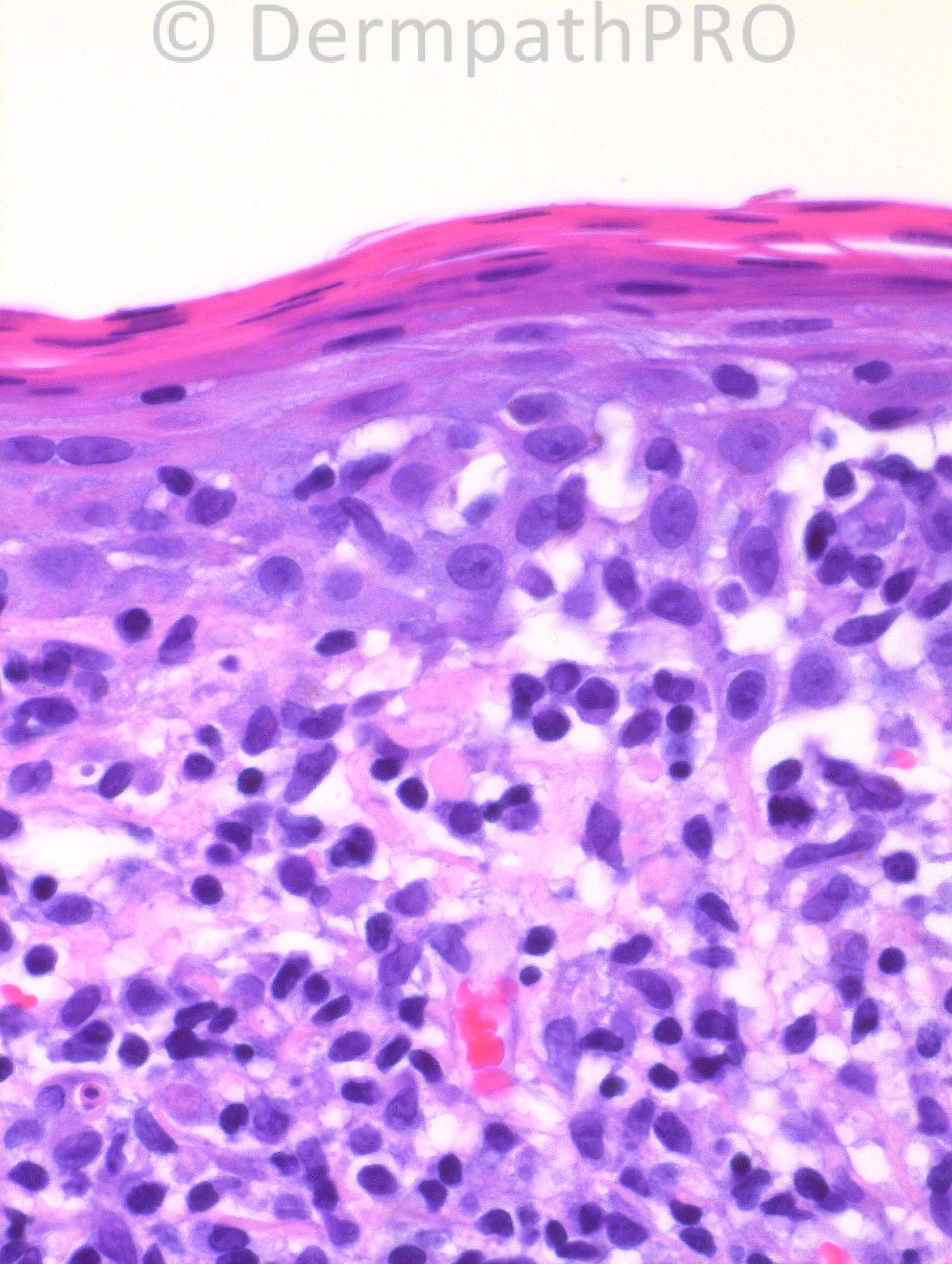
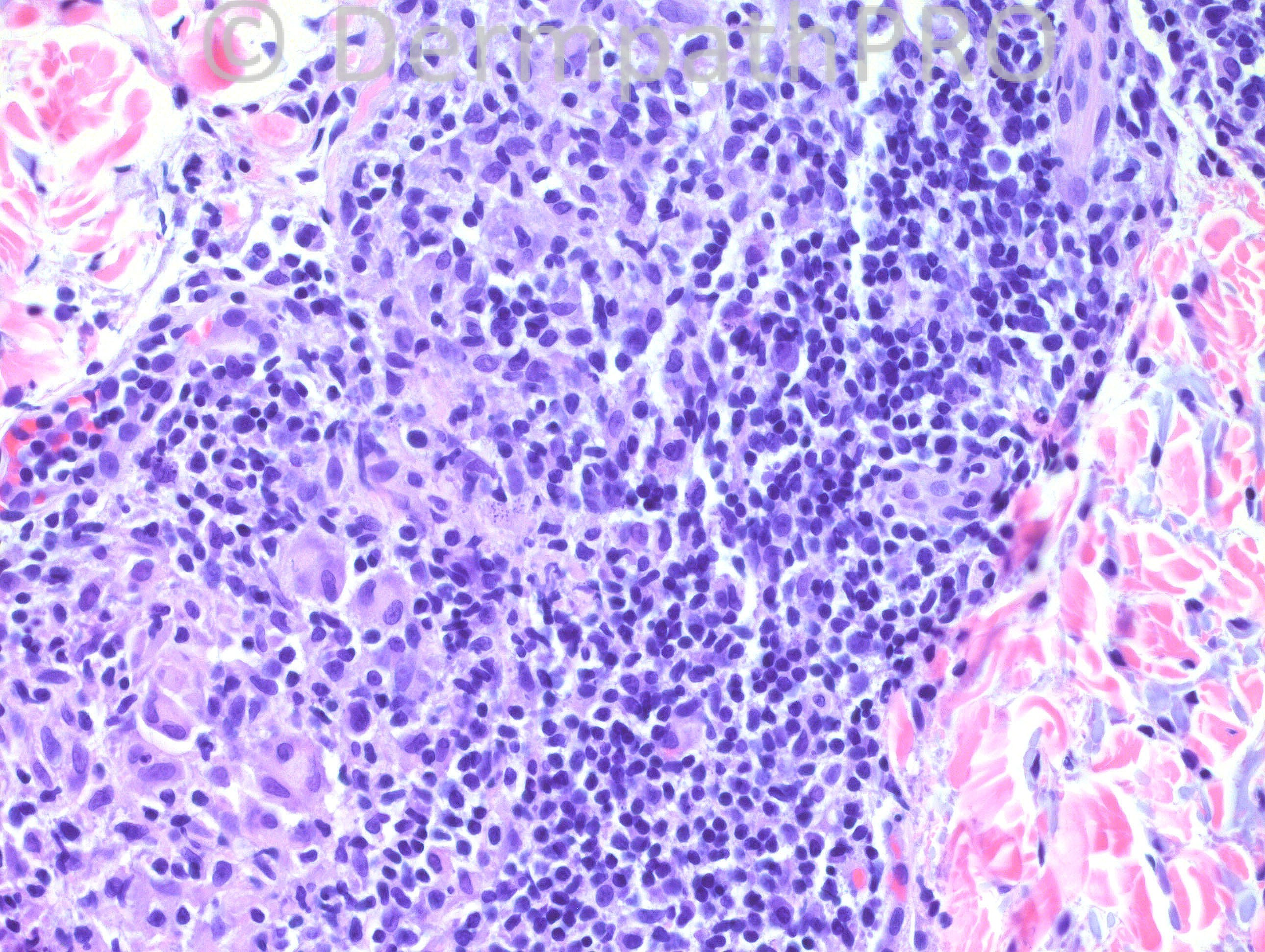
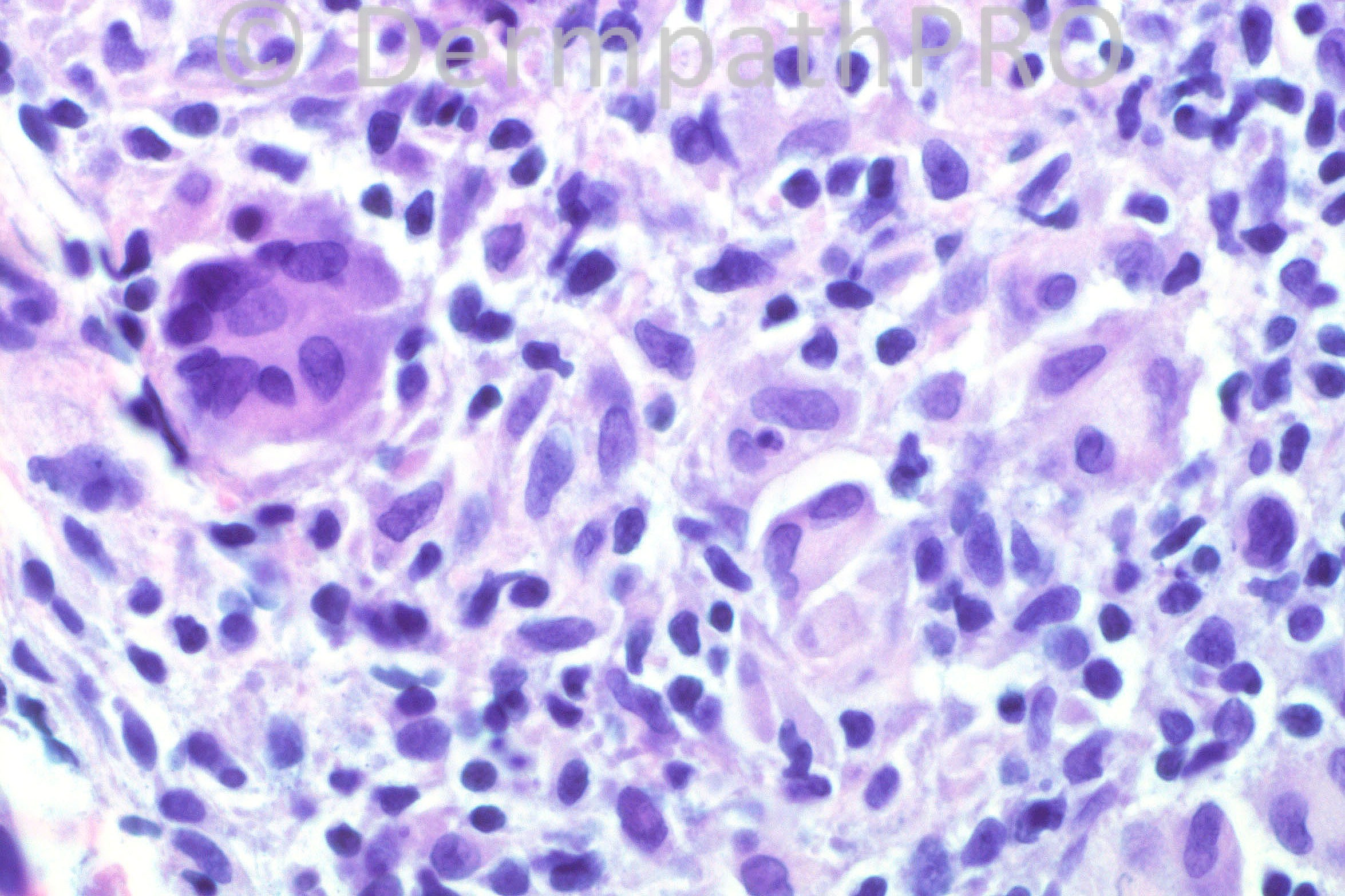
73-year-old male with a history of recurring granulomatous and necrotic lesions of uncertain etiology. A thigh biopsy from 2008 was read as “lichenoid and granulomatous dermatitis with eosinophils,” and other biopsies (from arm, trunk, and thigh) from 2009 as spongiotic dermatitis with eosinophils. On 1/11/2013, two biopsies were performed: one from the back, which was a “mature” lesion, and another from the right arm, which was a “new” lesion. The one that is shown here is the new lesion from the arm. The “mature” lesion showed ulceration, and had a more dense lymphocytic and histiocytic proliferation.

Case posted by Dr. Hafeez Diwan


  Report Record

User Feedback


Guest Hazem Hamed

Posted

[color=#403838][font=Arial][size=3]This is a difficult case. Given the patient’s clinical history, I would be concerned about the features present in the biopsy and I would like to perform a panel of immuno (CD30, [/size][/font][/color][color=#403838][font=Arial][size=3]CD3, CD8, granzyme B, perforin, TIA-1, CD2, CD4, CD5, CD7) and possibly to do a T cell receptor gene arrangement to exclude[b][font=Arial][b][i] primary cutaneous aggressive epidermotropic CD8[sup]+[/sup] cytotoxic T-cell lymphoma (though epidermotropism is not very prominent) before considering any other differential.[/i][/b][/font][/b][/size][/font][/color]

Share this comment


Link to comment
share_externally

? granulomatous MF. Needs immunohistochemical studies as suggested above.

Share this comment


Link to comment
share_externally

Eman El-Nabarawy

Posted

Granulomatous MF for Immunos.

Share this comment


Link to comment
share_externally

Guest Dr Engin Sezer

Posted

MF/dug-induced pseudolymphoma

Share this comment


Link to comment
share_externally

Robledo F. Rocha

Posted

The history of recurrent necrotic lesions suggests lymphomatoid papulosis. The microscopic feature of epidermotropic cerebriform lymphocytes makes me think of Type B (mycosis fungoides-like).

Share this comment


Link to comment
share_externally

Dr. Mona Abdel-Halim

Posted

I would have loved to c the scanning power. Recurrent necrotic lesions will make me think of LyP. Granulomatous reaction of different kinds have been reported with LyP. Agree with Robeldo, that this could be type B because of the focal epidermotropism. Will ask for CD30.

Share this comment


Link to comment
share_externally

Dr. Hafeez Diwan

Posted

I called this granulomatous mycosis fungoides. There was predominance of CD4 over CD 8. CD30 was increased. Both lesions had the identical T cell receptor gamma rearrangement.

Share this comment


Link to comment
share_externally


×
×
  • Create New...