Jump to content
In this section we have spot diagnoses posted on a daily basis since June 2010, now over 4000! You can review the archived cases and read the suggested diagnoses by users and the final comment by the contributors.
Case are uploaded each week day by 10 am UK time with the correct diagnosis will generally be posted at 8 pm UK time. Why not view the most recent spot diagnosis and proffer a diagnosis?

Case Number : Case 702 - 22 Feb Posted By: Guest

Please read the clinical history and view the images by clicking on them before you proffer your diagnosis.
Submitted Date :
   (0 reviews)

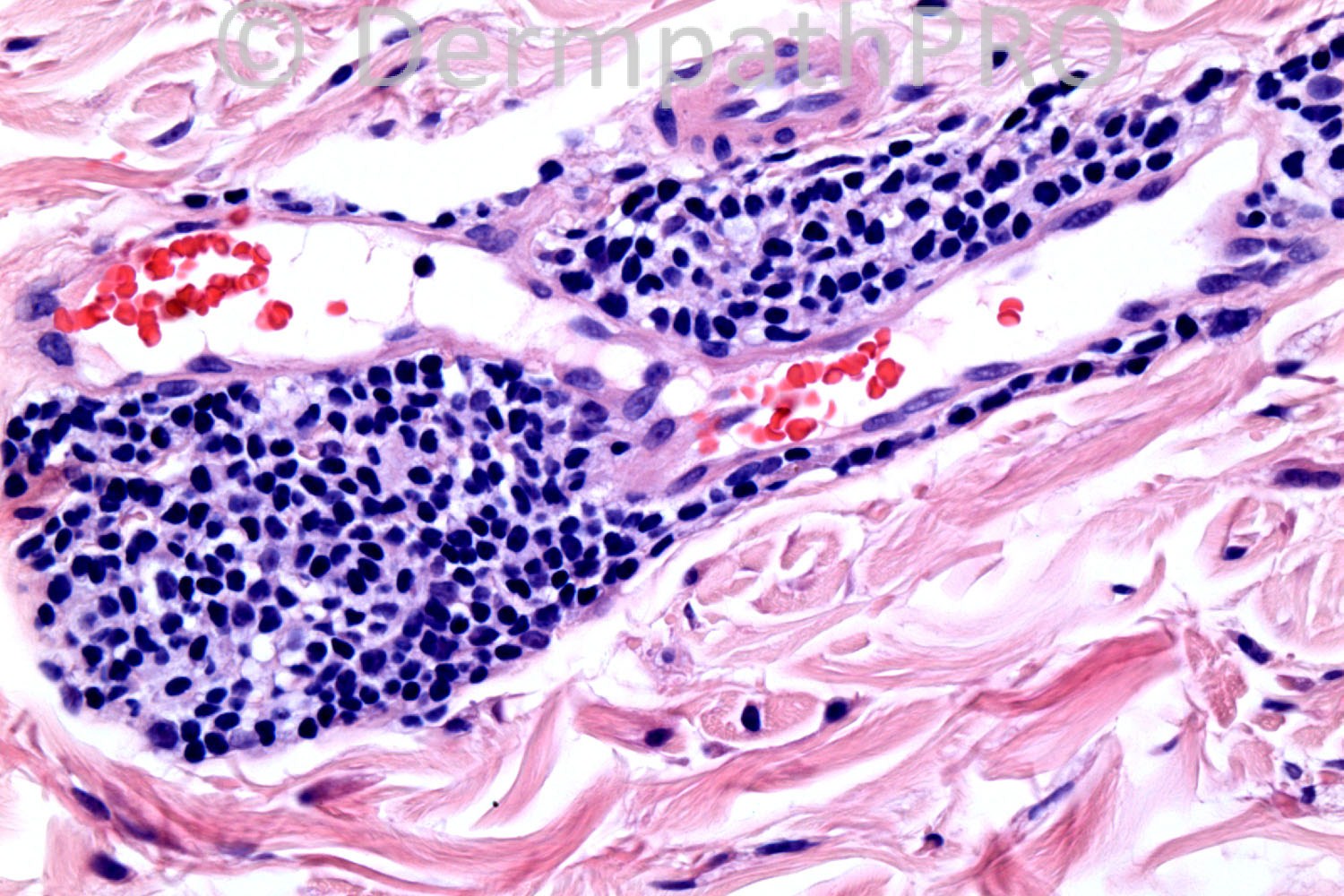
61 years-old male. Left clavicular. yellowish flat plaques with some erythema Previous biopsy in 2009 from lower eyelid suggestive of xanthelasma ?amyloidosis, ?xanthelasma ?plane xanthoma.

Case posted by Dr. Richard Carr, courtesy of Dr. Saleem Taibjee.


  Report Record

User Feedback


Eman El-Nabarawy

Posted

I think there r extracellular lipids (photo 4) suggestive of planar xanthomata. I think there r atypical lymphocytes (photo 4) so search for associated multiple myeloma is required.

Share this comment


Link to comment
share_externally

Dr. Mona Abdel-Halim

Posted

Planar xanthoma..

Share this comment


Link to comment
share_externally

Guest Bansal_

Posted

Planar xanthoma and ?atypical lymphoid infiltrate.

Share this comment


Link to comment
share_externally

Robledo F. Rocha

Posted

Plane xanthoma is an excellent diagnosis, mainly because, in addition to discrete interstitial foamy macrophages in papillary dermis, perivascular lymphocytic infiltrate is also found.
The patient is unlikely to have elevated serum lipids, but it’s advisable a careful search for associated lymphoproliferative disorder.

Share this comment


Link to comment
share_externally

nick turnbull

Posted

Agree, Plane xanthoma. would look for sytemic disease including lymphoproliferative disorders especially as multiple plaques, on the trunk. I also might check for diabetes, dylipidemia, FMHx of premature CAD and cholestasis disorders.

Share this comment


Link to comment
share_externally

Hanan Vaknine MD

Posted

[font="Revival565BT-Roman"][size="2"][font="Revival565BT-Roman"][size="2"][font="Revival565BT-Roman"][size="2"][font="Revival565BT-Roman"][size="2"]diffuse (generalized) plane xanthoma[/size][/font][/size][/font][/size][/font][/size][/font]
[font="Revival565BT-Roman"][size="2"][font="Revival565BT-Roman"][size="2"] [/size][/font][/size][/font]

Share this comment


Link to comment
share_externally

Hanan Vaknine MD

Posted

In addition to xanthoma cells and perivascular infiltration by lymphocytes one can also see fibroblast proliferation (figure 2), an additional histologic clue to the diagnosis of diffuse plane xanthoma

Share this comment


Link to comment
share_externally

Dr. Richard Carr

Posted

The diagnosis is plane xanthomatosis and CLL (B-cells on IHC)
Regards to all and enjoy the weekend.

Share this comment


Link to comment
share_externally


×
×
  • Create New...