Jump to content
In this section we have spot diagnoses posted on a daily basis since June 2010, now over 4000! You can review the archived cases and read the suggested diagnoses by users and the final comment by the contributors.
Case are uploaded each week day by 10 am UK time with the correct diagnosis will generally be posted at 8 pm UK time. Why not view the most recent spot diagnosis and proffer a diagnosis?

Case Number : Case 815 - 1st August Posted By: Guest

Please read the clinical history and view the images by clicking on them before you proffer your diagnosis.
Submitted Date :
   (0 reviews)

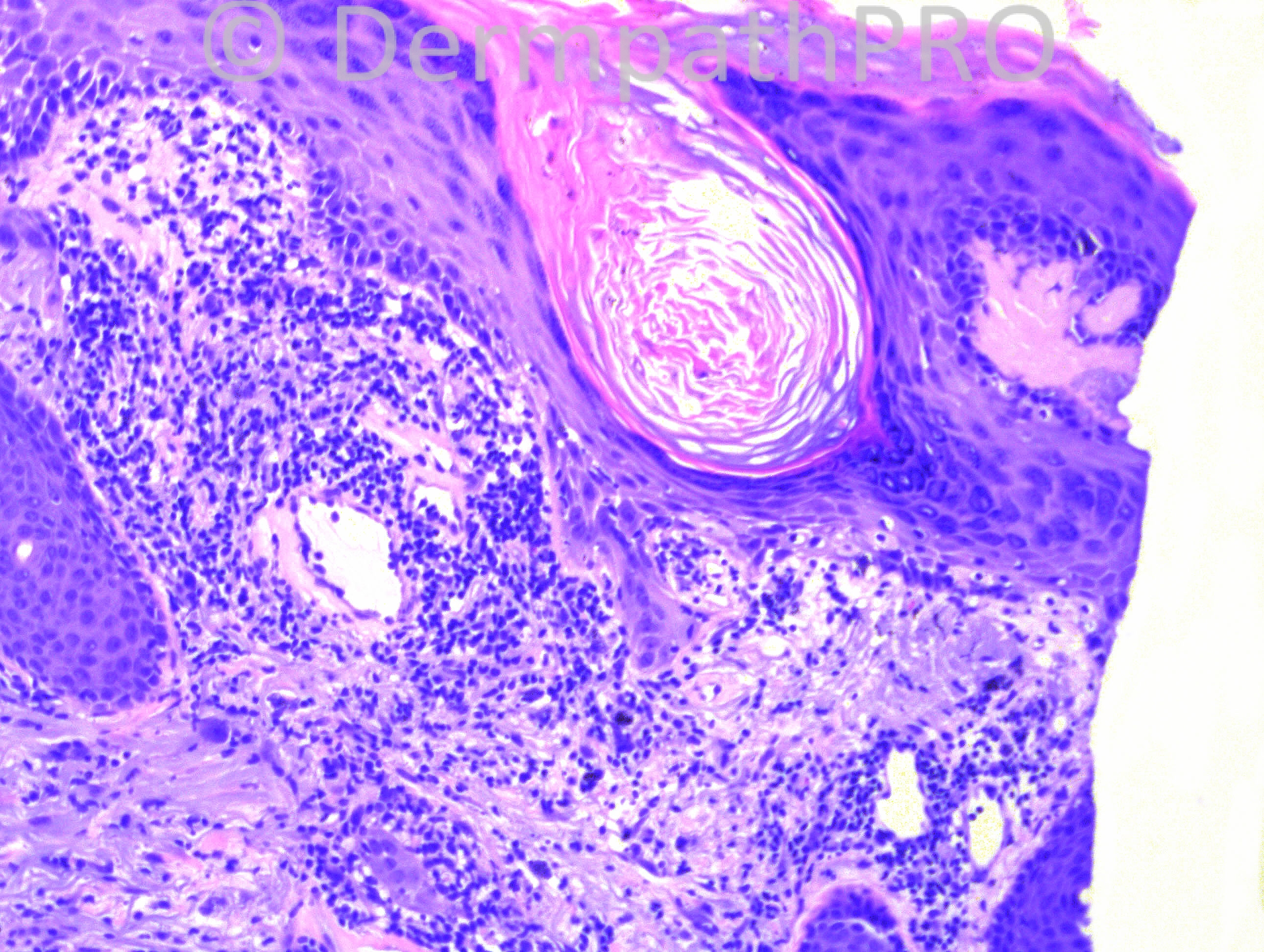
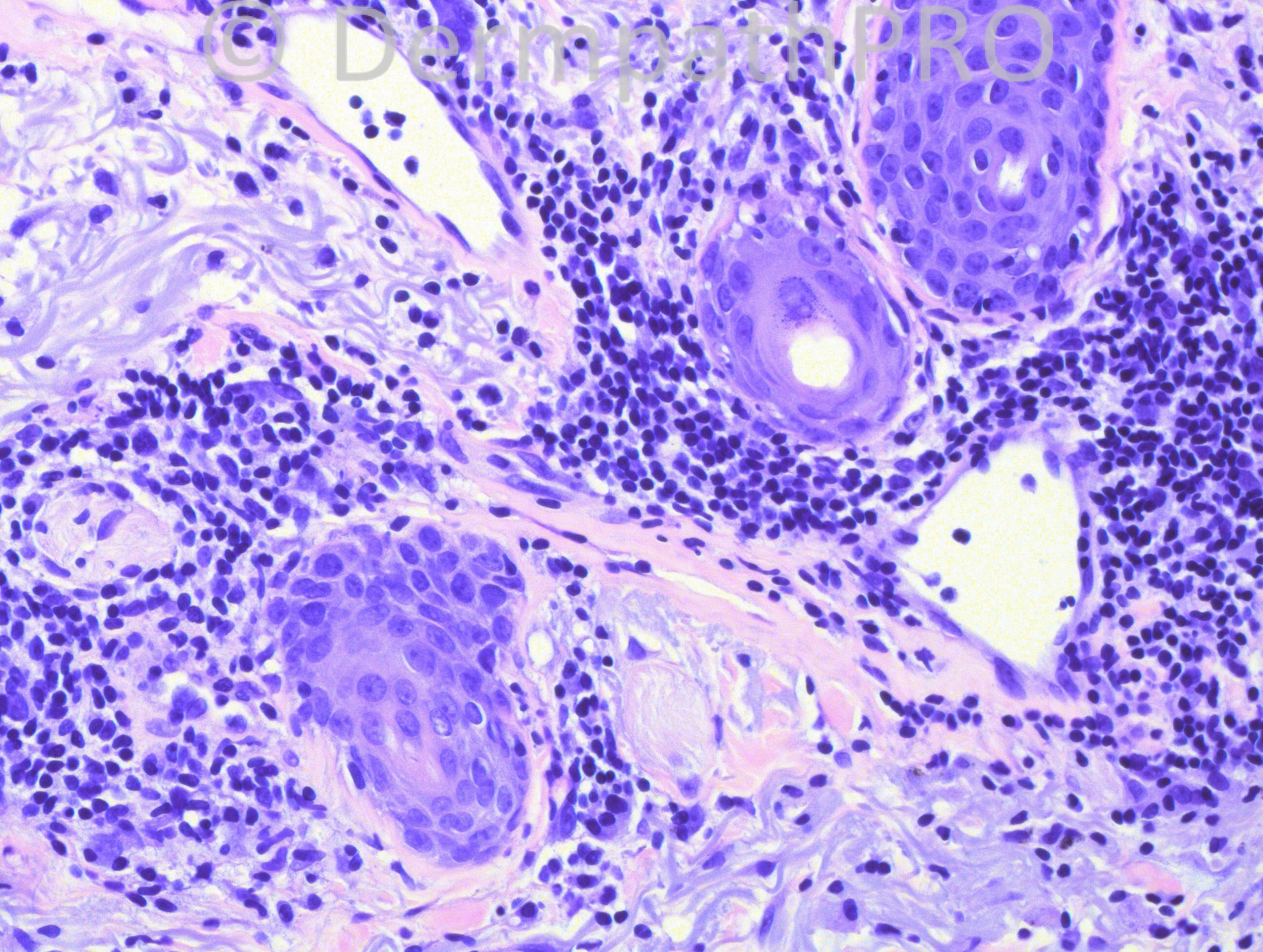
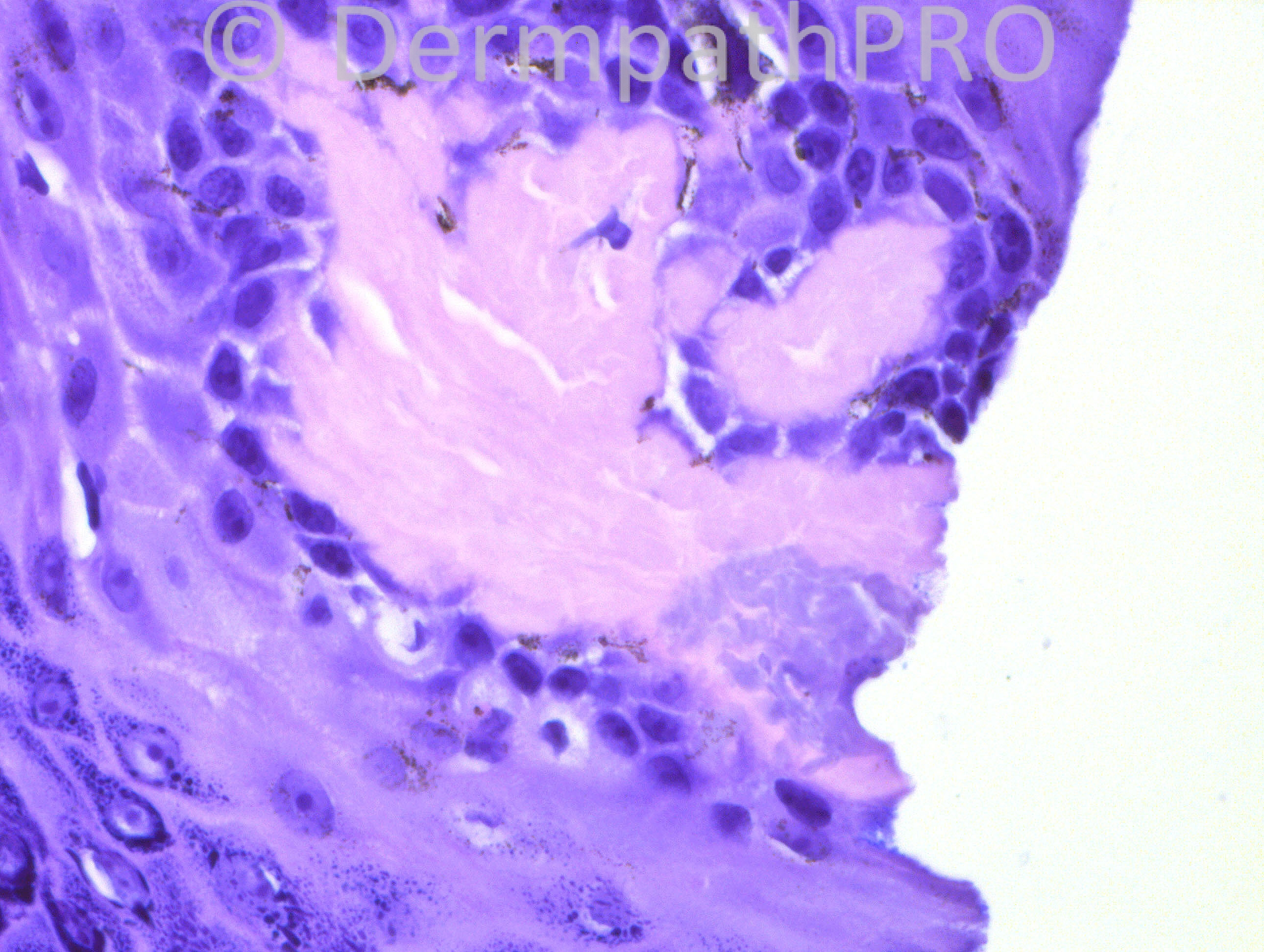
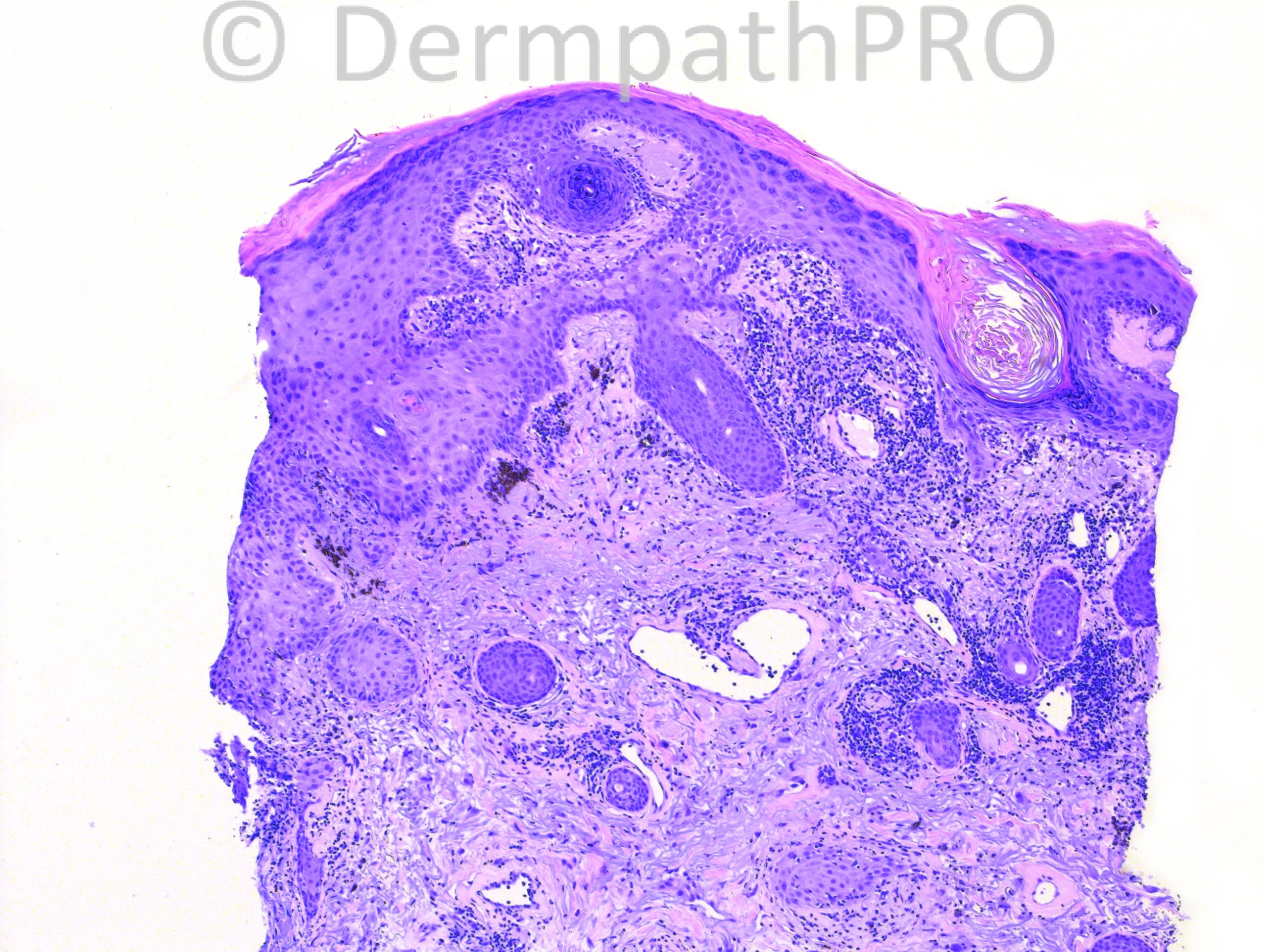
63 year old Hispanic female with lesion on her upper lip.

Case posted by Dr. Hafeez Diwan.

  Report Record

User Feedback


Robledo F. Rocha

Posted

Chronically sun-damaged skin with a large and clefted homogeneous hyaline deposit widening a dermal papilla. I favor colloid milium of the adult until Congo red and PAS stains become available.

Share this comment


Link to comment
share_externally

Guest Romualdo

Posted

Discoid lupus erythematosus with secondary amyloidosis.

Share this comment


Link to comment
share_externally

Guest Jim Davie MD

Posted

[size=2][i]I think there is something wrong with access to this case. It shows up on the right side in the SPOT DIAGNOSIS [b]Categories folder [/b](Main level) as the latest entry, but doesn't show up in the subfolder for [b]August 2013[/b].[/i][/size]

Share this comment


Link to comment
share_externally

Guest Jim Davie MD

Posted

Hypertrophic DLE seems a reasonable consideration, given photo limitations, but would obviously need clinical context/history.

The features of prurigo nodularis, nodular solar elastosis, and tangential orientation complicate the interpretation. The hyaline changes in the papillary dermis seem accentuated by the tangential section, and may represent either thickened BM (going along the DLE / chronic interface dermatitis pathway), localized amyloidosis, or just fibrosis secondary to prurigo changes. The notched basal aspect of the keratinocytes, immediately adjacent solar elastosis, and the absence of melanophages inside the hyaline areas imply the latter (fibrosis) to me. There is postinflammatory hyperpigmentation and focal atrophy of epidermis.

Share this comment


Link to comment
share_externally

Dr. Hafeez Diwan

Posted

Yes, I called it discoid lupus erythematosus as well. This went along with the clinical impression.

Share this comment


Link to comment
share_externally

Robledo F. Rocha

Posted

An oblique section played a trick on me.

Share this comment


Link to comment
share_externally


Join the conversation

You can post now and register later. If you have an account, sign in now to post with your account.

Guest
Add a comment...

×   Pasted as rich text.   Paste as plain text instead

  Only 75 emoji are allowed.

×   Your link has been automatically embedded.   Display as a link instead

×   Your previous content has been restored.   Clear editor

×   You cannot paste images directly. Upload or insert images from URL.

Loading...

×
×
  • Create New...