Jump to content
In this section we have spot diagnoses posted on a daily basis since June 2010, now over 4000! You can review the archived cases and read the suggested diagnoses by users and the final comment by the contributors.
Case are uploaded each week day by 10 am UK time with the correct diagnosis will generally be posted at 8 pm UK time. Why not view the most recent spot diagnosis and proffer a diagnosis?

Case Number : Case 816 - 2nd August Posted By: Guest

Please read the clinical history and view the images by clicking on them before you proffer your diagnosis.
Submitted Date :
   (0 reviews)

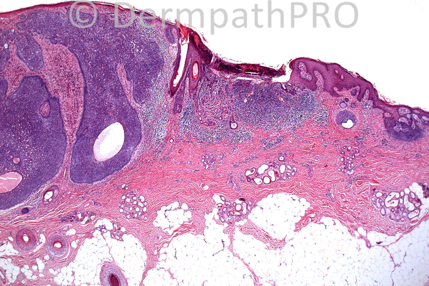
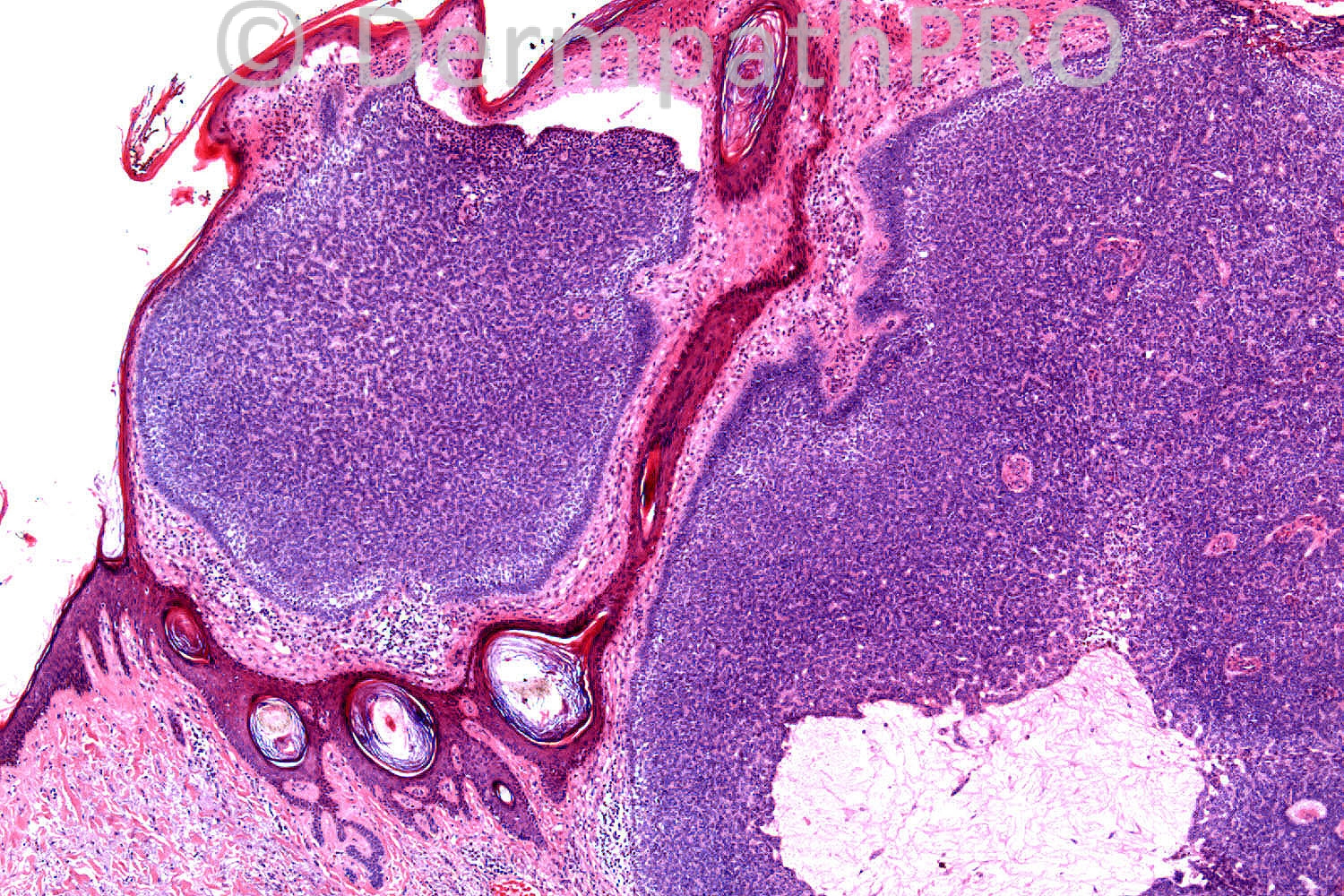
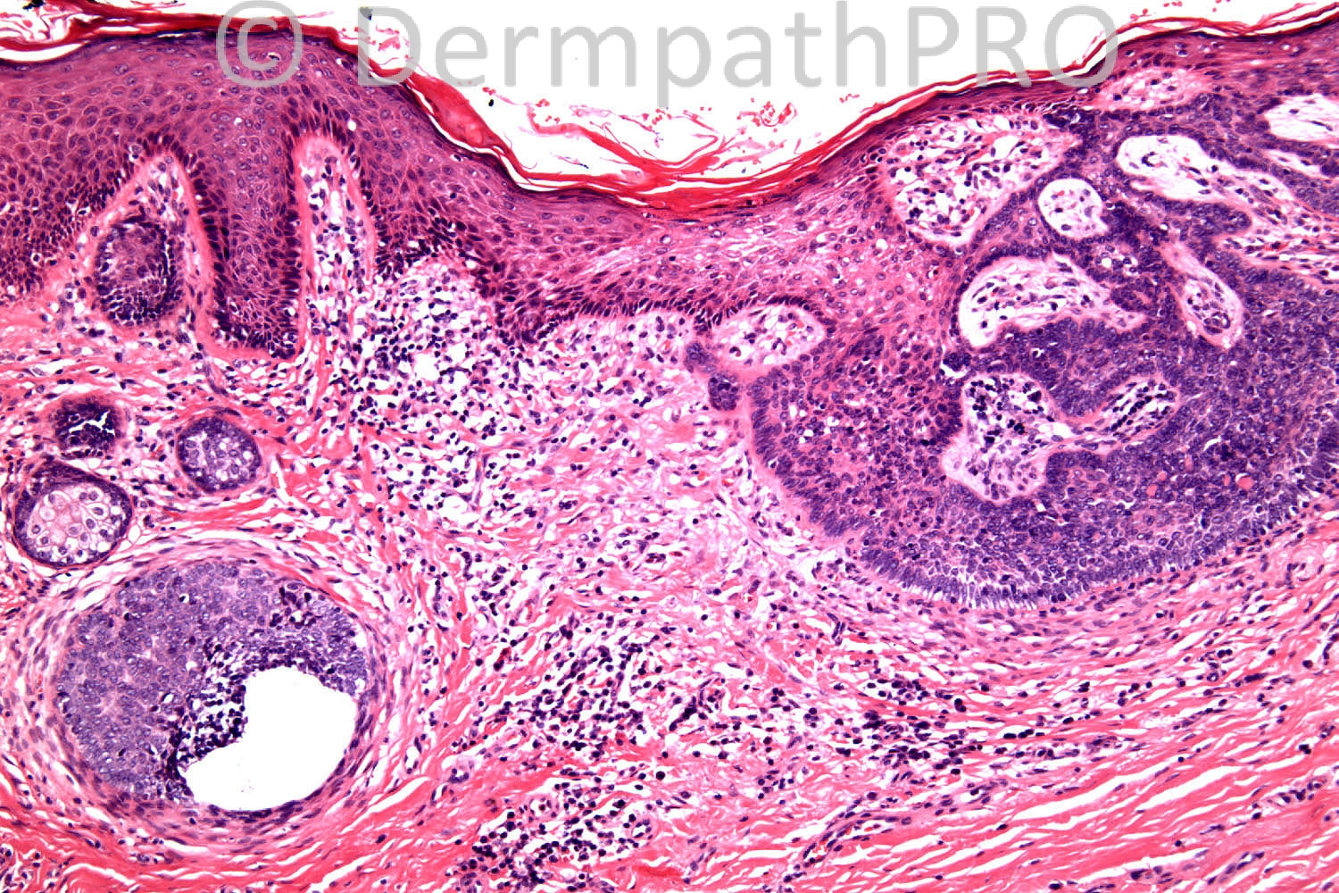
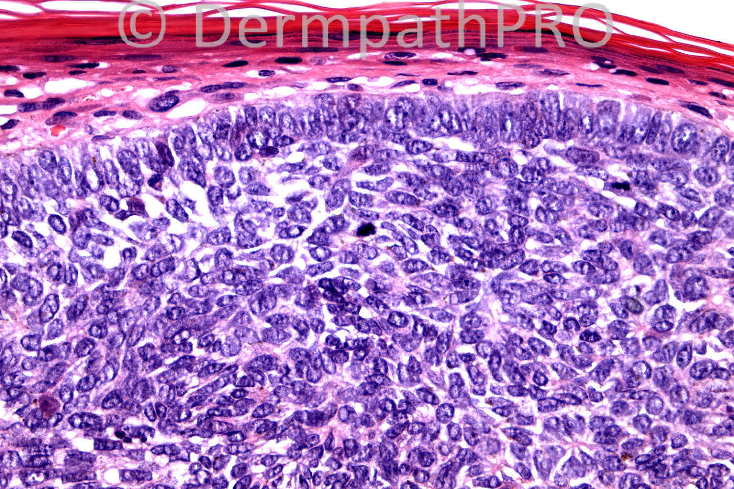
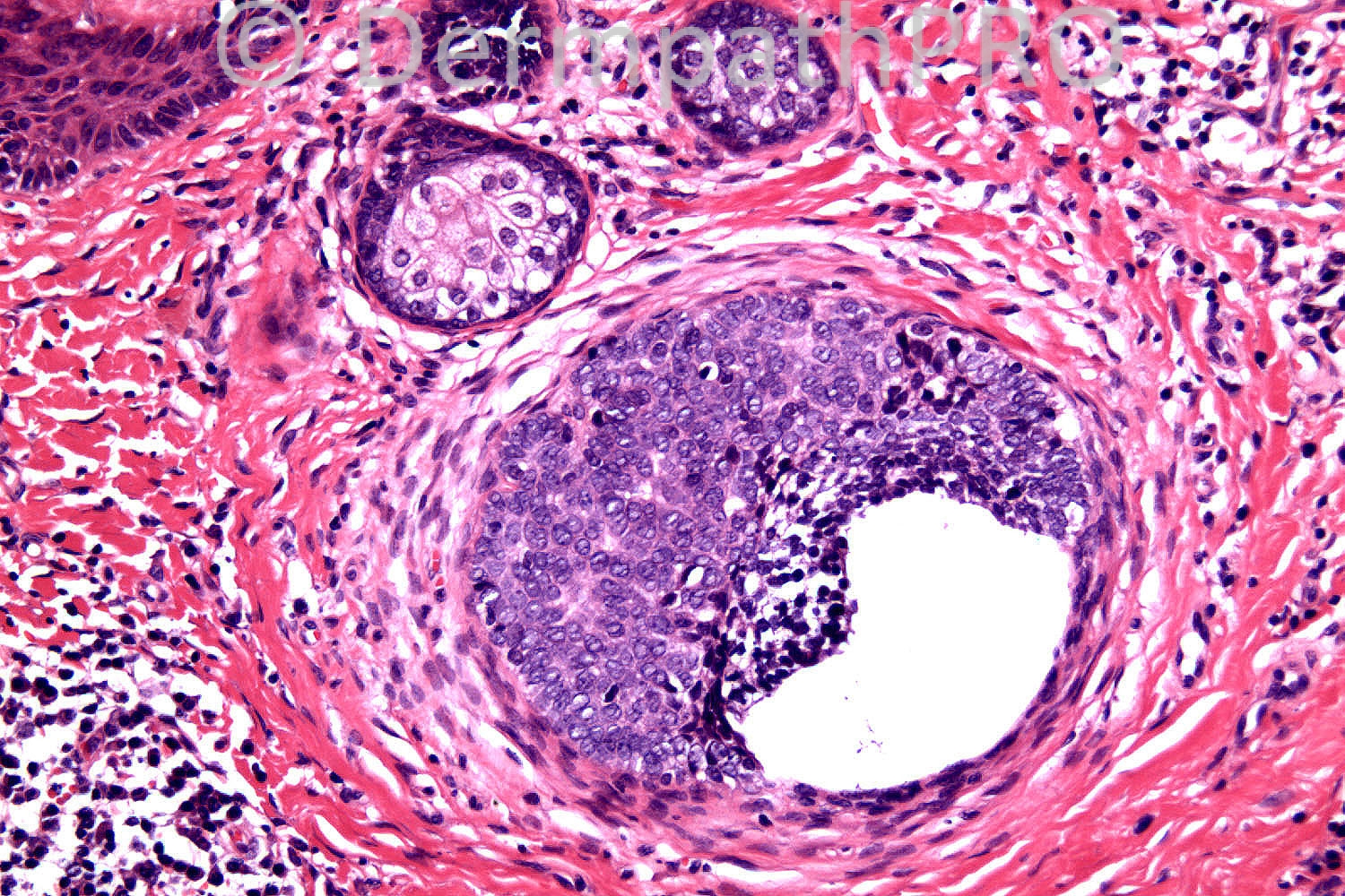
60 years old female. 2years duration, 15x10mm clinically bodular BCC.

Case posted by Dr. Richard Carr.


  Report Record

User Feedback


Guest Maria George

Posted

Hidraacanthoma simplex/poroma simulating both clinically and histopathologically BCC.Amazing.

Share this comment


Link to comment
share_externally

Sasi Attili

Posted

I would have quickly reported this as a BCC. But am a bit puzzled because you wouldn't put up this case if it was just an ordinary BCC. But I am not sure what is special about this tumour. The cells around the basaloid nest in the 5th image look a bit misplaced. ?Nerve i.e. intraneural BCC invasion?

Share this comment


Link to comment
share_externally

Robledo F. Rocha

Posted

I think it’s a collision tumor. On the left of the picture #1, a basal cell carcinoma showing typical peripheral peripheral palisading (picture #6), mucin-filled cyst and slit-like retraction artifact (picture #2), but an unusual arrangement of the basaloid cells within the neoplastic lobules reminiscent of a sebaceous tumor (picture #4). On the right of the picture #1, an early fibroepithelioma of Pinkus consisting of narrow strands of basaloid cells connected to the overlying epidermis (picture #3) and surrounded by a cellular stroma (picture #6).

Share this comment


Link to comment
share_externally

Dr. Richard Carr

Posted

Getting a little warmer. Excision was from the temple.

Share this comment


Link to comment
share_externally

Wolter Mooi

Posted

I would consider trichoblastoma arising in naevus sebaceus. Are the large-caliber sweat glands (left top picture) apocrine ones? How about CK20 (for Merkel cells), EMA, CD34 and BerEp4 immunoreactivities? Or do you feel the H&E is distinctive enough :)?

Share this comment


Link to comment
share_externally

Guest Romualdo

Posted

I completely agree with Dr. Mooi: nodular trichoblastoma arising in a sebaceous nevus. The small superficial basaloid proliferations seen in some images and the superficial location of some sebaceous lobules are sugestive of a sebaceous nevus.

Share this comment


Link to comment
share_externally

Dr. Richard Carr

Posted

This could be a double bluff. Also no-one commented on the non-basaloid central superficial lesion in Image 1 so far (you may need to imagine a high power though).

Share this comment


Link to comment
share_externally

Guest Jim Davie MD

Posted

Agree: Nevus sebaceus. The terminal hair follicles are replaced one-for-one with evenly spaced apocrine coils (a favourite clue for nevus sebaceus in scalp). Superficial sebaceous lobules and epidermal nevus changes in epidermis. A basal cell carcinoma (or trichoblastoma simulant) may arise in this context. I think it is valid BCC given high mitotic activity (in upper right image), size, and reasons cited above. If mitotic activity is an illusion, I will readily favor trichoblastoma.

The non-basaloid central superficial lesion Richard was asking about, is heavily ulcerated, but the lichenoid inflammation and hint of pseudovillous architecture might be a syringocystadenoma papilliferum in the context of a nevus sebaceus.

Share this comment


Link to comment
share_externally

Dr. Richard Carr

Posted

Okay you have got there now - thanks Jim for putting the icing on the cake. There was also a basaloid tricholemmomatous area not discernible in these images but staining with CD34 and PAS (basement membrane) that blended with both the superficial trichoblastoma (right of image 1, image 3 and image 5). In image 5 the retraction is between the germinative basaloid epithelium and the subtle compressed papillary mesenchymal cells (note no mucin). For me the larger expansile basaloid lobules (left of image 1, image 4 and 6) is morphologically indistinguishable from BCC (I will refer to is as BCC-like areas). There is indeed a little syringocystadenoma in image 1 (central) and it was much nicer in other blocks but I deliberately did not make it easy for you. You should also note the epidermal hyperplasia / acanthosis and as has been pointed out absence of terminal folliles and presence of abnormal sweat glands and even effete / odd sebaceous units as the clues to naevus sebaceous.

Regarding immunos it was interesting as there appeared to be two distint patterns with Merkel cells, CD10 stromal / papillary mesenchymal staining and peripheral Bcl2 in the superficial trichoblastoma but surprisingly widespread epithelial CD10 (stroma absent and no PMCs), CK20 negative for MC and diffuse Bcl2 in the nodular BCC-like areas. On that basis immunos favour a BCC for the latter areas.

Prior to 2000 BCC was believed to be the commonest basaloid tumour in NS. But in the 2000's it was considered that the vast majority of lesions interpreted as BCC previously were in fact trichoblastomas. I have now seen several cases in which morphologically and immunophenotypically the case favoured BCC but to me is morphologically indistinguishable from some large nodular trichoblastomas also seen in NS (with nice papillary mesenchymal cells). I was therefore interested in the following reference (but I am a sceptic as I have not seen any really nasty BCC in naevus sebaceous so far):

Sellheyer K, Cribier B, Nelson P, Kutzner H, Rütten A. Basaloid tumors in nevus sebaceus revisited: the follicular stem cell marker PHLDA1 (TDAG51) indicates that most are basal cell carcinomas and not trichoblastomas. J Cutan Pathol. 2013 May;40(5):455-62. doi: 10.1111/cup.12107. Epub 2013 Mar 14.

So I guess you can all decide for yourselves which hypothesis you prefer. Provided the lesion is excised I guess it is entirely immaterial. You might want to check out the adidtional papers on PHLDA1 discussing DTE! and TE v's BCC.

Enjoy your weekends.

Share this comment


Link to comment
share_externally

So how was this case finally signed out as? could we know? did the patient report the presence of a lesion since birth or childhood consistent with a nevus sebaceus?

Share this comment


Link to comment
share_externally


Join the conversation

You can post now and register later. If you have an account, sign in now to post with your account.

Guest
Add a comment...

×   Pasted as rich text.   Paste as plain text instead

  Only 75 emoji are allowed.

×   Your link has been automatically embedded.   Display as a link instead

×   Your previous content has been restored.   Clear editor

×   You cannot paste images directly. Upload or insert images from URL.

Loading...

×
×
  • Create New...