Jump to content
In this section we have spot diagnoses posted on a daily basis since June 2010, now over 4000! You can review the archived cases and read the suggested diagnoses by users and the final comment by the contributors.
Case are uploaded each week day by 10 am UK time with the correct diagnosis will generally be posted at 8 pm UK time. Why not view the most recent spot diagnosis and proffer a diagnosis?

Case Number : Case 817 - 5th August Posted By: Guest

Please read the clinical history and view the images by clicking on them before you proffer your diagnosis.
Submitted Date :
   (0 reviews)

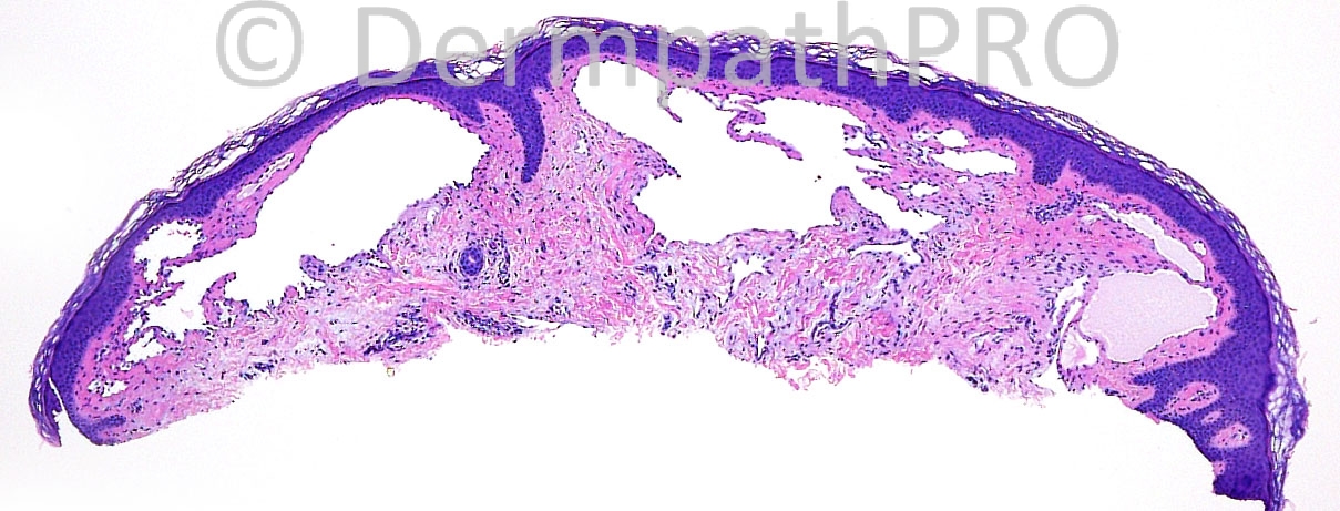
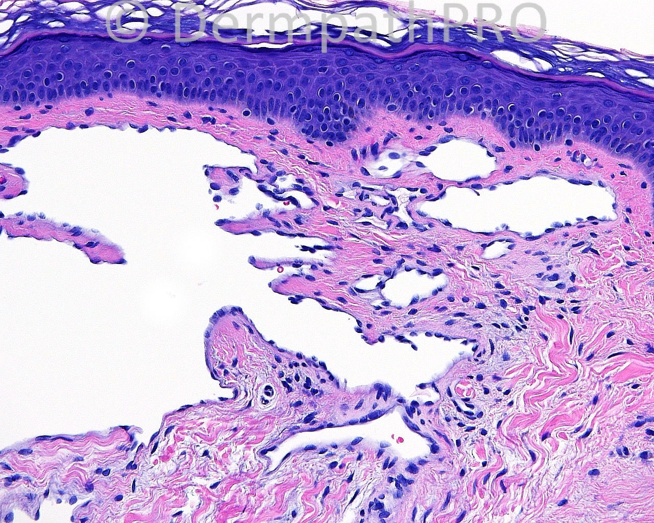
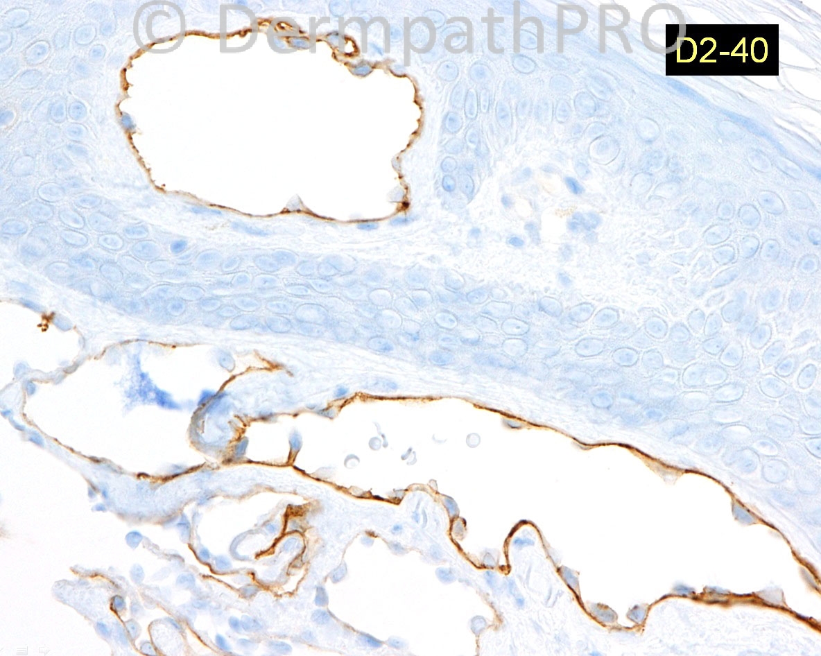
26 years old female with a shave biopsy of a 1cm skin colored, multi-focal, vaguely vesicular papule present since childhood, taken from the right buttock.

Case posted by Dr. Mark Hurt.


  Report Record

User Feedback


Dr. Mona Abdel-Halim

Posted

Lymphangioma circumscriptum

Share this comment


Link to comment
share_externally

Robledo F. Rocha

Posted

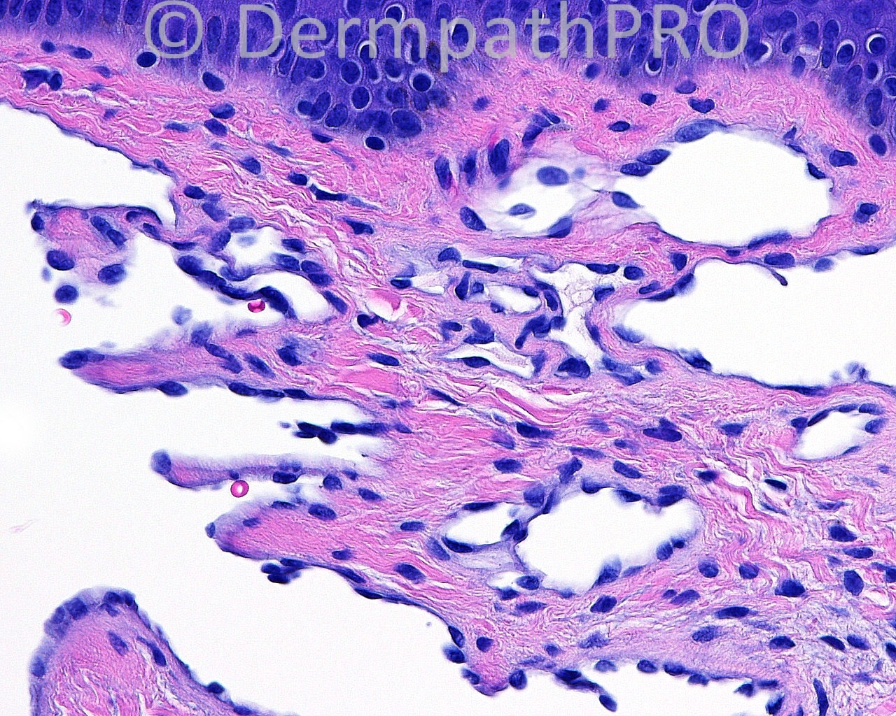
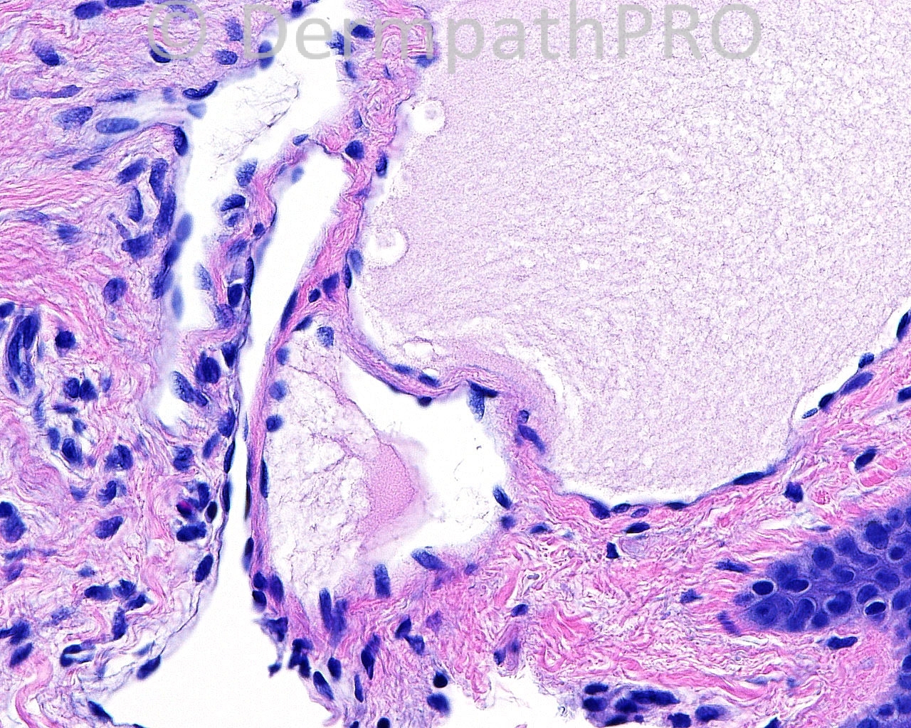
Superficial dermis shows thin-walled vascular spaces with slender papillary projections, but lacking erythrocytes in their dilated lumina. I would call this lymphangioma circumscriptum.

Share this comment


Link to comment
share_externally

Guest Graham Reilly

Posted

Agree lymphangioma circumscriptum- D2-40 stains lymphatic endothelium.

Share this comment


Link to comment
share_externally

Guest Rodrigo Restrepo

Posted

Lymphangioma circumscriptum

Share this comment


Link to comment
share_externally

Guest Maria George

Posted

As usual in medicine , there is ongoing debate for the nomenclature.
Some people prefer to call this as superficial lymphatic malformation.As the word ''circumscriptm'' is misleading because many of such lesions has no identifiable margin.
As it is known to every one deep lymphatic malformation is term given to ''cystic hygroma''.It seems that the [color=#1C2837][font=arial, verdana, tahoma, sans-serif][size=4][background=rgb(241, 246, 249)]buttock is a common site because I saw a case in this location also.[/background][/size][/font][/color]

Share this comment


Link to comment
share_externally

Sasi Attili

Posted

[color=#1C2837][font=arial, verdana, tahoma, sans-serif][size=4]Lymphangioma circumscriptum[/size][/font][/color]

Share this comment


Link to comment
share_externally

Guest Romualdo

Posted

I see some papillary projections, hobnailed cells, a tendency to horizontal orientation of the vascular spaces, some of them dissecting the dermis. I would like to suggest the diagnosis of acquired progressive lynphangioma.

Share this comment


Link to comment
share_externally

Guest Jim Davie MD

Posted

Lymphangioma circumscriptum.
This case has a classic clinical history that points to the most likely diagnosis, which is supported by the histology.
There is involvement of the papillary dermis by D2-40 positive thin walled vessels with no muscular component (although deep biopsies or excisions may show a muscular component in deeper vessels), proteinaceous fluid lacking significant red cells, and elevation of epidermal contour.

Share this comment


Link to comment
share_externally


Join the conversation

You can post now and register later. If you have an account, sign in now to post with your account.

Guest
Add a comment...

×   Pasted as rich text.   Paste as plain text instead

  Only 75 emoji are allowed.

×   Your link has been automatically embedded.   Display as a link instead

×   Your previous content has been restored.   Clear editor

×   You cannot paste images directly. Upload or insert images from URL.

Loading...

×
×
  • Create New...