Jump to content
In this section we have spot diagnoses posted on a daily basis since June 2010, now over 4000! You can review the archived cases and read the suggested diagnoses by users and the final comment by the contributors.
Case are uploaded each week day by 10 am UK time with the correct diagnosis will generally be posted at 8 pm UK time. Why not view the most recent spot diagnosis and proffer a diagnosis?

Case Number : Case 818 - 6th August Posted By: Guest

Please read the clinical history and view the images by clicking on them before you proffer your diagnosis.
Submitted Date :
   (0 reviews)

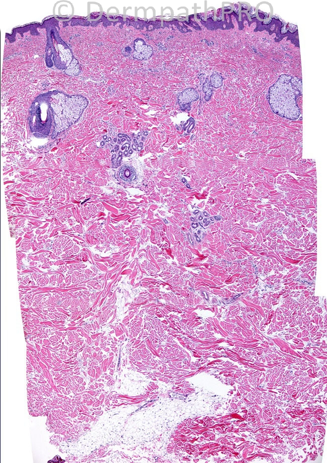
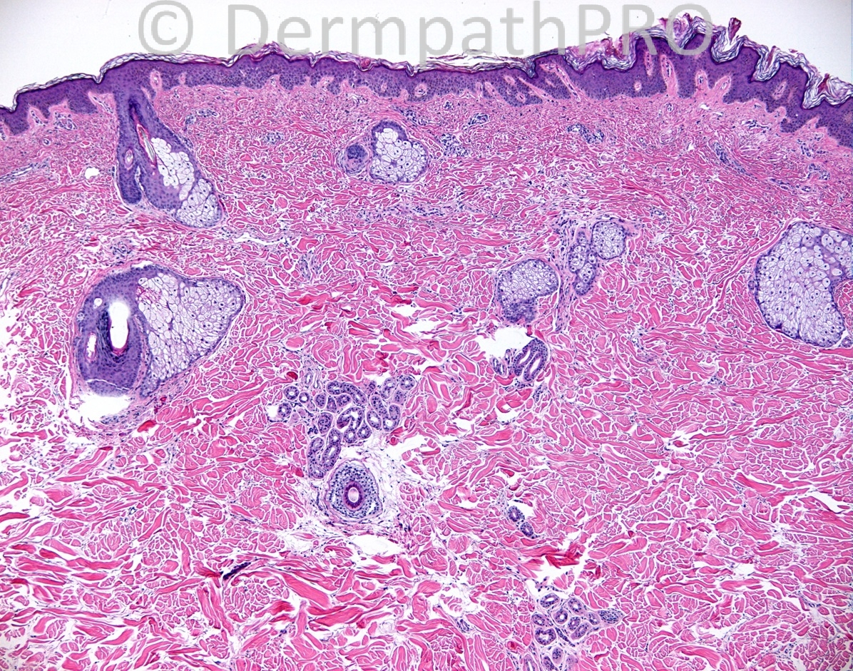
34 years old white man, carries a diagnosis of non-Hodgkin's lymphoma, and has been on chemotherapy for 3 months. He has a 1-2 years history of "tight" skin and dysphagia.

Case posted by Dr. Mark Hurt.


  Report Record

User Feedback


Arti Bakshi

Posted

[i]Agree[/i]
[i]Thickening of dermis with increased dermal mucin (appears to be more in deeper dermis), consistent with Scleredema.[/i]

Share this comment


Link to comment
share_externally

Guest Maria George

Posted

Increased fibroblast, mucin and fibrosis ends in a diagnosis of Sclermexedema which is associated with paraproteinemia.Patients usually have cardiac , neurologic and hematological complications like this patient who has lymphoma.

Share this comment


Link to comment
share_externally

Robledo F. Rocha

Posted

[font="Palatino Linotype, serif"][size="4"]Non-inflammatory dermal thickening characterized by morpheaform entrapment of eccrine sweat glands and fenestrations between sclerotic collagen bundles filled with abundant mucin deposits, highlighted by colloidal iron, in the absence of fibroblastic proliferation.[/size][/font]
[font="Palatino Linotype, serif"][size="4"]Given the case history, I would call this monoclonal gammopathy-associated scleredema.[/size][/font]

Share this comment


Link to comment
share_externally

Scleromyxedema should appear with increased fibroblasts and It also does not affects the tongue or the esophageal musculature, which could explain the dysphagic symptom. I don´t see increased fibroblasts.
I think this is a Scleredema of Buschke with its increased dermal thickness and mucin, no or few fibroblast proliferation and no inflammatory cells. Scleredema can also appear with dysphagic symptoms and can be secondary to malignant lymphoid diseases.

Share this comment


Link to comment
share_externally

Guest Rodrigo Restrepo

Posted

[color=#1C2837][font=arial, verdana, tahoma, sans-serif][size=4]Scleredema[/size][/font][/color]

Share this comment


Link to comment
share_externally

Guest Graham Reilly

Posted

Scleroedema - Thickened dermis,replacement of fat by coarse collagen bundles and mucin.I couldn`t see any fibroblast proliferation - are you supposed to see this in scleroedema?

Share this comment


Link to comment
share_externally

Dr. Mona Abdel-Halim

Posted

I do not perceive considerable fibroblast proliferation, so I also favor this to be scleredema.. Could be paraneoplastic.

Share this comment


Link to comment
share_externally

Sasi Attili

Posted

[quote name='Dr. Mona Abdel Halim' timestamp='1375793544']
I do not perceive considerable fibroblast proliferation, so I also favor this to be scleredema.. Could be paraneoplastic.
[/quote]
agree

Share this comment


Link to comment
share_externally

Guest Jim Davie MD

Posted

Scleredema.

The biopsy shows thickening of the reticular dermis (with illusion of an upwards displacement of eccrine coils from deep to mid-reticular dermis) and increased dermal mucin. No significant fibroblast increase is seen, which would be expected for the differential dx consideration of scleromyxedema.

I agree that the history of non-Hodgkin's lymphoma (with presumed absent history of infection or diabetes) and long history of sclerodermoid symptoms would favor monoclonal gammopathy as the likely cause of scleredema. Dysphagia can be seen in both scleredema and scleromyxedema.

Frequently, scleredema will present several years (median 2.5 years) before a monoclonal gammopathy can be detected by serum protein electrophoresis (SPEP) testing. A negative SPEP test therefore does not exclude gammopathy-related scleredema. (This delay may not apply to patients with previous lymphoma diagnosis).
Did this patient have a positive SPEP?

Share this comment


Link to comment
share_externally

Guest Romualdo

Posted

I agree with most of my colleagues: scleredema.

Share this comment


Link to comment
share_externally


Join the conversation

You can post now and register later. If you have an account, sign in now to post with your account.

Guest
Add a comment...

×   Pasted as rich text.   Paste as plain text instead

  Only 75 emoji are allowed.

×   Your link has been automatically embedded.   Display as a link instead

×   Your previous content has been restored.   Clear editor

×   You cannot paste images directly. Upload or insert images from URL.

Loading...

×
×
  • Create New...