Jump to content
In this section we have spot diagnoses posted on a daily basis since June 2010, now over 4000! You can review the archived cases and read the suggested diagnoses by users and the final comment by the contributors.
Case are uploaded each week day by 10 am UK time with the correct diagnosis will generally be posted at 8 pm UK time. Why not view the most recent spot diagnosis and proffer a diagnosis?

Case Number : Case 819 - 7th August Posted By: Guest

Please read the clinical history and view the images by clicking on them before you proffer your diagnosis.
Submitted Date :
   (0 reviews)

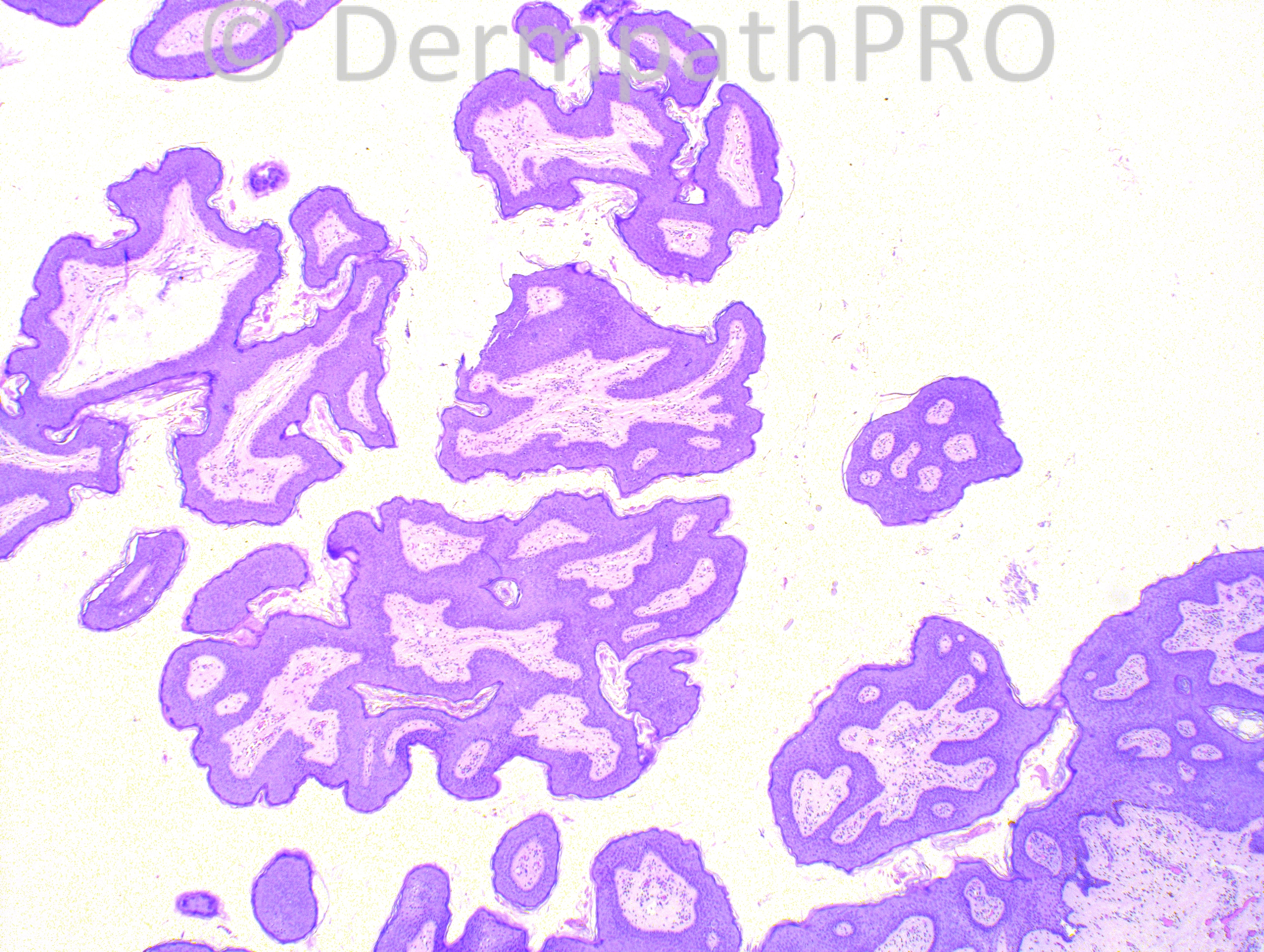
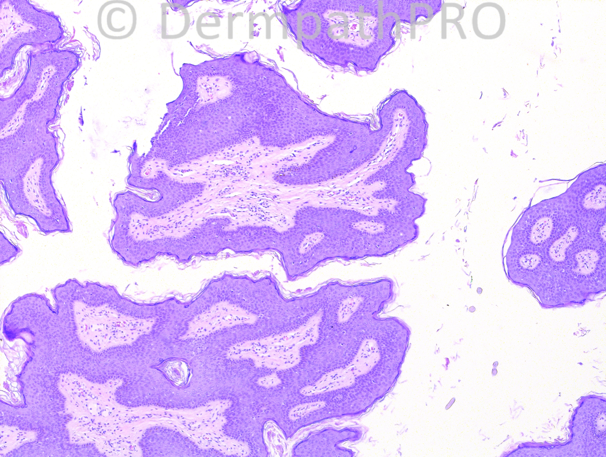
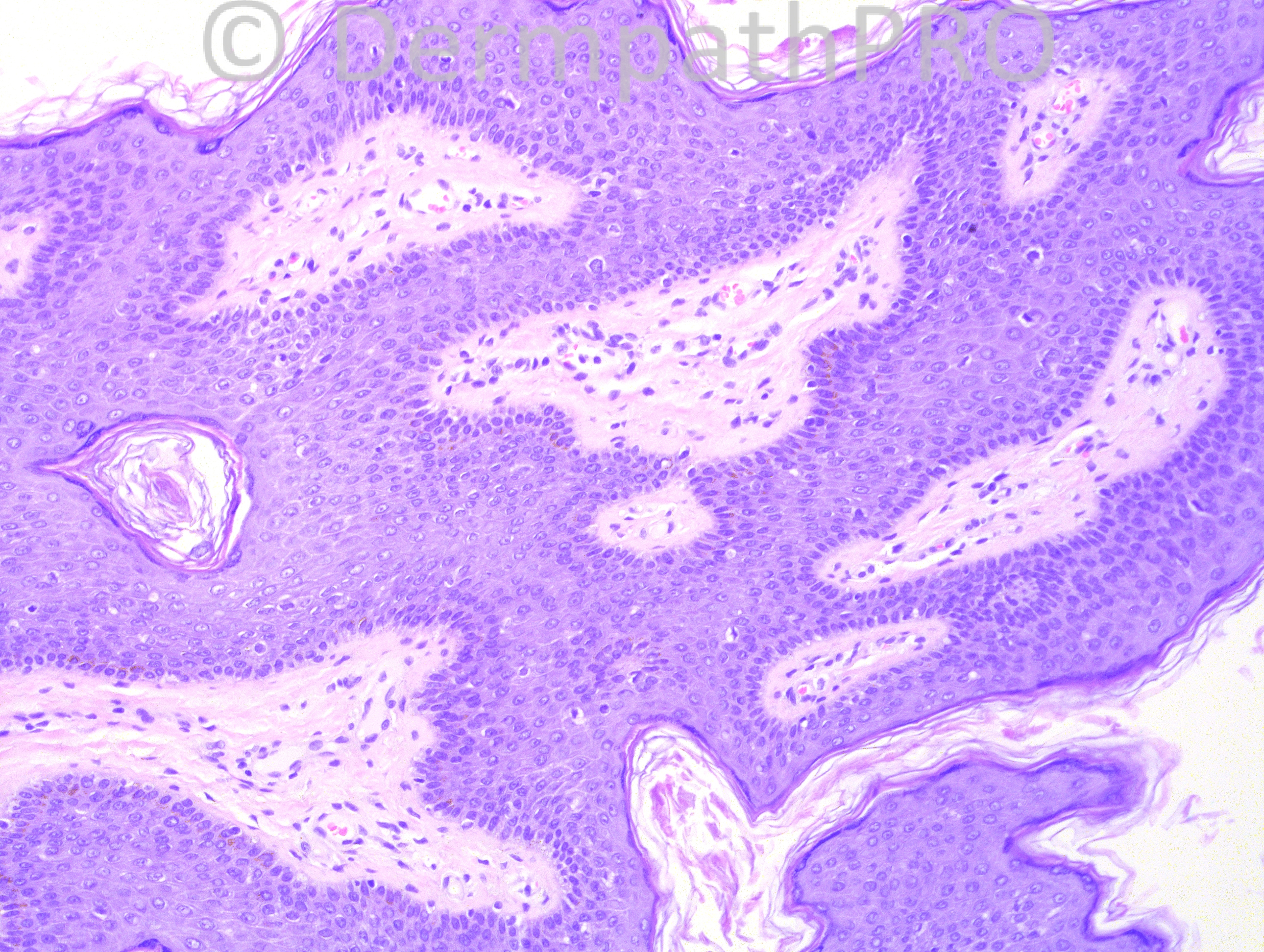
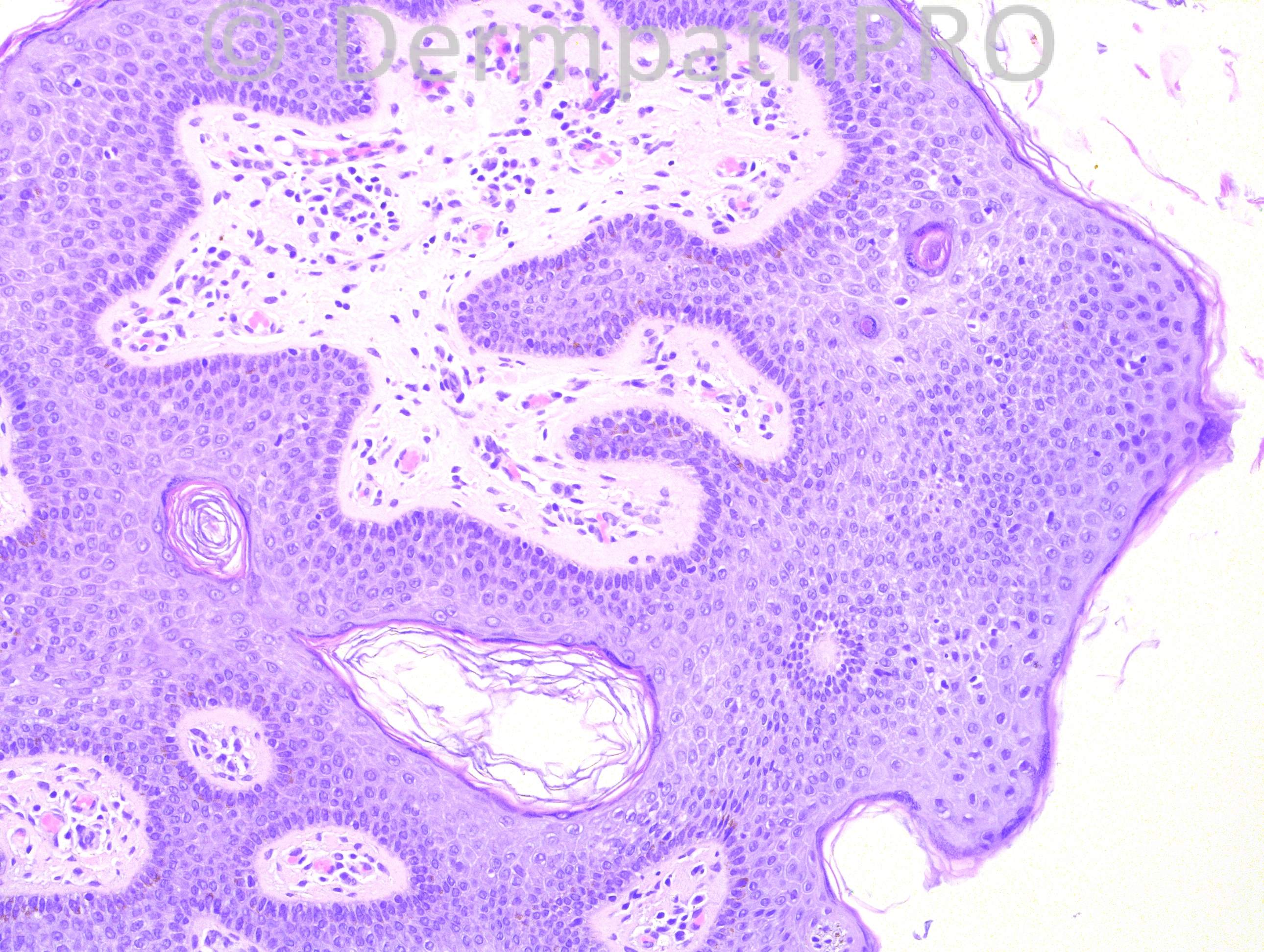
26-year-old female with a lesion in her right axilla.

Case posted by Dr. Hafeez Diwan.


  Report Record

User Feedback


My first impression was a large Acrochordon. I don´t think that there´s nothing more about this case, so or I am wrong or this is an unusual case, maybe associated with Birt-Hogg-Dubé syndrome or Diabetes mellitus. I´m looking forward to the answer.

Share this comment


Link to comment
share_externally

Sasi Attili

Posted

[quote name='IgorSC' timestamp='1375879288']
My first impression was a large Acrochordon. I don´t think that there´s nothing more about this case, so or I am wrong or this is an unusual case, maybe associated with Birt-Hogg-Dubé syndrome or Diabetes mellitus. I´m looking forward to the answer.
[/quote]

Indeed, was a bit puzzled to see what is apparently a simple case. Might be surprised!!

Share this comment


Link to comment
share_externally

Robledo F. Rocha

Posted

Agree with Igor’s diagnosis. I think the problem here is the presence of fronds artifactually apart from each others, giving the false impression that they are floating in the air.

Share this comment


Link to comment
share_externally

Guest Romualdo

Posted

Maybe Dr. Hafeez Diwan wants to show us the similarities between some polypoid seborrheic keratoses, especially when located on axillae, and fibroepitelial polyps (acrochordons). I think the papillomatosis, acanthosis and the ocasional horn cysts point to the diagnosis of papillomatous seborrheic keratosis.

Share this comment


Link to comment
share_externally

Guest Maria George

Posted

In addition to the above , I want to add Epidermal nevus .Doweling Dego is a good to think about it in axilla but for sure not in the above images.

Share this comment


Link to comment
share_externally

Arti Bakshi

Posted

What about 'Confluent and Reticulated Papillomatosis' ? (in addition to above)

Share this comment


Link to comment
share_externally

Dr. Mona Abdel-Halim

Posted

I thought of confluent and reticulated papillomatosis but I think it is not a unilateral condition. So my second possibility is a papillomatous seb K.

Share this comment


Link to comment
share_externally

Guest Jim Davie MD

Posted

I agree with the opinion of pigmented polypoid seborrheic keratosis, tangentially cut.
The differential includes acrochordon, as I think the dividing line between polypoid SK and acrochordon is somewhat subjective. I agree with Maria that epidermal nevus is a consideration, given the peculiar endophytic reticular architecture and basaloid changes (in the right-bottom corner of first photo). Tangential orientation in this case makes the differentiation between these more challenging. Doubt acanthosis nigricans (given a presumed solitary lesion, keratin cysts,and young adult age group).

Share this comment


Link to comment
share_externally

Guest Romualdo

Posted

Epidermal nevus is an excellent possibility, depending on how old the patient was when the lesion appeared. As Dr. McKee said: " It is the age of the patient that distinguishes the lesion (an epidermal nevus) from a seborrheic keratosis".

Share this comment


Link to comment
share_externally

Iskander H. Chaudhry

Posted

[font=arial, sans-serif][size=3]Thank you for the diagnoses. Dr Hafeez asked me to post the diagnosis as he is away. I note the insightful points made. The diagnosis, as suggested by Mark, is Epidermal Nevus. As you all rightly say it could easily have been an FEP or seb K if there was no relevant history given. [/size][/font]

[font=arial, sans-serif][size=3]Thank you for your contributions.[/size][/font]

Share this comment


Link to comment
share_externally


Join the conversation

You can post now and register later. If you have an account, sign in now to post with your account.

Guest
Add a comment...

×   Pasted as rich text.   Paste as plain text instead

  Only 75 emoji are allowed.

×   Your link has been automatically embedded.   Display as a link instead

×   Your previous content has been restored.   Clear editor

×   You cannot paste images directly. Upload or insert images from URL.

Loading...

×
×
  • Create New...