Jump to content
In this section we have spot diagnoses posted on a daily basis since June 2010, now over 4000! You can review the archived cases and read the suggested diagnoses by users and the final comment by the contributors.
Case are uploaded each week day by 10 am UK time with the correct diagnosis will generally be posted at 8 pm UK time. Why not view the most recent spot diagnosis and proffer a diagnosis?

Case Number : Case 820 - 8th August Posted By: Guest

Please read the clinical history and view the images by clicking on them before you proffer your diagnosis.
Submitted Date :
   (0 reviews)

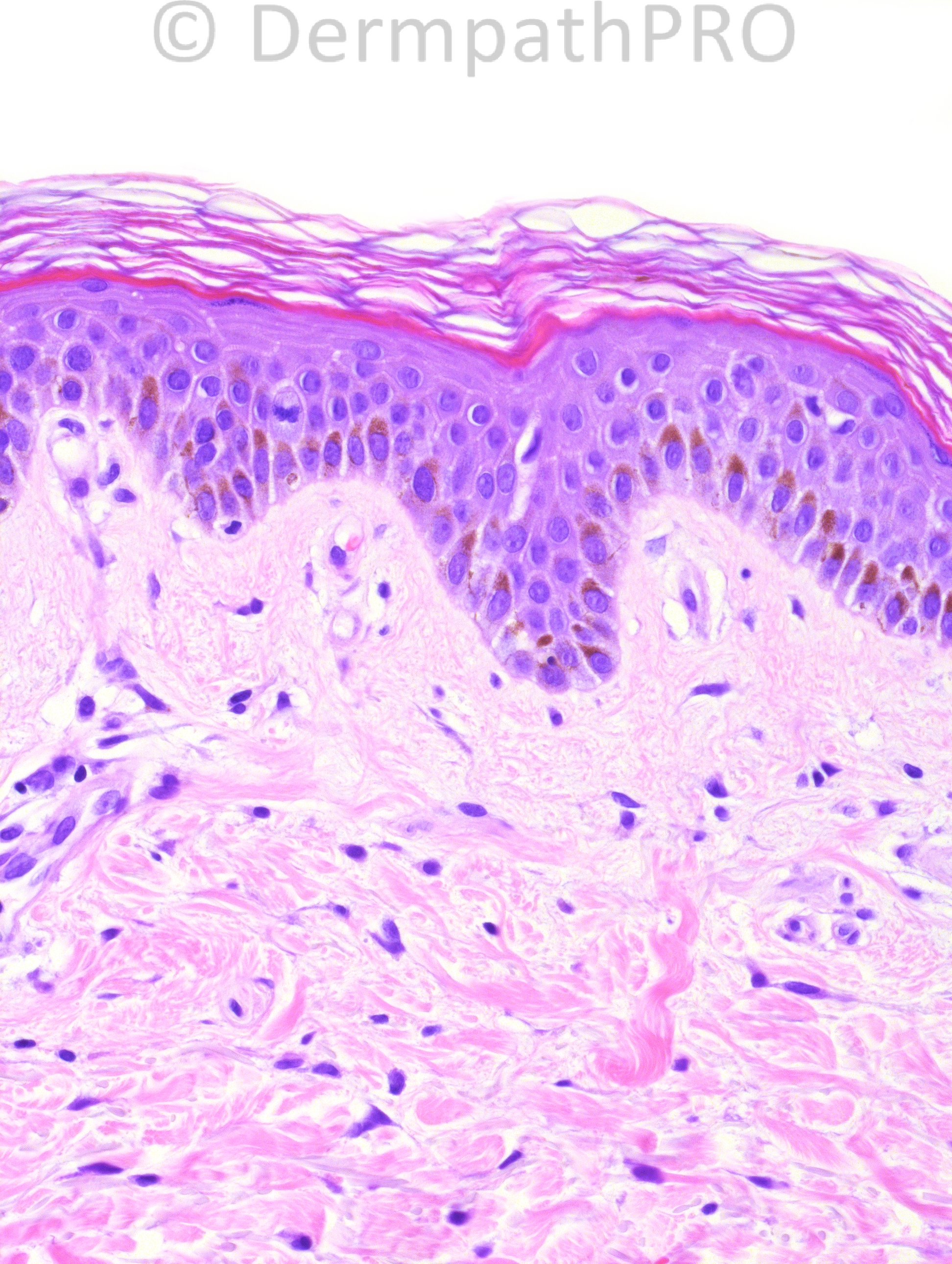
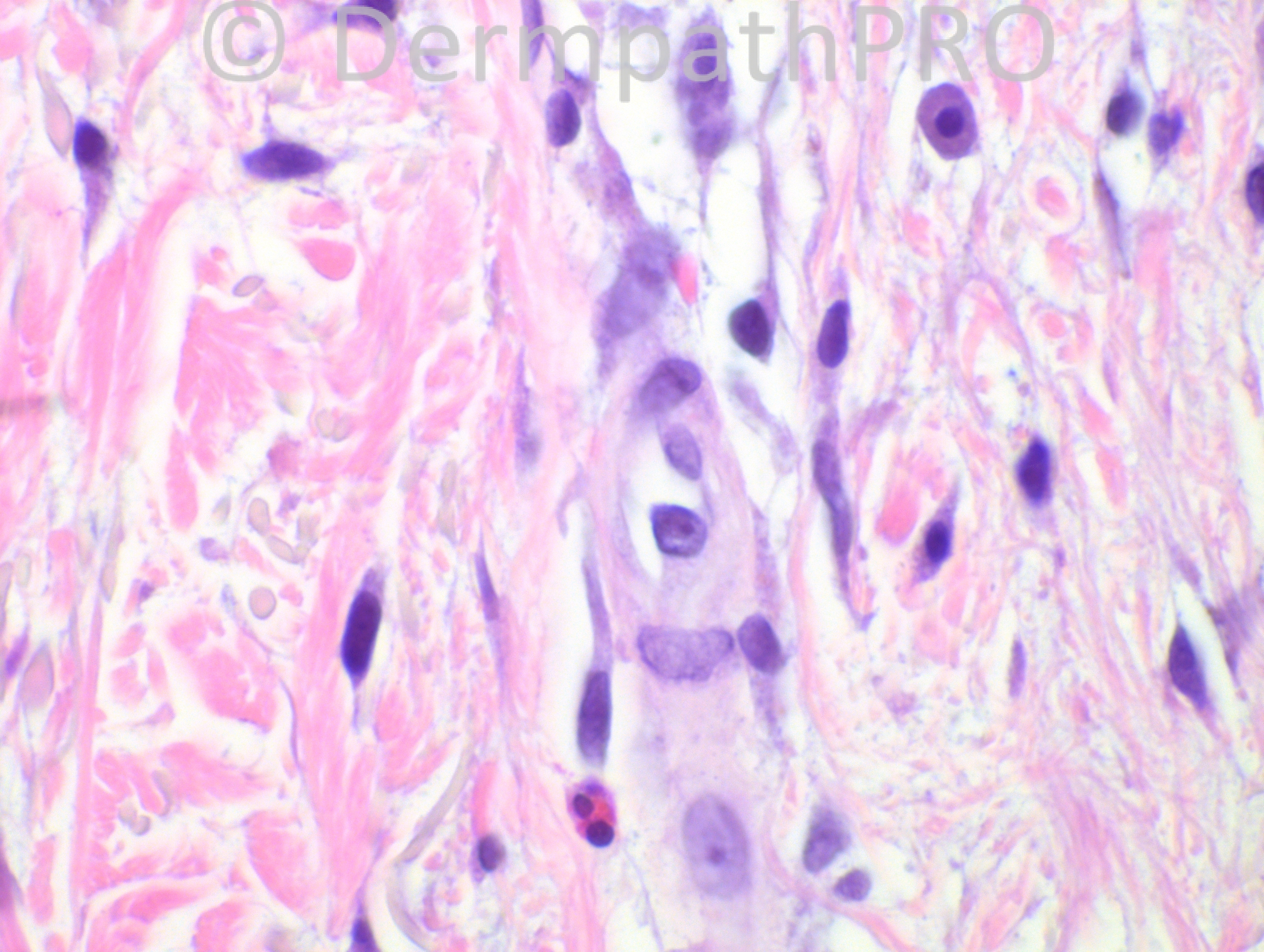
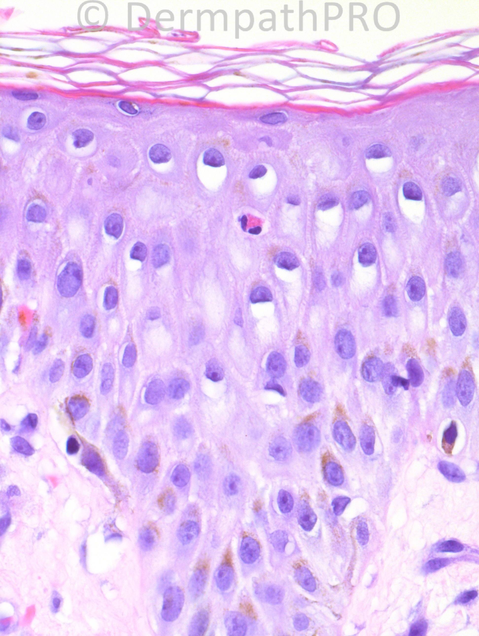
65 year old male with new onset rash, vesicles, and confluent erythema. Two biopsies from the left anterior forearm and left abdomen were performed. The biopsy shown is from the left abdomen.

Case posted by Dr. Hafeez Diwan.

  Report Record

User Feedback


I see an epidermis with mild spongiosis accompanied by eosinophil exocitosis. There are some eosinophils also in the dermis together with neutrophils (fig. 3). I think this could be a initial Bullous pemphigoid or a drug reaction too, even without interface reaction. History of drug intake is important.

Share this comment


Link to comment
share_externally

Robledo F. Rocha

Posted

At first glance, it simulates a normal skin, but this apparent normality is debunked by a subtle interstitial infiltrate of scattered eosinophils, neutrophils and mast cells in a slightly edematous dermis. Bearing in mind the information that this biopsy was taken from abdomen, the presence of some stellate fibroblasts and gray-blue elastic fibers makes me think of physical urticaria induced by sun exposure.

Share this comment


Link to comment
share_externally

Sasi Attili

Posted

I think this is cutaneous mastocytosis.....

Share this comment


Link to comment
share_externally

Guest Maria George

Posted

Nothing exciting.Drug eruption.

Share this comment


Link to comment
share_externally

Guest Romualdo

Posted

I can see only one intraepidermal eosinophil. Maybe most of the cells that look like fibroblasts are, in reality, mast cells. So I agree with Dr. Sasi: cutaneous mastocytosis.

Share this comment


Link to comment
share_externally

Guest Jim Davie MD

Posted

Agree with Maria and Igor...acute/early drug eruption.

Clinical of sudden onset confluent erythema and vesicles, without bullae, would support this slightly over BP.

Looking at the biopsy, I see a mixed sparse eosinophilic and neutrophilic interstitial infiltrate with subtle edema, telangiectasia, spongiosis, rare necrotic basal keratinocytes, and minimal eosinophil exocytosis. No parakeratosis is seen.

The differential would include an early (spongiotic phase) biopsy of bullous pemphigoid, but the urticarial pattern inflammation seems too sparse, too neutrophil predominant, and lacking in subtle subepidermal clefting to be an early BP, in my opinion.

Share this comment


Link to comment
share_externally

Dr. Mona Abdel-Halim

Posted

Drug vs. urticarial stage of BP

Share this comment


Link to comment
share_externally

Iskander H. Chaudhry

Posted

Dear All the diagnosis was urticarial bullous pemphigoid. Direct IMF confirmed the diagnosis. Differential would have included a drug eruption on morphological grounds.

Share this comment


Link to comment
share_externally


Join the conversation

You can post now and register later. If you have an account, sign in now to post with your account.

Guest
Add a comment...

×   Pasted as rich text.   Paste as plain text instead

  Only 75 emoji are allowed.

×   Your link has been automatically embedded.   Display as a link instead

×   Your previous content has been restored.   Clear editor

×   You cannot paste images directly. Upload or insert images from URL.

Loading...

×
×
  • Create New...