Jump to content
In this section we have spot diagnoses posted on a daily basis since June 2010, now over 4000! You can review the archived cases and read the suggested diagnoses by users and the final comment by the contributors.
Case are uploaded each week day by 10 am UK time with the correct diagnosis will generally be posted at 8 pm UK time. Why not view the most recent spot diagnosis and proffer a diagnosis?

Case Number : Case 821 - 9th August Posted By: Guest

Please read the clinical history and view the images by clicking on them before you proffer your diagnosis.
Submitted Date :
   (0 reviews)

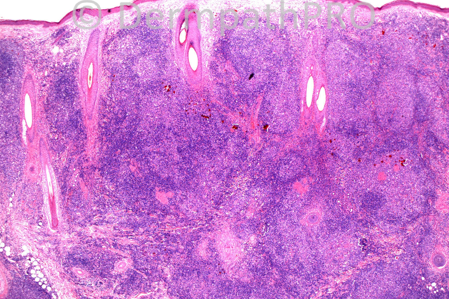
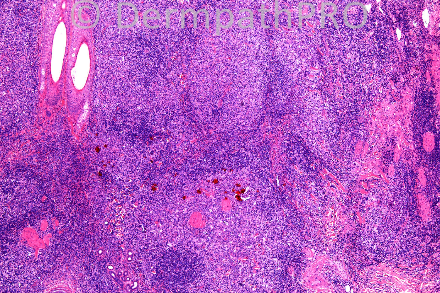
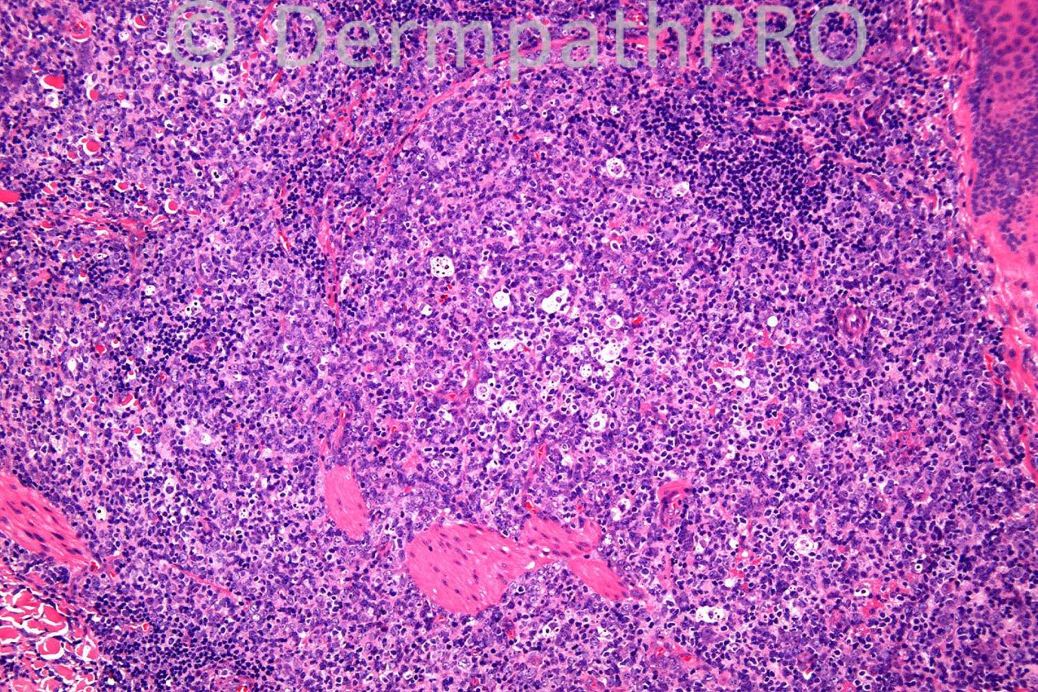
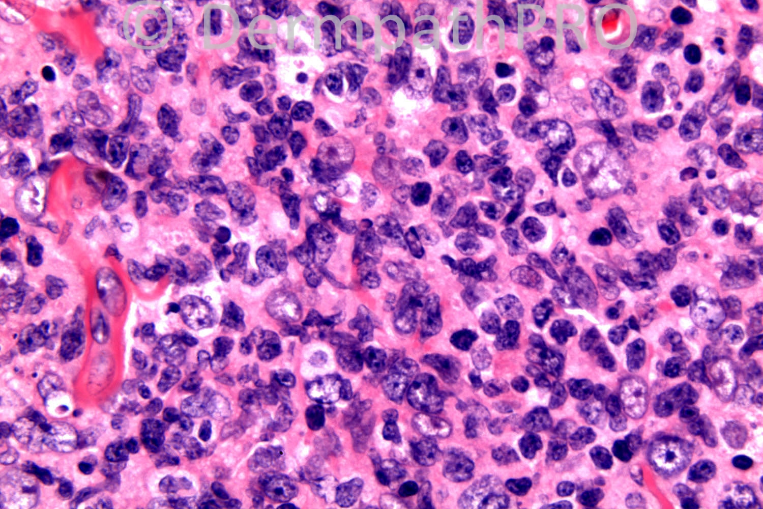
70 years old male, 12/12 hx of papular lesion 5cm x 2.5cm (posterior scalp). Granulomatous
inflammation - ?granuloma faciale? granulomatous rosacea? sarcoidosis?deep granuloma annulare??BCC.

Case posted by Dr. Richard Carr


  Report Record

User Feedback


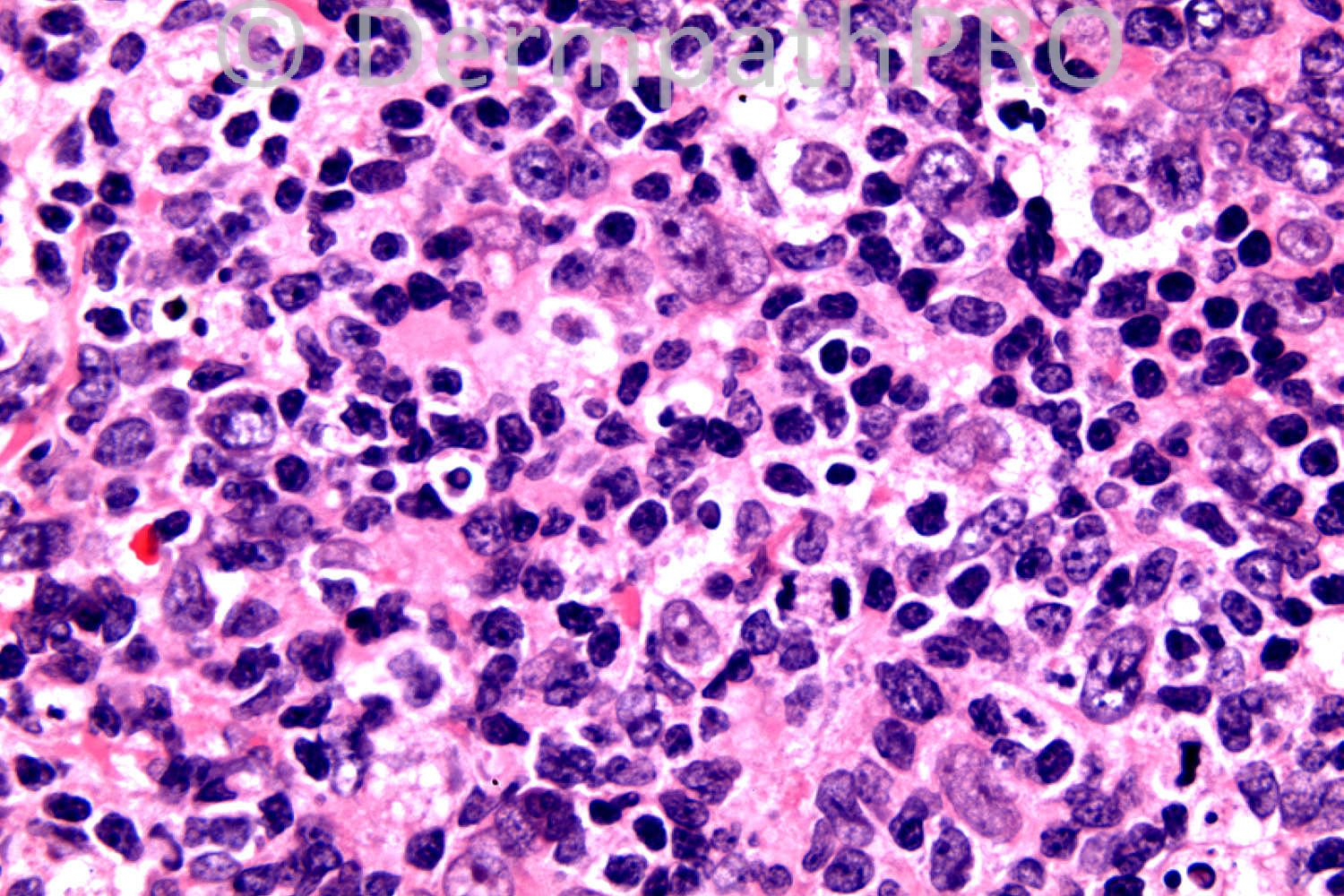
I see an intense lymphoid proliferation compromising superficial and deep dermis, composed of several lymphoid nodules. These nodules show centrofollicular hyperplasia with lots of tingible body macrophages, immunoblasts and centroblasts.
Despite the adnexial involvement by the infiltrate, I think this is a Benign cutaneous B-cell hyperplasia. I would probably perform immunohistochemistry hoping no change in my diagnosis. CD30 would be probably positive, but at reactive immunoblasts. Sory for bad english.

Share this comment


Link to comment
share_externally

Dr. Richard Carr

Posted

In case anyone is confused this is to replace the case posted initially (which was one already used and was a collision of melanoma and AFX). Thanks Iskander and team.

Share this comment


Link to comment
share_externally

Robledo F. Rocha

Posted

I think this is a pseudolymphomatous folliculitis. There are dilated hair follicles surrounded by dense nodular lymphocytic infiltrate.

Share this comment


Link to comment
share_externally

Guest Maria George

Posted

pseudolymphoma versus lymphphoma.But I prefer follicular lymphoma.IHC will make the final word about this lesion.

Share this comment


Link to comment
share_externally

Guest Romualdo

Posted

I agree with Igor and Robledo: pseudolymphoma, possibly secondaty to folliculitis.

Share this comment


Link to comment
share_externally

Arti Bakshi

Posted

B cell cutaneous lymphoid hyperplasia vs Follicular lymphoma.
IHC needed, favour hyperplasia on images.

Share this comment


Link to comment
share_externally

Dr. Richard Carr

Posted

Yes this was an unusually florid cutaneous follicular lymphoid hyperplasia. Flow cytometry was performed and showed polyclonal plasma cells / no aberrant profile. Immunostains confirmed high Ki67 in the germinal centres of the follilcles (Bcl2-ve). Patient has been prescribed potent topical steroids and follow-up awaited.

Share this comment


Link to comment
share_externally

Guest Jim Davie MD

Posted

Agree with my colleagues. I favor B-cell pseudolymphoma, given the high mitotic rate, mixed centroblastic/centrocytic follicular center population, relative sparing of epidermis and hair follicles, and numerous tingible body macrophages within the poorly formed geographic follicular centers.

Kappa and lambda light chain immunostaining would be indicated for confirmation of absence of light chain restriction.

Share this comment


Link to comment
share_externally


Join the conversation

You can post now and register later. If you have an account, sign in now to post with your account.

Guest
Add a comment...

×   Pasted as rich text.   Paste as plain text instead

  Only 75 emoji are allowed.

×   Your link has been automatically embedded.   Display as a link instead

×   Your previous content has been restored.   Clear editor

×   You cannot paste images directly. Upload or insert images from URL.

Loading...

×
×
  • Create New...