Jump to content
In this section we have spot diagnoses posted on a daily basis since June 2010, now over 4000! You can review the archived cases and read the suggested diagnoses by users and the final comment by the contributors.
Case are uploaded each week day by 10 am UK time with the correct diagnosis will generally be posted at 8 pm UK time. Why not view the most recent spot diagnosis and proffer a diagnosis?

Case Number : Case 822 - 12th August Posted By: Guest

Please read the clinical history and view the images by clicking on them before you proffer your diagnosis.
Submitted Date :
   (0 reviews)

40 years old female, Mid-Back


Case posted by Dr. Mark Hurt.


  Report Record

User Feedback


Guest Maria George

Posted

Nevus Spilus (lentigine +compound nevus) containing ballon cell nevus.Nice case.
There lentiginous proliferation of melanocyte in the basal layer in addition to a compound nevus which shows clear cell features.So Nevus spilus.

Share this comment


Link to comment
share_externally

Arti Bakshi

Posted

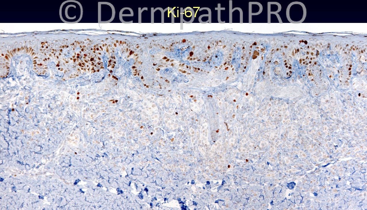
Symmetrical compound melanocytic proliferation with clear/balloon cell change of intradermal component.
Good maturation, no dermal mitoses and no epidermal consumption.
Agree with 'Balloon cell naevus'.

Share this comment


Link to comment
share_externally

Robledo F. Rocha

Posted

I go with balloon cell melanoma. Even though maturation can be discerned with both HE and melan-A, there are some alarming findings like mitotic activity in the deeper reaches, some nested cells persisting in the deeper reaches surrounded by diffuse cells, and some pleomorphic cells scattered throughout the lesion.

Share this comment


Link to comment
share_externally

Sasi Attili

Posted

I think this is a benign Balloon Cell Nevus....

Share this comment


Link to comment
share_externally

Guest Jim Davie MD

Posted

Regarding my experiences with balloon cell change, I associate it most frequently in nevi (both acquired and congenital) that have other senescent features. In this case, the senescent atypia in the balloon-cell nuclei (smudgy nuclear hyperchromasia and enlargement, large nuclear pseudoinclusions, rare multinucleation) seem more prominent in the upper dermal component, which also shows the greatest balloon cell changes.

Share this comment


Link to comment
share_externally


Join the conversation

You can post now and register later. If you have an account, sign in now to post with your account.

Guest
Add a comment...

×   Pasted as rich text.   Paste as plain text instead

  Only 75 emoji are allowed.

×   Your link has been automatically embedded.   Display as a link instead

×   Your previous content has been restored.   Clear editor

×   You cannot paste images directly. Upload or insert images from URL.

Loading...

×
×
  • Create New...