Jump to content
In this section we have spot diagnoses posted on a daily basis since June 2010, now over 4000! You can review the archived cases and read the suggested diagnoses by users and the final comment by the contributors.
Case are uploaded each week day by 10 am UK time with the correct diagnosis will generally be posted at 8 pm UK time. Why not view the most recent spot diagnosis and proffer a diagnosis?

Case Number : Case 825 - 15th August Posted By: Guest

Please read the clinical history and view the images by clicking on them before you proffer your diagnosis.
Submitted Date :
   (0 reviews)

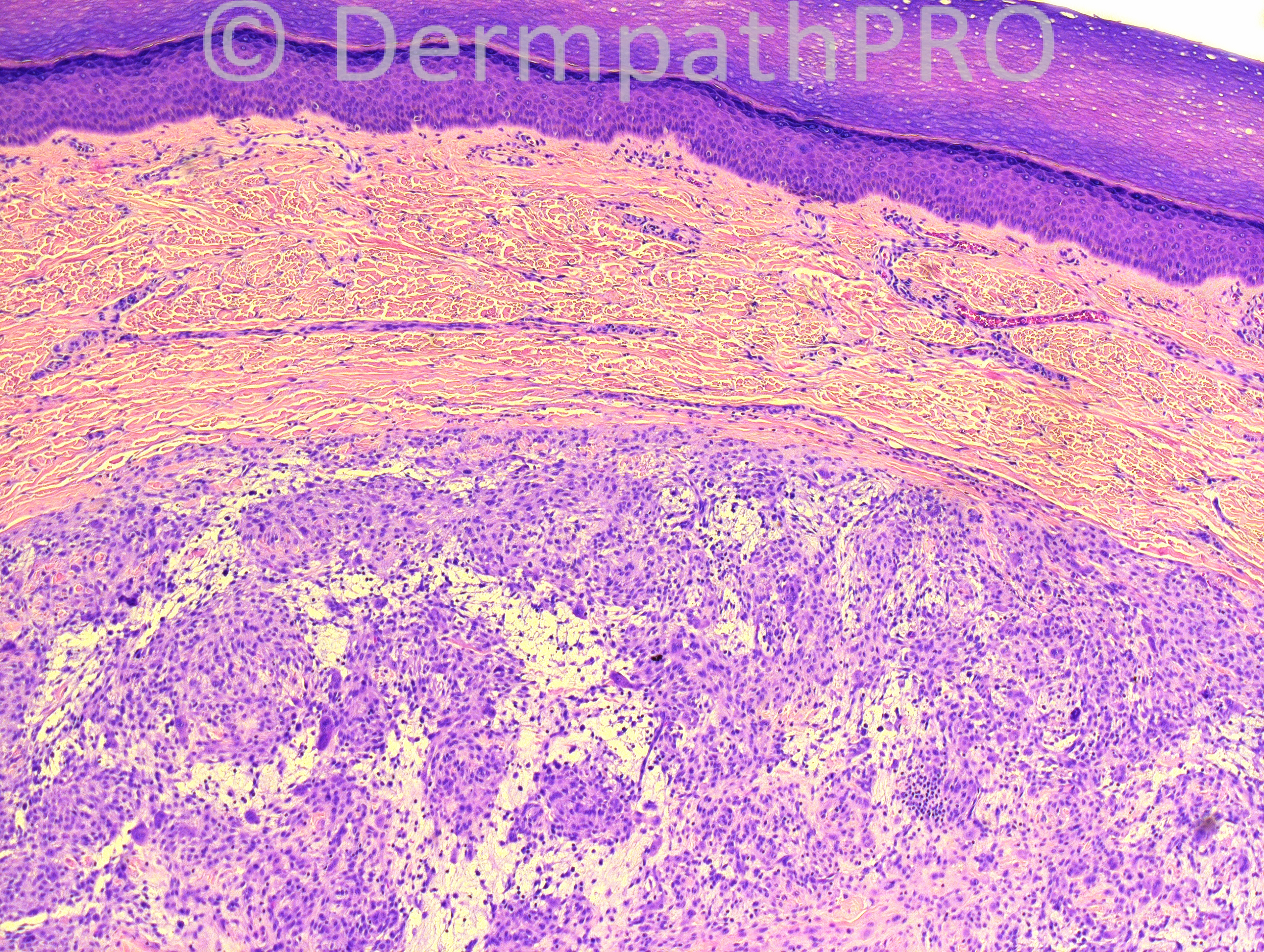
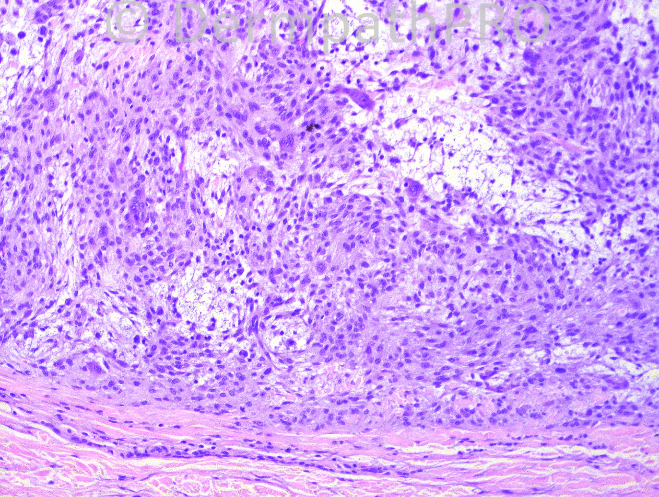
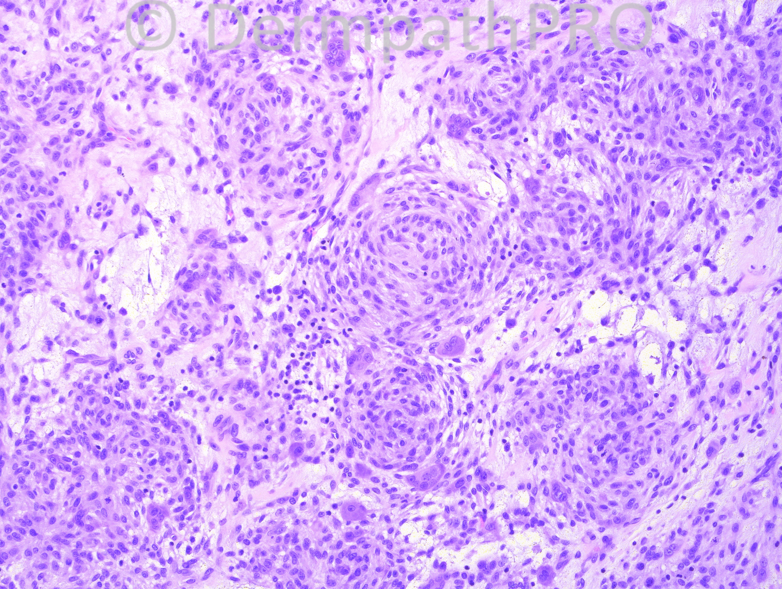
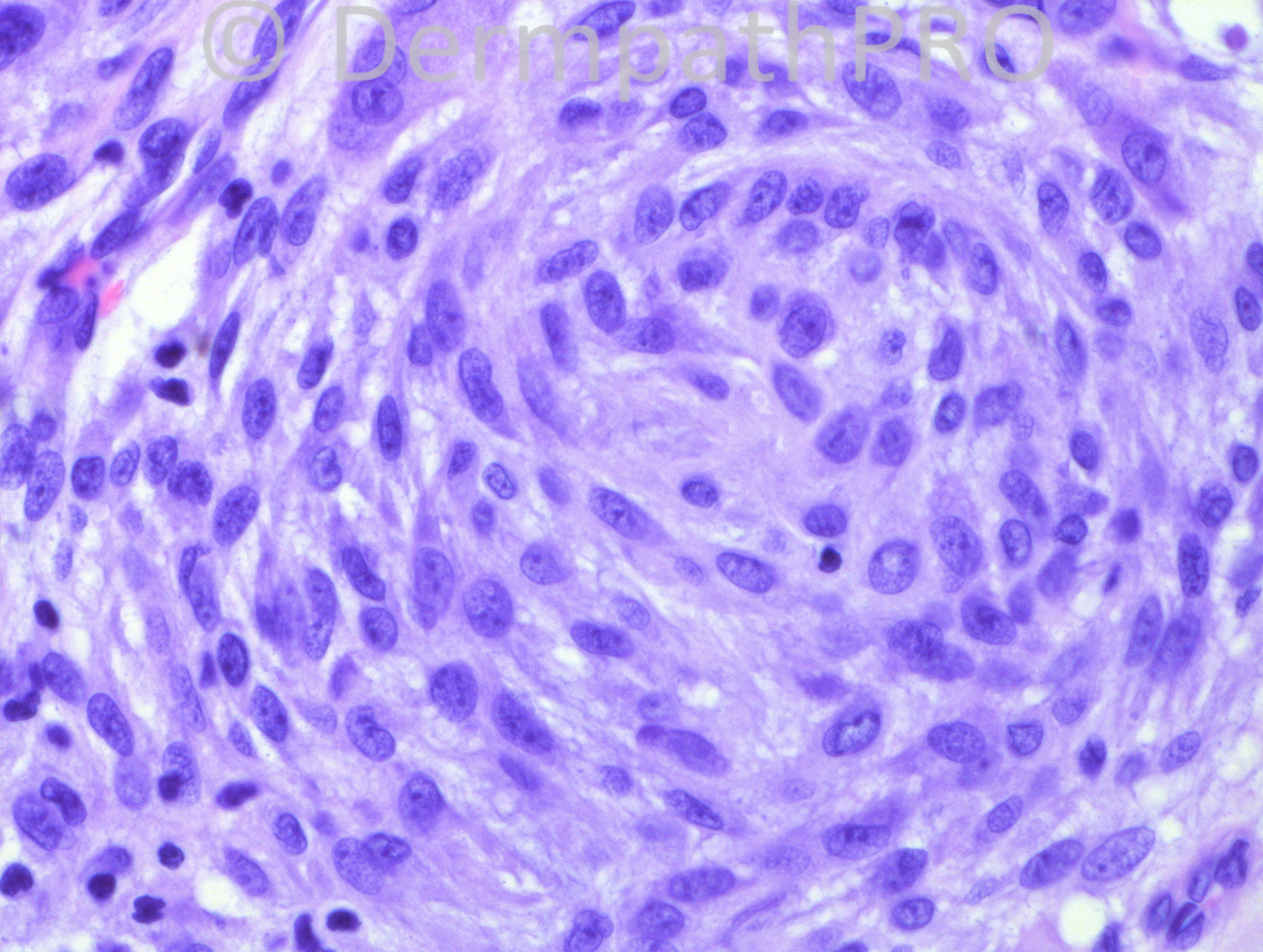
8-year-old Hispanic male with a right great toe lesion.

Case posted by Dr. Hafeez Diwan.

  Report Record

User Feedback


Guest Yüksel OKUMUŞ, MD

Posted

Plexiform fibrous histiocytoma

Share this comment


Link to comment
share_externally

Robledo F. Rocha

Posted

I don’t feel it is plexiform fibrohistiocytic tumor. Myxoid component is not present in this tumor and circumscribedness is not a feature of it which, conversely, emanates tentacle-like extensions into the subcutaneous fat as a typical characteristic.
I go with cellular neurothekeoma.

Share this comment


Link to comment
share_externally

Guest Jim Davie MD

Posted

[font=tahoma,geneva,sans-serif][size=4]I agree this resembles cellular neurothekeoma, given overall good circumscription, internal whorled nodular architecture, and especially the variable, focally extensive mucinous stroma; there is absence of significant pigmentation, vascular stroma, hemorrhage, or fibrosis. The cells are large, oval to spindled with frequent osteoclast-like multinucleation, and absence of pleomorphism or high-grade atypia.
I am concerned, however, by the moderate mitotic activity with aberrant spindles. Staining may be helpful.[/size][/font]

Share this comment


Link to comment
share_externally

Guest Romualdo

Posted

I agree this could represent cellular neurothekeoma, although according to Dr. Miettinen's book location at the hands and feet is distinctly uncommon. In my opinion, another possibility is nodular fasciitis. The irregular tears in the tissue containing mucin, the non-atypical spindle cells and the presence of osteoclast giant cells suggest this diagnosis, although location in the dermis is also very uncommon.

Share this comment


Link to comment
share_externally

Dr. Hafeez Diwan

Posted

I called this cellular neurothekeoma.

Share this comment


Link to comment
share_externally


Join the conversation

You can post now and register later. If you have an account, sign in now to post with your account.

Guest
Add a comment...

×   Pasted as rich text.   Paste as plain text instead

  Only 75 emoji are allowed.

×   Your link has been automatically embedded.   Display as a link instead

×   Your previous content has been restored.   Clear editor

×   You cannot paste images directly. Upload or insert images from URL.

Loading...

×
×
  • Create New...