Jump to content
In this section we have spot diagnoses posted on a daily basis since June 2010, now over 4000! You can review the archived cases and read the suggested diagnoses by users and the final comment by the contributors.
Case are uploaded each week day by 10 am UK time with the correct diagnosis will generally be posted at 8 pm UK time. Why not view the most recent spot diagnosis and proffer a diagnosis?

Case Number : Case 826 - 16th August Posted By: Guest

Please read the clinical history and view the images by clicking on them before you proffer your diagnosis.
Submitted Date :
   (0 reviews)

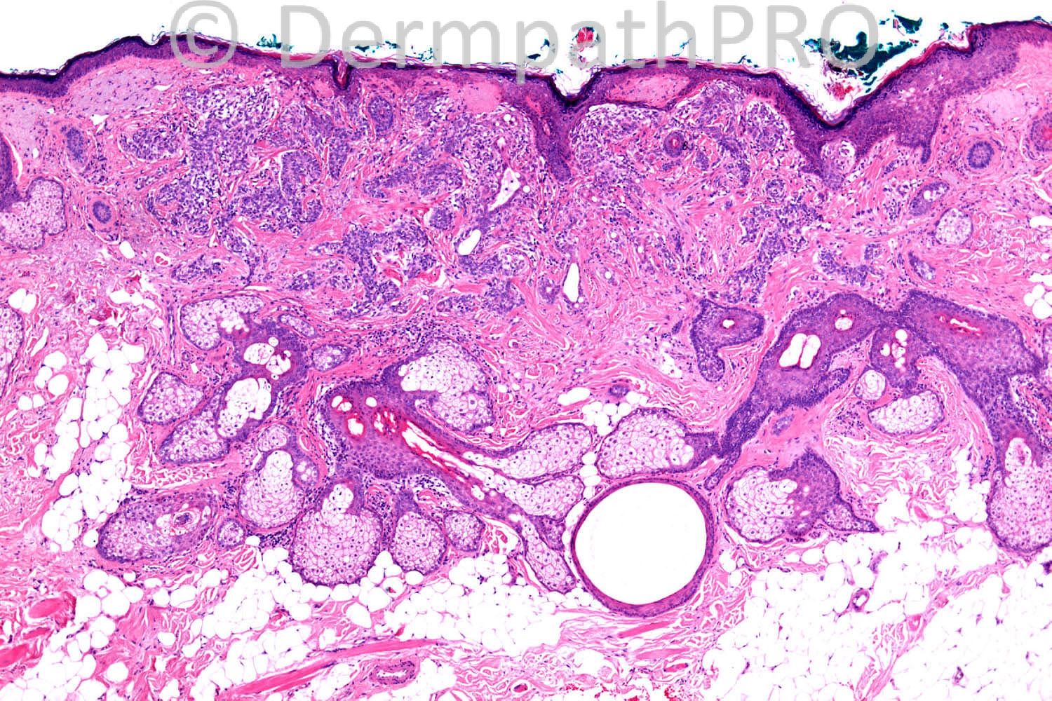
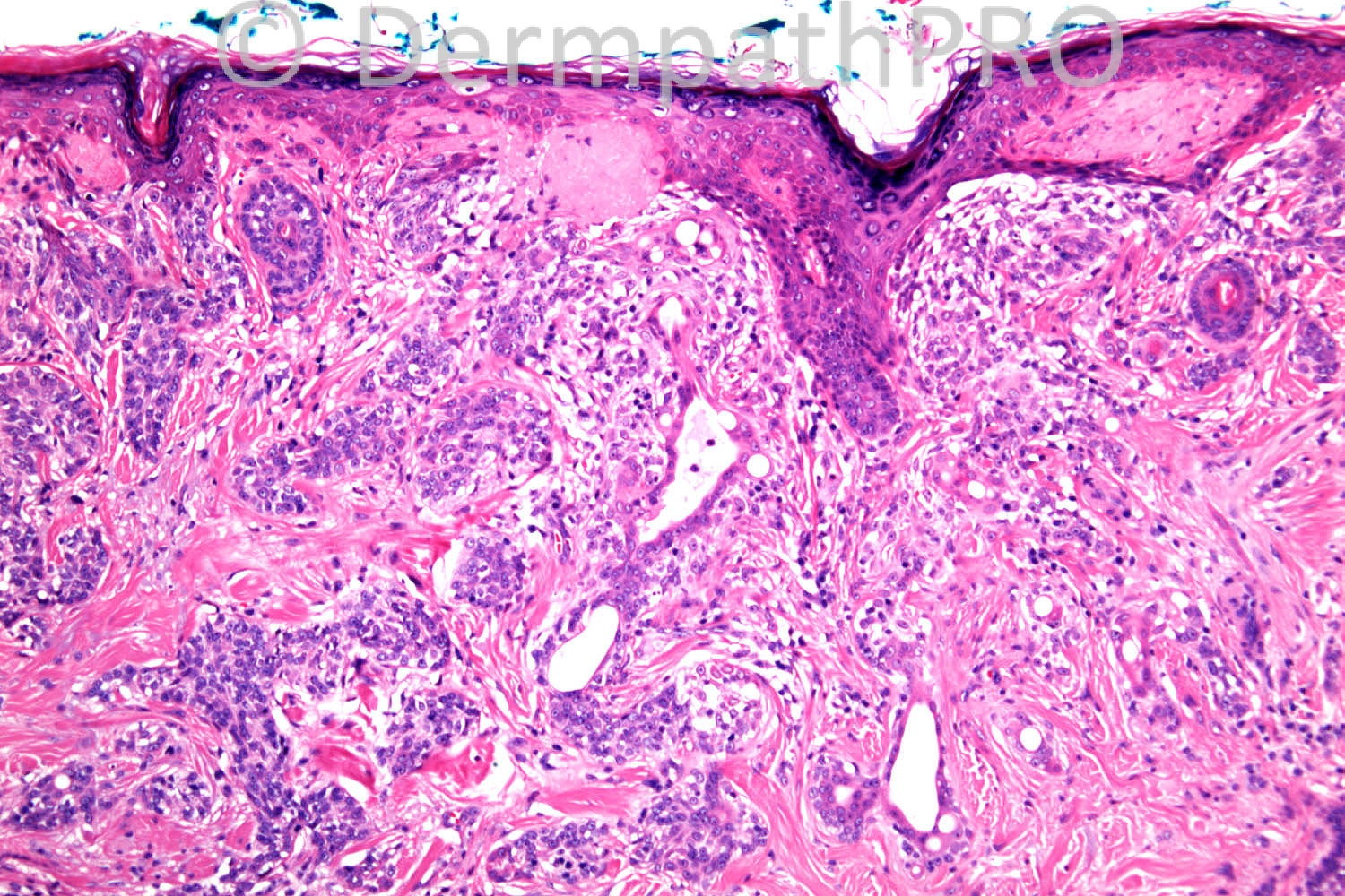
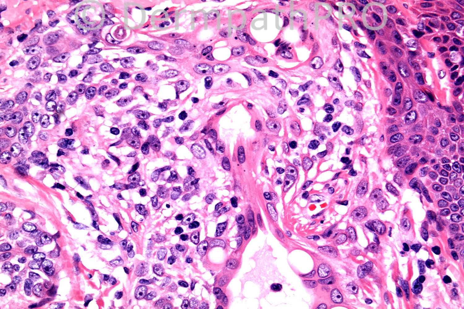
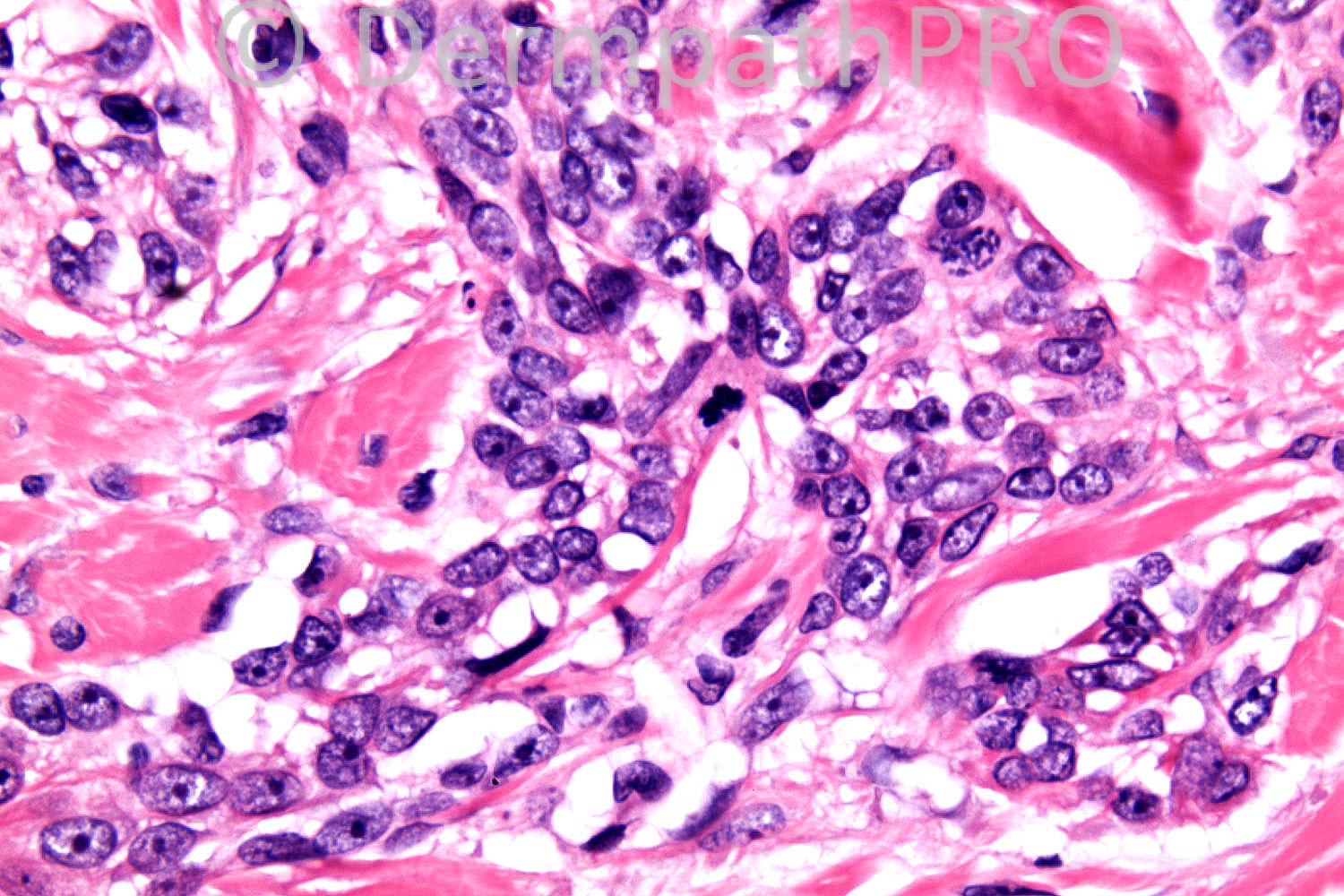
81 years old female. ?BCC Nasal side wall


Case posted by Dr. Richard Carr.

  Report Record

User Feedback


Hanan Vaknine MD

Posted

BCC with ductal differentiation? entrapment?

Share this comment


Link to comment
share_externally

Guest Maria George

Posted

[color=#444444][font=arial, sans-serif][size=1][right]adamantinoid [/right][/size][/font][/color][color=#444444][font=arial, sans-serif][size=1][b][right]trichoblastoma[/right][/b][/size][/font][/color][color=#444444][font=arial, sans-serif][size=1][right] ([/right][/size][/font][/color][color=#444444][font=arial, sans-serif][size=1][right]lymphadenoma).[/right][/size][/font][/color]

Share this comment


Link to comment
share_externally

Robledo F. Rocha

Posted

I think micronodular basal cell carcinoma is a better hypothesis since there’s no specific follicular stroma which would permit to construe a diagnosis of trichoblastoma.
The micronodular morphological subtype of basal cell carcinoma typically displays minimal peripheral palisading and lacks retraction clefts, but in picture #4 the surrounding myxoid stroma drafts spaces between it and the small nests of basaloid cells. Asymmetric silhouette (picture #1), apoptotic cells (picture #3), and mitotic figures (picture #6) are additional findings that bear out my hypothesis.
Eccrine ducts entrapment and microcystic change due to mucin accumulation impart a pseudoglandular appearance to the tumor.

Share this comment


Link to comment
share_externally

Dr. Mona Abdel-Halim

Posted

I think it is BCC with ductal differentiation...

Share this comment


Link to comment
share_externally

Wolter Mooi

Posted

Perhaps a small eccrine porocarcinoma (continuity with epidermis; intra- and intercellular lumina; absence of palissading and clefting; mitotic activity and invasive growth)?

Share this comment


Link to comment
share_externally

Guest Jim Davie MD

Posted

I agree with BCC with adnexal differentiation. Clear-cell porocarcinoma may also be in the differential.
There is multifocal colonization of the epidermis and superficial dermis by mitotically active squamoid to vesiculated cells (two mitoses in last photo). I am suspicious that there is some peculiar vacuolation and nuclear indenting, and sebaceous duct-like features (crenelated cuticle visible in the larger ductal structures) that would suggest sebaceous differentiation...although in the absence of convincing sebocytes, I admit this is more likely eccrine differentiation with glycogenated features, such as seen in clear-cell syringoma. PAS/PASD and EMA/CEA would be helpful in this case.

Share this comment


Link to comment
share_externally

Guest Romualdo

Posted

Ductal lumens lined by cuticular cells with abundant eosinophilic cytoplasm are present. Some intracytoplasmic lumens are seen in these same cells. In the absence of well defined peripheral palisades and clefting I favor the diagnosis of porocarcinoma over that of a CBC with eccrine diferentiation.

Share this comment


Link to comment
share_externally

Guest Dr.ebtisam FAIDA

Posted

??b.c.c with ductal differentiation

Share this comment


Link to comment
share_externally

Arti Bakshi

Posted

I think there is eccrine differentiation and the cells look atypical. D/D- Eccrine porocarcinoma vs eccrine syringoid carcinoma.

Share this comment


Link to comment
share_externally

Dr. Richard Carr

Posted

I think all the suggestions of BCC with glandular differentiation are probably the best wrong responses - based on probability and appearances that should have been the diagnosis in this case although remember if a tumour lacks absolutely classic peripheral palisading and stromal mucin (in retraction spaces) always confirm (or refute) your diagnosis with IHC (BerEP4 with glandular will be diffusely strongly BerEP4 in the basaloid epithelium and almost more importantly completely EMA negative in the basaloid epithelium. In this case the tumour was BerEP4 negative in the basaloid epithelium with EMA staining squamoid and squamoid cuticular ductal areas of differentiation. I found this particular lesion difficult to classify (?unique case)! Of the remaining responses I do like the mention of adamantinoid trichoblastoma because there is some resemblance to trichadenoma with the lymphocytes pepperring the tumour although the infiltrative pattern and features of a low grade carcinoma have been pointed about above. We might consider a low-grade malignant mixed follicular-ductal (bearing in mind there is increasing recognition of mixed folliculo-sebaceous-ductal-apocrine unit origins in adnexal tumours). Indeed the tumour does seems to drop of adnexa ?follicles or acrosyringia and reminds me (in that respect) of the origin of some follicular squamous cell carcinomas that I have seen. However so far in none of the (infundibular/ishmic-pilar) FSQC previously have I seen ductal differentiation. In summary there is some resemblance to trichadenoma (adamantinoid trichoblastoma) but in setting of severe elastosis and with occasional mitotic figures and mild cellular pleomorphism in my opinion this is a low-grade carcinoma with squamoid and squamoid cuticular ductal differentiation. Tumour was BerEP4 negative in the basaloid epithelium which in the absence of typical palisading and stromal mucin (excludes BCC for practical purposes) with EMA in ducts and squamoid cells. My original report is pasted below with the pragmatic label "low-grade infiltrative squamoid ductal carcinoma" but I guess some would be happy to lump this with a low-grade porocarcinoma. I think the so-called eccrine squamoid ductal carcinomas are in a spectrum with syringoid carcinomas and MACs the important point being that they are deceptively bland lesions that can easily be labelled as benign but may be extensive at the time of (final) diagnosis due to long indolent growth and mis-diagnosis on prior biopsies. There is discussion that some of these lesions (e.g. MAC) are follicular-ductal-apocrine-sebaceous unit lesions with mixed differentiation. Management would be similar to other infiltrative low-grade tumour i.e. complete excision with clear margins that may require margin control surgery (not this case which was unusually small and fully excised with clear margins on first excision).
Histological type: Low grade infiltrative squamoid ductal carcinoma
Diameter 5 mm
Thickness: 1.3 mm
Clark Level: 4
Perineural invasion: No
Lymphovascular invasion: No
Type of invasive border: Discohesive/infiltrative
Background benign adnexal tumour: No
Radial margin: 2.5 mm
Deep margin: 3 mm
Stage (AJCC7): pT1. Apologies for delay in posting my thoughts on the case (just come back off holidays at Lake Garda, Italy. My advice don't go to Venice with teenage children in the height of summer on August 15th which is a national holiday in Italy!! Verona was lovely and good for shopping for said female off-spring).

Share this comment


Link to comment
share_externally

John Zhang

Posted

Can we entertain the idea of lymphoepithelioma-like carcinoma in this case?

Share this comment


Link to comment
share_externally


Join the conversation

You can post now and register later. If you have an account, sign in now to post with your account.

Guest
Add a comment...

×   Pasted as rich text.   Paste as plain text instead

  Only 75 emoji are allowed.

×   Your link has been automatically embedded.   Display as a link instead

×   Your previous content has been restored.   Clear editor

×   You cannot paste images directly. Upload or insert images from URL.

Loading...

×
×
  • Create New...