Jump to content
In this section we have spot diagnoses posted on a daily basis since June 2010, now over 4000! You can review the archived cases and read the suggested diagnoses by users and the final comment by the contributors.
Case are uploaded each week day by 10 am UK time with the correct diagnosis will generally be posted at 8 pm UK time. Why not view the most recent spot diagnosis and proffer a diagnosis?

Case Number : Case 831 - 23rd August Posted By: Guest

Please read the clinical history and view the images by clicking on them before you proffer your diagnosis.
Submitted Date :
   (0 reviews)

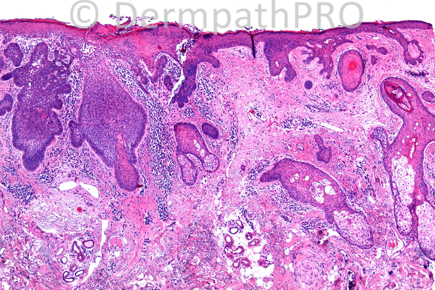
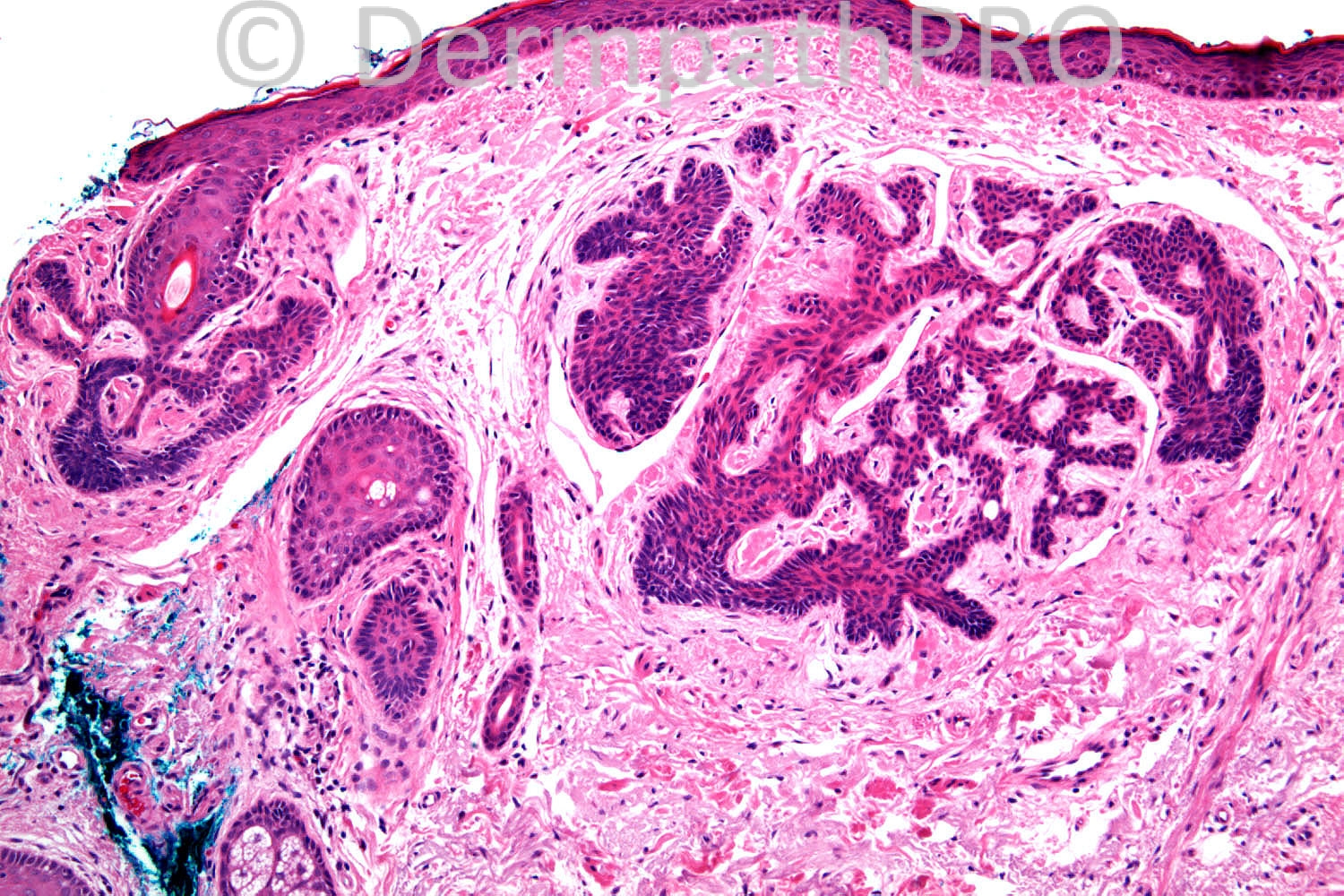
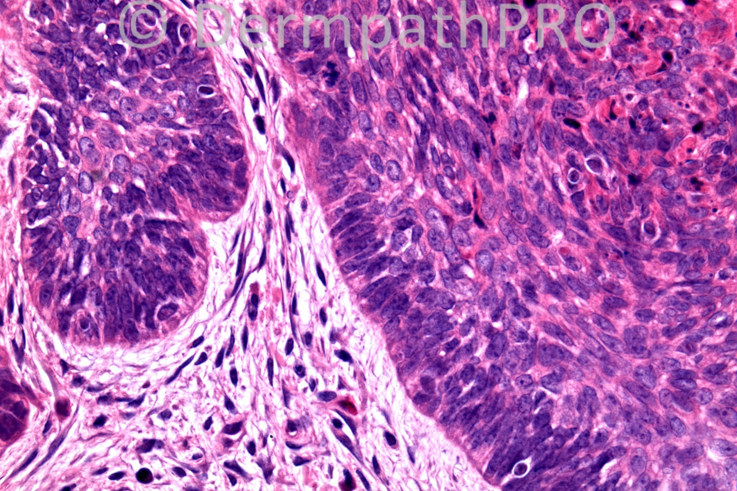
75 years old female Upper Lip. x4 BCC on face.

Case posted by Dr. Richard Carr


  Report Record

User Feedback


Guest Yüksel OKUMUŞ, MD

Posted

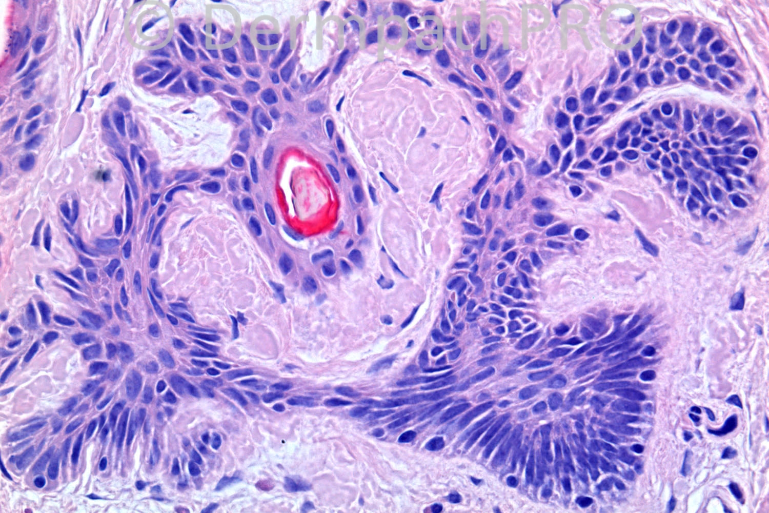
Basaloid follicular hamartoma + Basal cell carcinoma

Share this comment


Link to comment
share_externally

Guest Romualdo

Posted

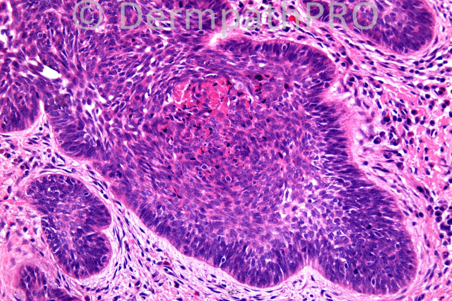
Agree. The absence of mitotic figures, apoptosis and nuclear atypia is shown in the small hamartomatous lesions, as contrasted with BCC. Immunostains with Ki-67, CD34 and bcl-2 will certainly emphasize the differences between the two conditions.

Share this comment


Link to comment
share_externally

Guest Graham Reilly

Posted

Agree with above comments - BFH and BCC

Share this comment


Link to comment
share_externally

Guest Jim Davie MD

Posted

I agree with the opinion of BFH and BCC (given aberrant mitoses and apoptotic figures [bottom right photo]). The lesion has been traumatized.
The differential would include a trichoepithelioma in conjunction with a basaloid follicular hamartoma.

Share this comment


Link to comment
share_externally

Dr. Richard Carr

Posted

I reported this as BFH (this is really a very typical example) and BCC. BFH lesions were present in multiple areas of the patients excision biopsy. Importantly BFH lesions are superficial (limited to upper dermis) and clearly limited to pre-existing (abnormal/hamartomatous) follicular units and mitoses are inconspicuous. This case was from afar and I do not know if it was part of a larger congenital unilateral case as I would suspect. In contrast to TE/TB, BFH lack stromal condensations of papillary mesenchymal cells but in my experience do have reactive Merkel cells (as were present in this case). Note the stromal clefts in the BFH that are common in benign follicular lesions. I have an exactly similar case in a patient with multiple BCC in the field of a large unilateral congenital BFH on the face. The latter patient was fair skinned and had significant solar damage.

Share this comment


Link to comment
share_externally


Join the conversation

You can post now and register later. If you have an account, sign in now to post with your account.

Guest
Add a comment...

×   Pasted as rich text.   Paste as plain text instead

  Only 75 emoji are allowed.

×   Your link has been automatically embedded.   Display as a link instead

×   Your previous content has been restored.   Clear editor

×   You cannot paste images directly. Upload or insert images from URL.

Loading...

×
×
  • Create New...