Jump to content
In this section we have spot diagnoses posted on a daily basis since June 2010, now over 4000! You can review the archived cases and read the suggested diagnoses by users and the final comment by the contributors.
Case are uploaded each week day by 10 am UK time with the correct diagnosis will generally be posted at 8 pm UK time. Why not view the most recent spot diagnosis and proffer a diagnosis?

Case Number : Case 833 - 27th August Posted By: Guest

Please read the clinical history and view the images by clicking on them before you proffer your diagnosis.
Submitted Date :
   (0 reviews)

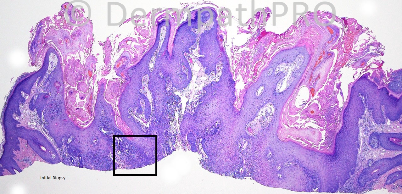
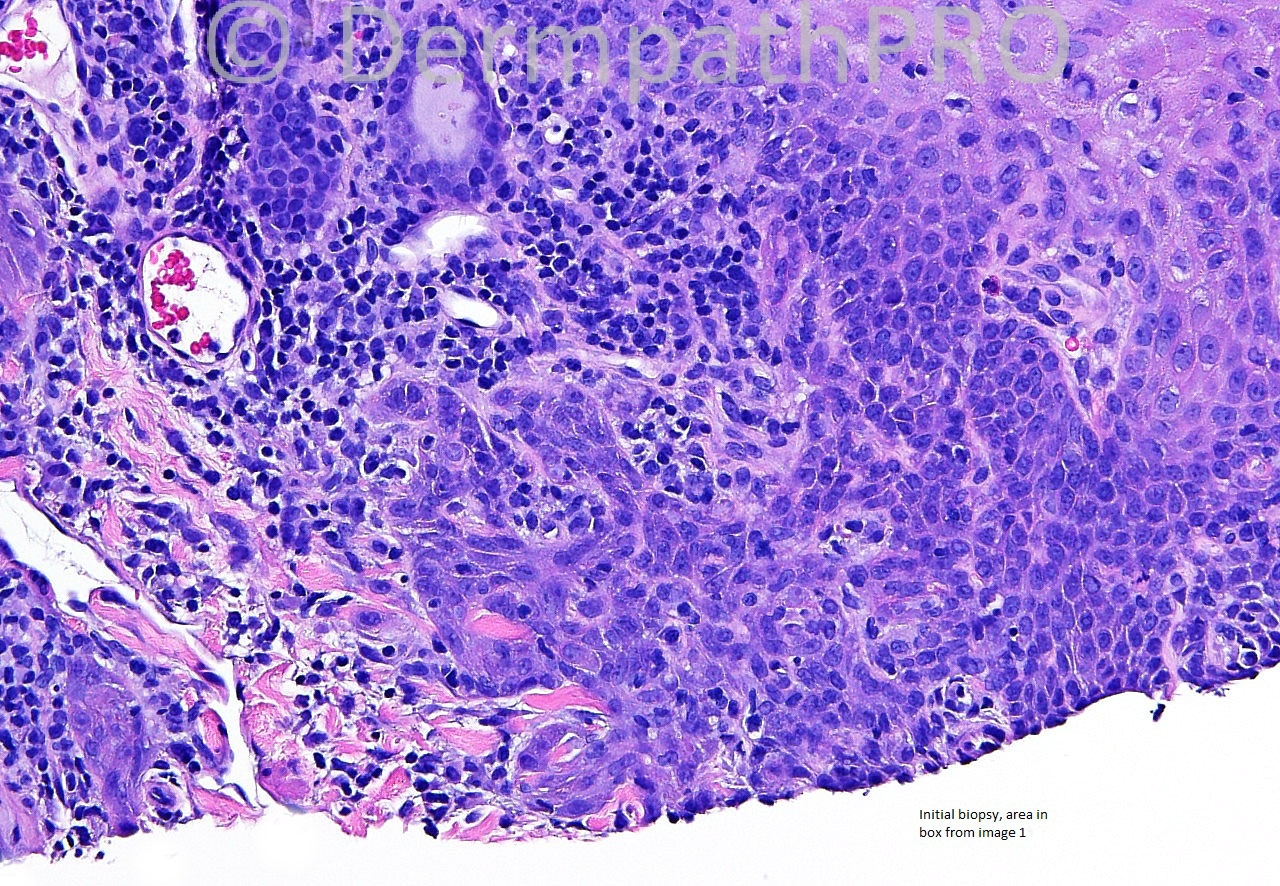
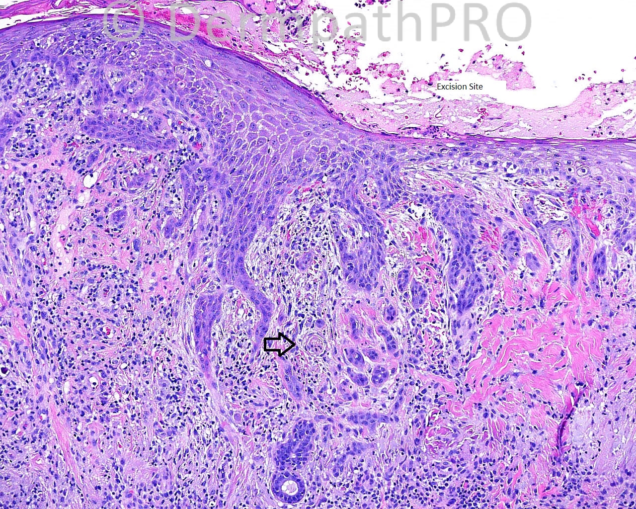
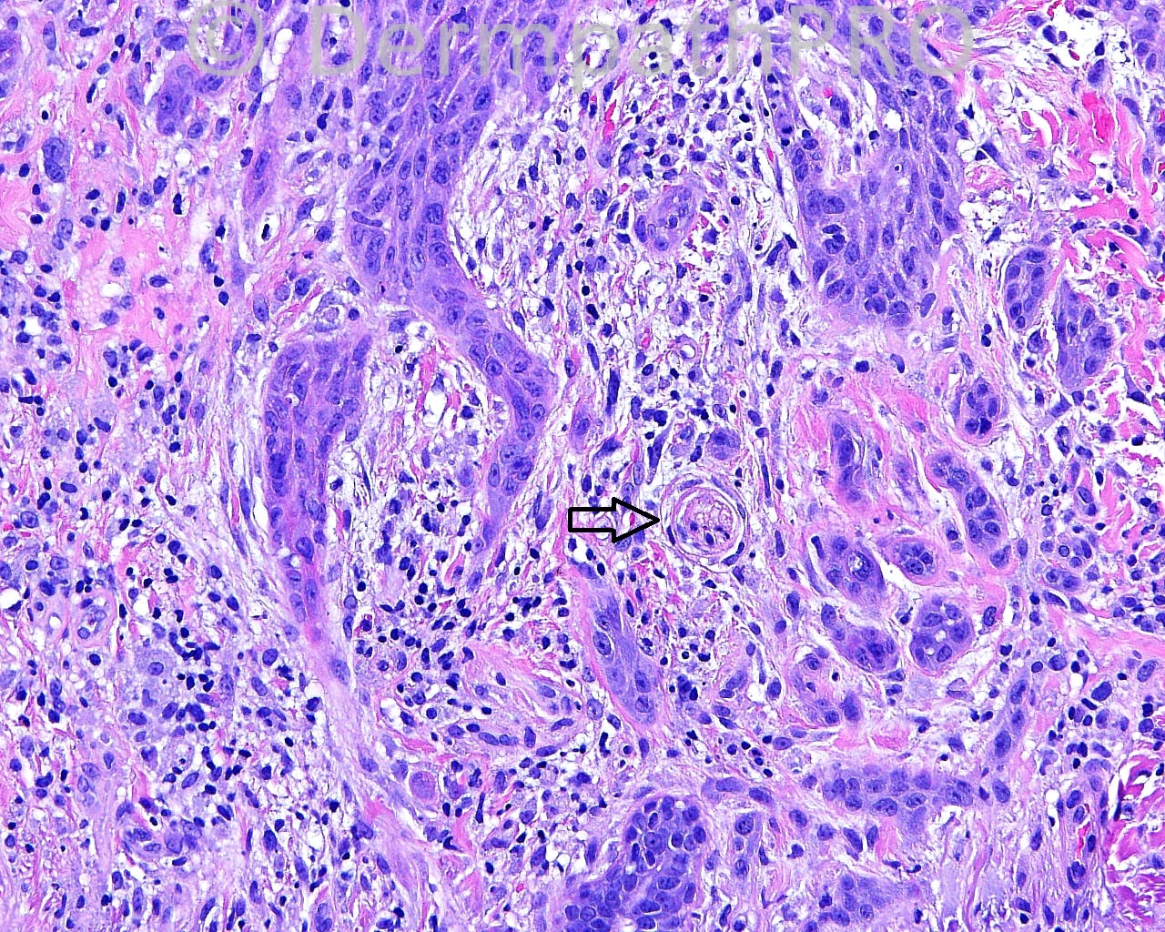
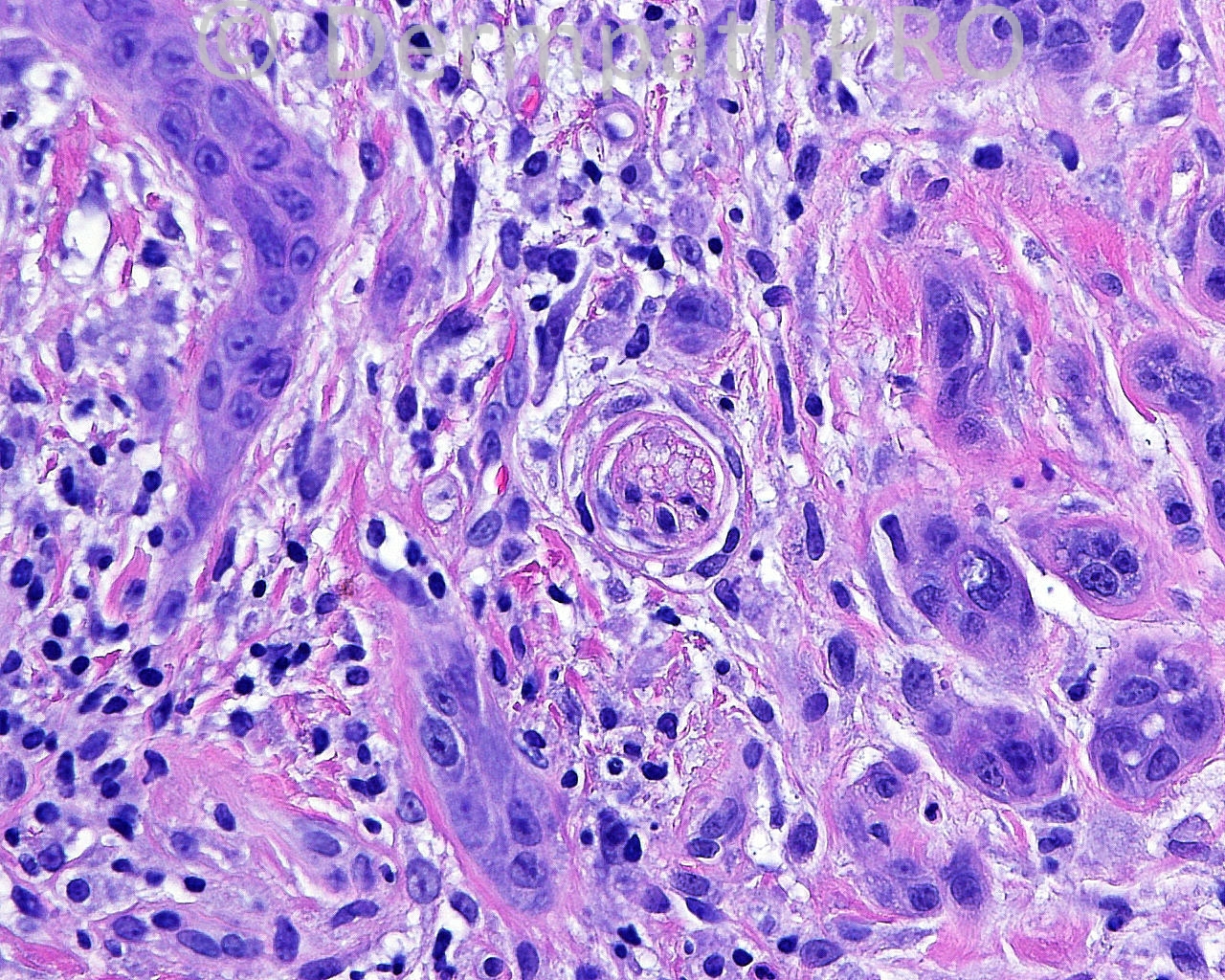
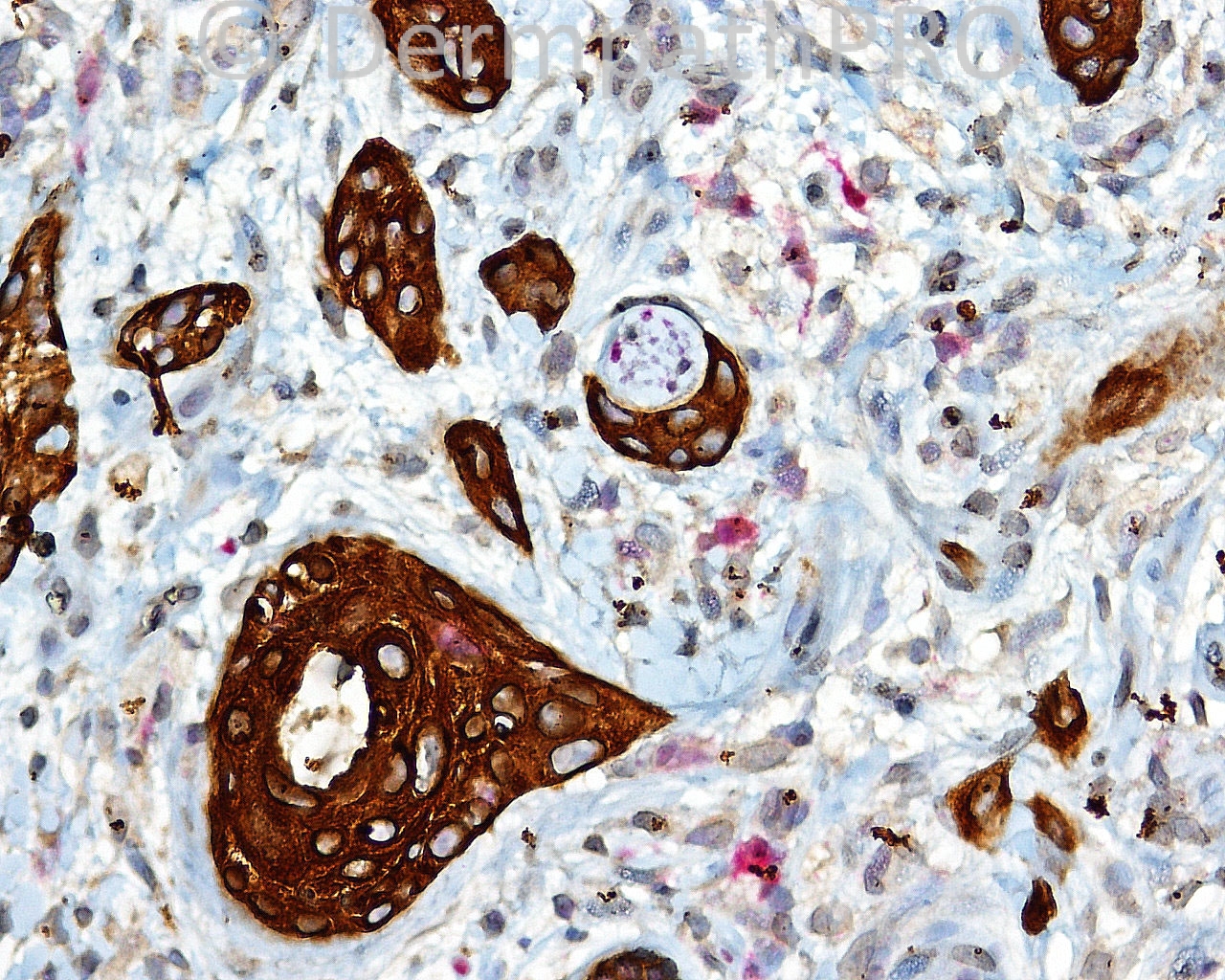
The patient is a 23 year old white woman with shave biopsies of a lesion on the left cheek.

Case posted by Dr. Mark Hurt.


  Report Record

User Feedback


Guest Yüksel OKUMUŞ, MD

Posted

SCC or microcystic adnexal carcinoma arising in organoid nevus

Share this comment


Link to comment
share_externally

Robledo F. Rocha

Posted

I favor pseudoepitheliomatous hyperplasia of the epidermis and adnexal epithelium. A few mitotic figures can be found, but I’m not definitively persuaded about cytological atypia. The problem here is identify the hidden cause of this reactive reaction pattern.
I’m looking forward to learn if perineural invasion is enough to rule out the diagnosis of reactive hyperplasia.

Share this comment


Link to comment
share_externally

I agree with pseudoepitheliomatous hyperplasia with some squamous syringometaplasia. I don´t think that the perineural invasion by squamous cells has relationship with malignant behavior.
I don´t know the diagnosis, maybe an inflamed Epidermal nevus with reactive atypia.

Share this comment


Link to comment
share_externally

Guest Jim Davie MD

Posted

I agree with Robledo and Igor. Given the young patient age, I would think this most likely a badly abused verruca, given the hemorrhagic and serous crust, and evidence of dense prurigo nodularis changes in the flanking epidermis (on the far right of first photo). Traumatized epidermal nevus or verrucous SK is also a consideration; clinical history may allow differentiation.
1. [i]Shave biopsy[/i]: The top two photos (shave biopsy) seem tangentially oriented with en-face representation of a reactive basal layer. To me, this looks like reactive/regenerative pseudocarcinomatous hyperplasia secondary to trauma and prurigo changes.
2. [i]Wide excision[/i]: The infiltrative spindled nesting pattern of the lower wide-excision photos seems associated with a regenerating eccrine duct...a regenerative pattern common in wide excisions of previous shave biopsies. Perineural association seems incidental, and does not indicate malignancy in this regenerative context, in my opinion. The infiltrative component appears cytologically similar to the overlying reactive/regenerative epidermis, and lacking in high-grade atypia. The immunostain looks like a double stain for cytokeratin (brown) and S100 (red) with presumed negative staining for eccrine differentiation in the infiltrative component.

Share this comment


Link to comment
share_externally


Join the conversation

You can post now and register later. If you have an account, sign in now to post with your account.

Guest
Add a comment...

×   Pasted as rich text.   Paste as plain text instead

  Only 75 emoji are allowed.

×   Your link has been automatically embedded.   Display as a link instead

×   Your previous content has been restored.   Clear editor

×   You cannot paste images directly. Upload or insert images from URL.

Loading...

×
×
  • Create New...