Jump to content
In this section we have spot diagnoses posted on a daily basis since June 2010, now over 4000! You can review the archived cases and read the suggested diagnoses by users and the final comment by the contributors.
Case are uploaded each week day by 10 am UK time with the correct diagnosis will generally be posted at 8 pm UK time. Why not view the most recent spot diagnosis and proffer a diagnosis?

Case Number : Case 835 - 29th August Posted By: Guest

Please read the clinical history and view the images by clicking on them before you proffer your diagnosis.
Submitted Date :
   (0 reviews)

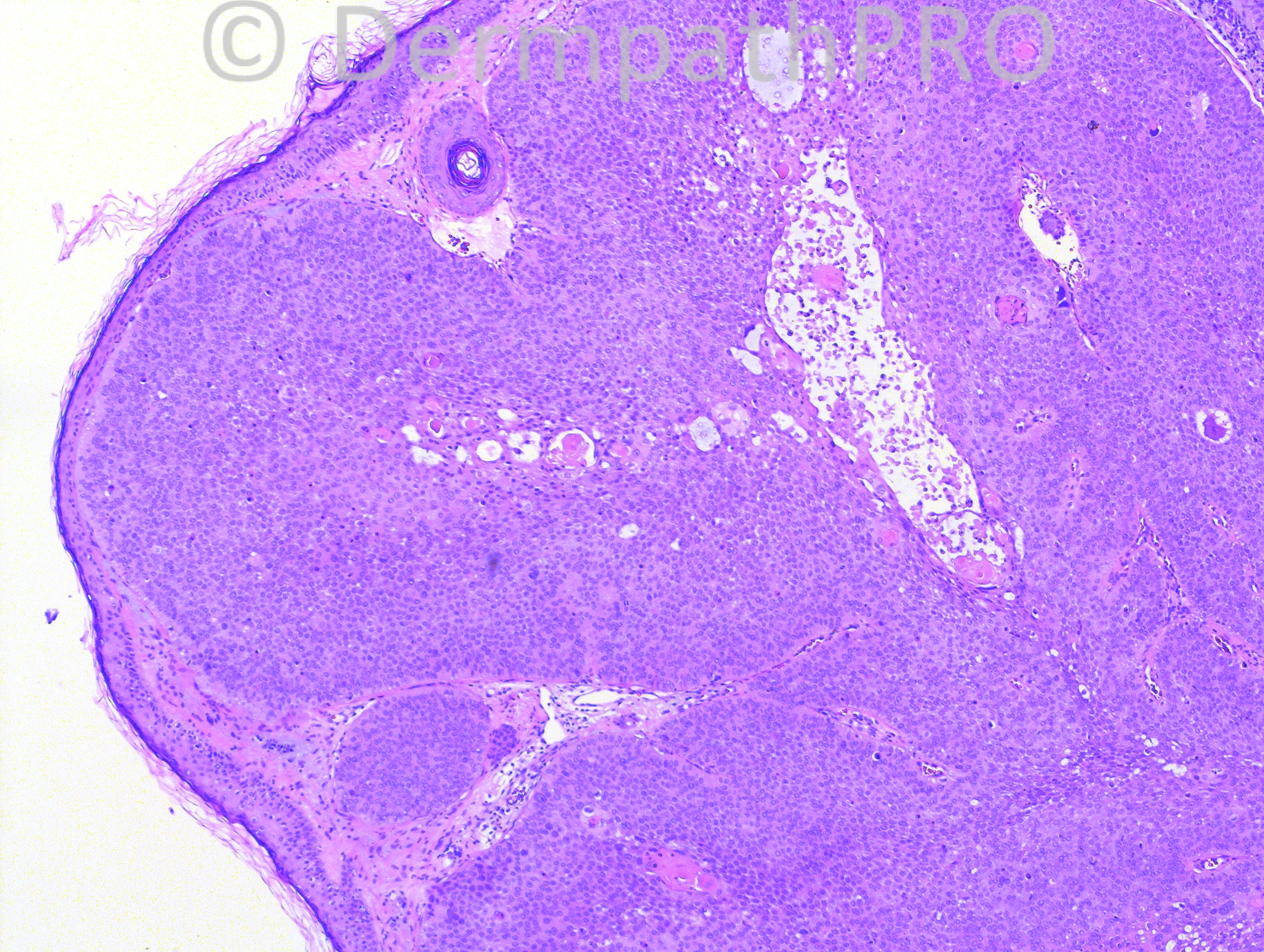
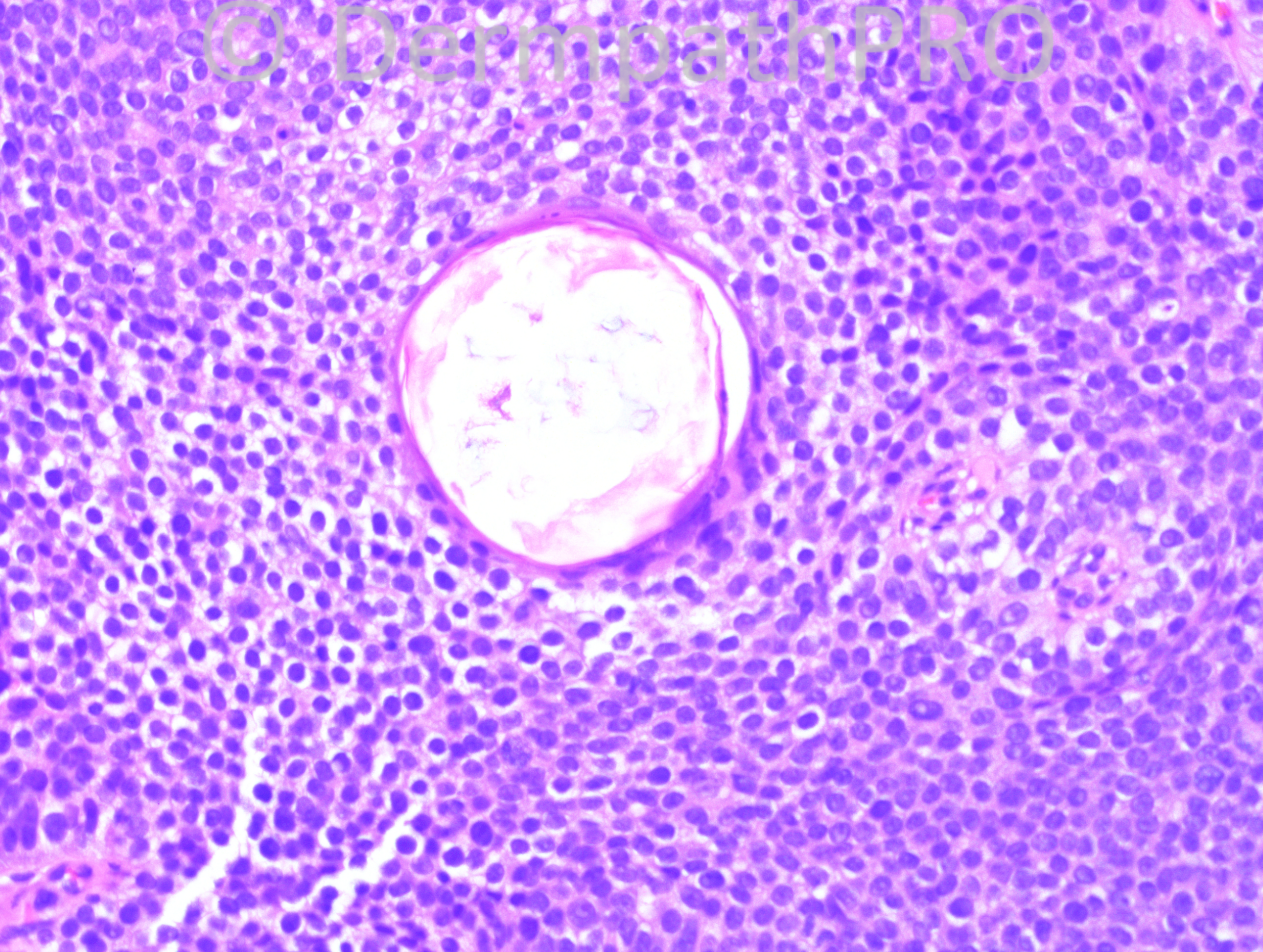
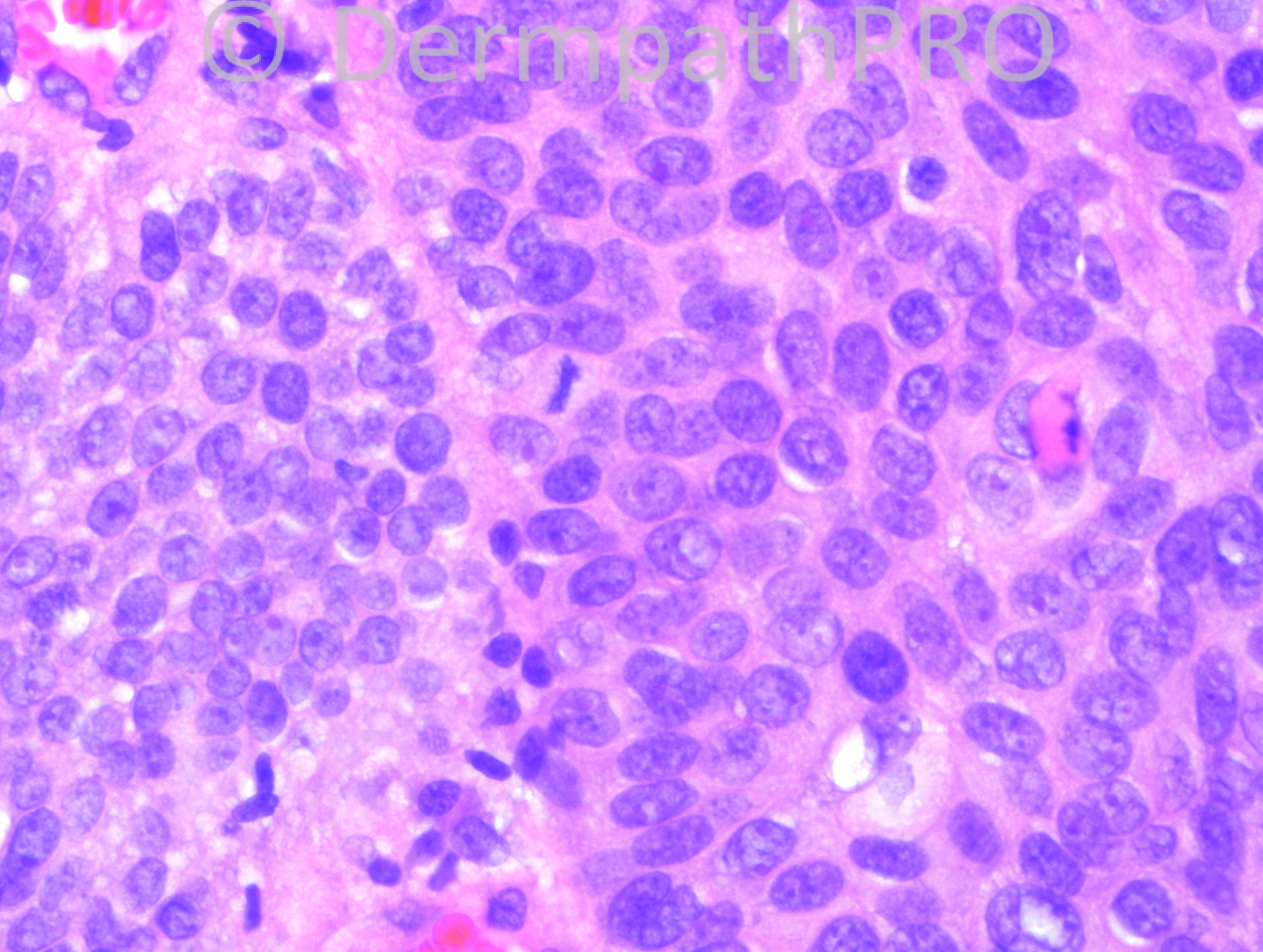
68-year-old male with “irritated” lesion on right pinna.


Case posted by Dr. Hafeez Diwan.


  Report Record

User Feedback


it looks poroid to me- most of it looks like a poroid hidradenoma-, but taking into account that the last images show convincing cytologic atypia and some necrosis and that hidradenocarcinomas can look quite bland, I would call this a carcinoma-poroid hidradenocarcinoma. it would be good to see the whole architecture of this lesion though( areas of infiltration etc).

Share this comment


Link to comment
share_externally

Guest Rodrigo Restrepo

Posted

[color=#1C2837][font=arial, verdana, tahoma, sans-serif][size=4]Porocarcinoma[/size][/font][/color]

Share this comment


Link to comment
share_externally

Porocarcinoma. Poroid cells with areas of clear cytoplasma, ductular formation, atypical cells and some mitoses.

Share this comment


Link to comment
share_externally

Guest Jim Davie MD

Posted

Favor Porocarcinoma (based on these images).[list]
[*]There is edematous vascular stroma, with focal retraction artifact, and ductal differentiation. The cells are medium to large, rounded, eosinophilic to focally glycogenated, with apoptosis and high mitotic activity. Some cells show possible intracytoplasmic lumens. There is subtle peripheral palisading of expansile, bosselated tumor nodules (best seen in first image), with squamous metaplasia.
[*]Hidradenocarcinoma is in the differential (we are not seeing the entire lesion here), but these are rarer; I like seeing a greater clear-cell component.
[*]Immunostains for EMA or CEA (+ in lumens), and BER-EP4 (negative, to r/o a metatypical BCC ) would be useful for confirmation, if H&E is equivocal.
[/list]

Share this comment


Link to comment
share_externally

Guest Romualdo

Posted

I think continuity with surface epidermis, at least focal, is necessary (definitional feature) to the diagnosis of porocarcinoma. I am not able to see any continuity, at least in the images shown, so I favor the diagnosis of hidradenocarcinoma.

Share this comment


Link to comment
share_externally

Robledo F. Rocha

Posted

I go with porocarcinoma just because non-anaplastic cells are reminiscent of poroid cells. I would expected a hidradenocarcinoma with prominent clefting between the neoplastic lobules and the desmoplastic stroma.

Share this comment


Link to comment
share_externally

Dr. Hafeez Diwan

Posted

Agreed. I called this porocarcinoma.

Share this comment


Link to comment
share_externally


Join the conversation

You can post now and register later. If you have an account, sign in now to post with your account.

Guest
Add a comment...

×   Pasted as rich text.   Paste as plain text instead

  Only 75 emoji are allowed.

×   Your link has been automatically embedded.   Display as a link instead

×   Your previous content has been restored.   Clear editor

×   You cannot paste images directly. Upload or insert images from URL.

Loading...

×
×
  • Create New...