Jump to content
In this section we have spot diagnoses posted on a daily basis since June 2010, now over 4000! You can review the archived cases and read the suggested diagnoses by users and the final comment by the contributors.
Case are uploaded each week day by 10 am UK time with the correct diagnosis will generally be posted at 8 pm UK time. Why not view the most recent spot diagnosis and proffer a diagnosis?

Case Number : Case 836 - 30th August Posted By: Guest

Please read the clinical history and view the images by clicking on them before you proffer your diagnosis.
Submitted Date :
   (0 reviews)

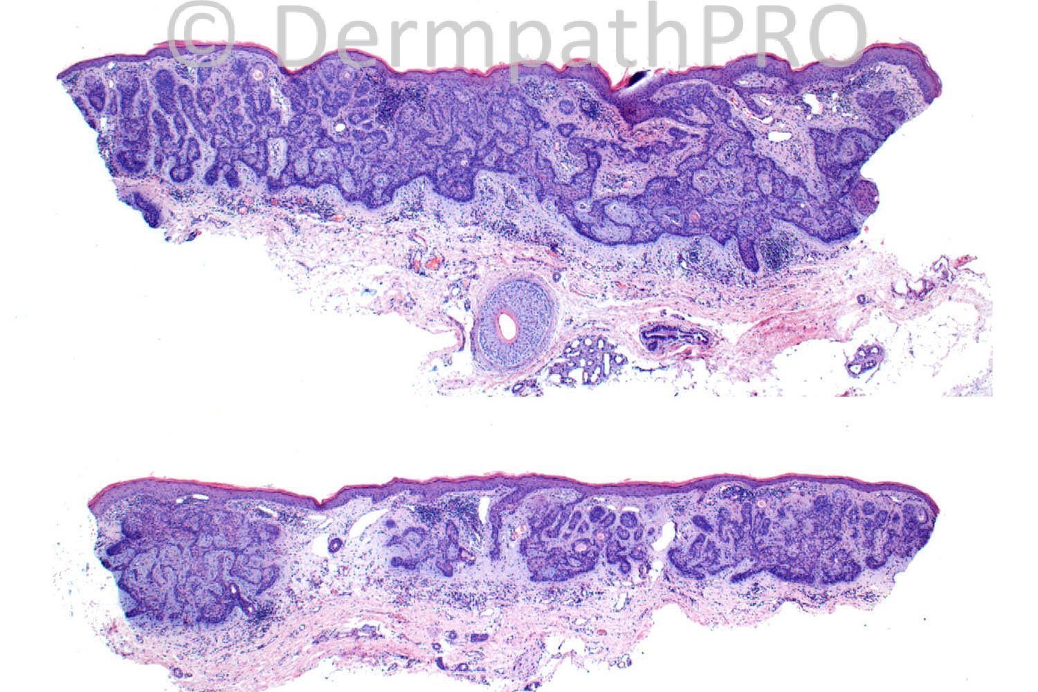
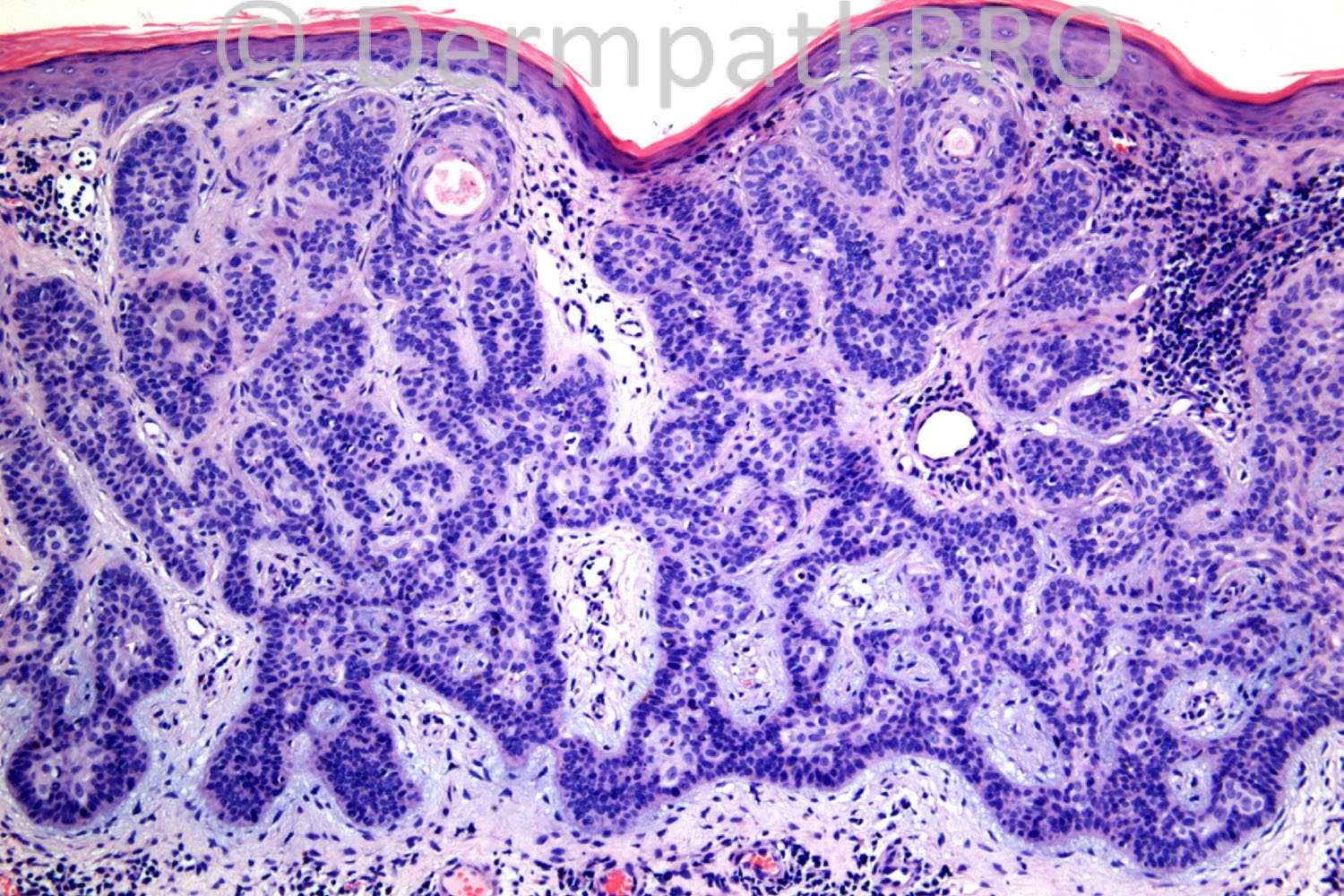
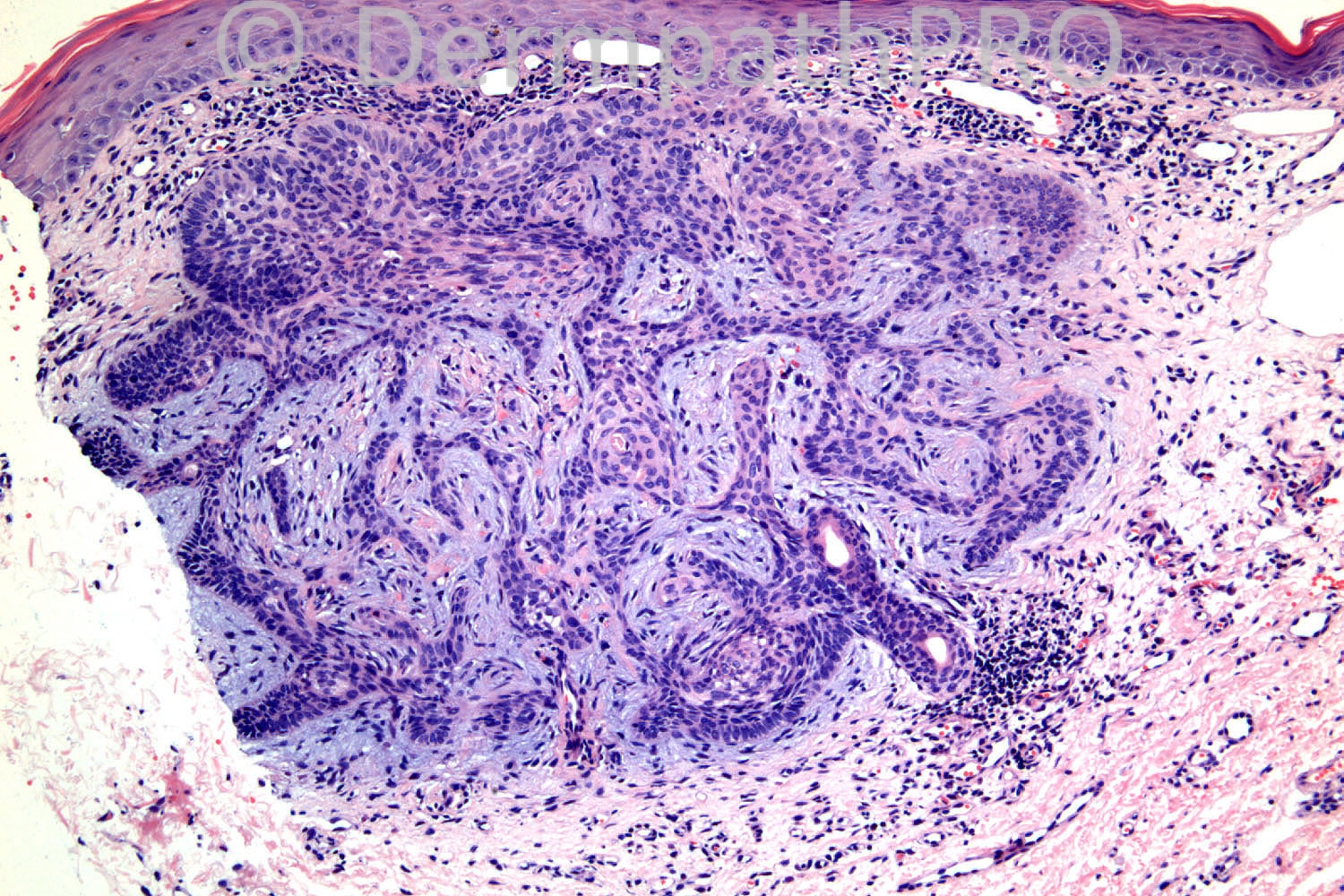
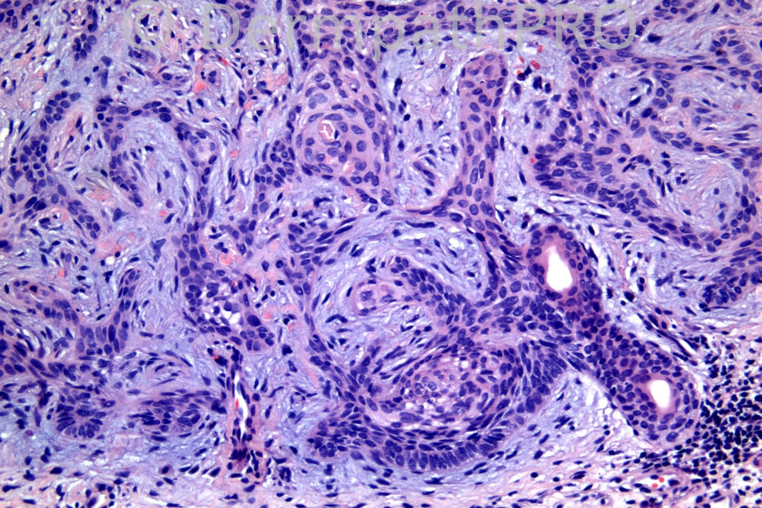
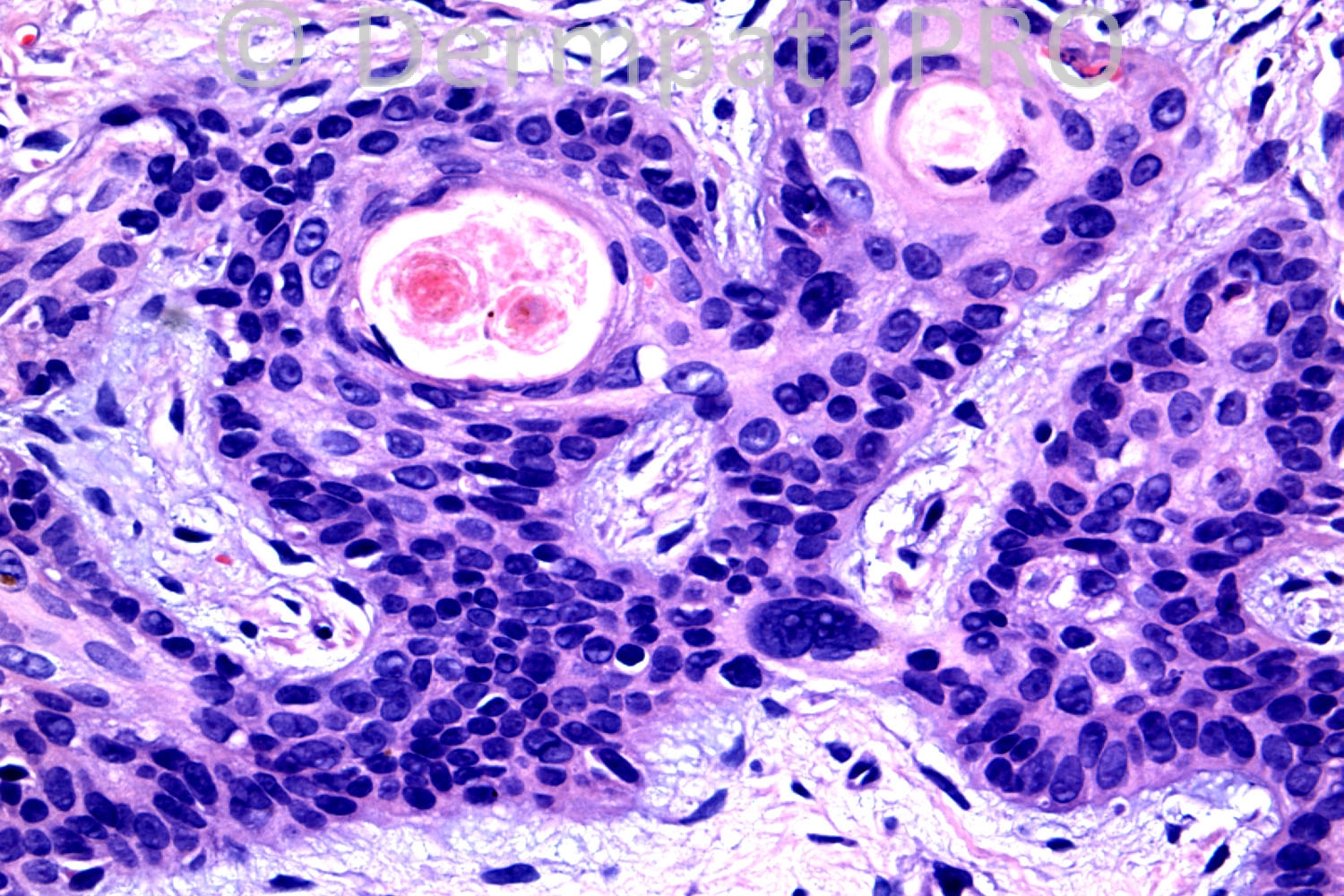
71 years old male. 25 year scaly, sore, purple, plaque in groin, gradual increase in size. Some red and indurated areas. Has psoriasis. ?MF

Case posted by Dr. Richard Carr


  Report Record

User Feedback


My differential diagnosis would be :
Fibroepithelioma of Pinkus
Tumor of the follicular infudibulum
Trichoepithelioma
clinically Fibroepithelioma of pinkus fits more ,
the connection to the epidermis would exlude trichoepithelioma
the proliferation of basalod cell and not keratinocytes would favor a diagnosis of Pinkus which the pt could have for years, any way this is a punch biopsy and complete excision would be more helpful with immunos.

Share this comment


Link to comment
share_externally

Guest Amira Tawdy

Posted

acrosyringial nevus vs syringocyctadenoma papilliferum vs fibroepithelioma of pinkus but i favour the first two because of the presence of ducts

Share this comment


Link to comment
share_externally

Guest Engin Sezer

Posted

FEP which is already a trichoblastoma

Share this comment


Link to comment
share_externally

Dr. Mona Abdel-Halim

Posted

Syringofibroadenoma (acrosyringeal nevus)

Share this comment


Link to comment
share_externally

Guest Giovanni Falconieri

Posted

Agree, favor Pinkus FE due to pronounced reticulated pattern plus fibromucinous surrounding stroma. Small cysts filled with keratin and lacking granular cell layer might suggest infundibulocystic BCC at an odd site.

Share this comment


Link to comment
share_externally

Guest Maria George

Posted

add chondroid syringioma (Mixed tumor of skin).

Share this comment


Link to comment
share_externally

Robledo F. Rocha

Posted

I found this lesion hard to classify, but I think three diagnosis are possible, all of them characterized by anastomosing epithelial strands connected to the undersurface of the epidermis.

[url="http://journals.lww.com/amjdermatopathology/Abstract/1994/12000/Fibroepithelioma_of_Pinkus__Eccrine_Duct_Spread_of.2.aspx"]Fibroepithelioma of Pinkus with eccrine duct spreading[/url].
Supportive findings: basaloid cells; peripheral palisading.
Adverse findings: lesion is not typically located on the trunk; there’s no persuasive specific follicular stroma; follicular papillae are lacking.

[url="http://www.ncbi.nlm.nih.gov/pmc/articles/PMC3120003/pdf/ad-23-70.pdf"]Syringofibroadenoma[/url].
Supportive findings: fibrovascular mucinous stroma with lymphocytic infiltrate; ductal elements within strands; areas of squamous cells.
Adverse findings: peripheral palisading.

[url="http://journals.lww.com/amjdermatopathology/pages/articleviewer.aspx?year=2009&issue=10000&article=00002&type=abstract"]Tumor of the follicular infundibulum with ductal differentiation[/url].
Supportive findings: peripheral palisading.
Adverse findings: lesion is not typically located on the face; cells have scant rather than abundant pale eosinophilic cytoplasm; strands are not parallel to the skin surface.

Share this comment


Link to comment
share_externally

Guest Jim Davie MD

Posted

BCC, FEP-type
The basaloid neoplasm focally involves a normal eccrine duct , has cytologic atypia, peripheral palisading, mucinous stroma and infundibulocystic squamous metaplasia.

Share this comment


Link to comment
share_externally

Guest Romualdo

Posted

Fenestrated trichoblastoma (fibroepithrlioma of Pinkus).

Share this comment


Link to comment
share_externally

Eman El-Nabarawy

Posted

Favor Fibroepithelioma of Pinkus with eccrine duct spreading.

Share this comment


Link to comment
share_externally

Dr. Richard Carr

Posted

I reported this as a fibroepithelioma-(Pinkus-)like BCC. The lesion lacks the stromal condensations of papillary mesenchymal cells and longer fenestrations of a bona fide Pinkus tumour. Immunos were helpful as the lesion lacked reactive Merkel cells and CD10 stromal cells that are always seen in Pinkus tumour (a fenestrated trichoblastoma). I would therefore agree with the designation of infundibulocystic BCC, Pinkus-like.

Share this comment


Link to comment
share_externally


Join the conversation

You can post now and register later. If you have an account, sign in now to post with your account.

Guest
Add a comment...

×   Pasted as rich text.   Paste as plain text instead

  Only 75 emoji are allowed.

×   Your link has been automatically embedded.   Display as a link instead

×   Your previous content has been restored.   Clear editor

×   You cannot paste images directly. Upload or insert images from URL.

Loading...

×
×
  • Create New...