Jump to content
In this section we have spot diagnoses posted on a daily basis since June 2010, now over 4000! You can review the archived cases and read the suggested diagnoses by users and the final comment by the contributors.
Case are uploaded each week day by 10 am UK time with the correct diagnosis will generally be posted at 8 pm UK time. Why not view the most recent spot diagnosis and proffer a diagnosis?

Case Number : Case 667 - 3 Jan Posted By: Guest

Please read the clinical history and view the images by clicking on them before you proffer your diagnosis.
Submitted Date :
   (0 reviews)

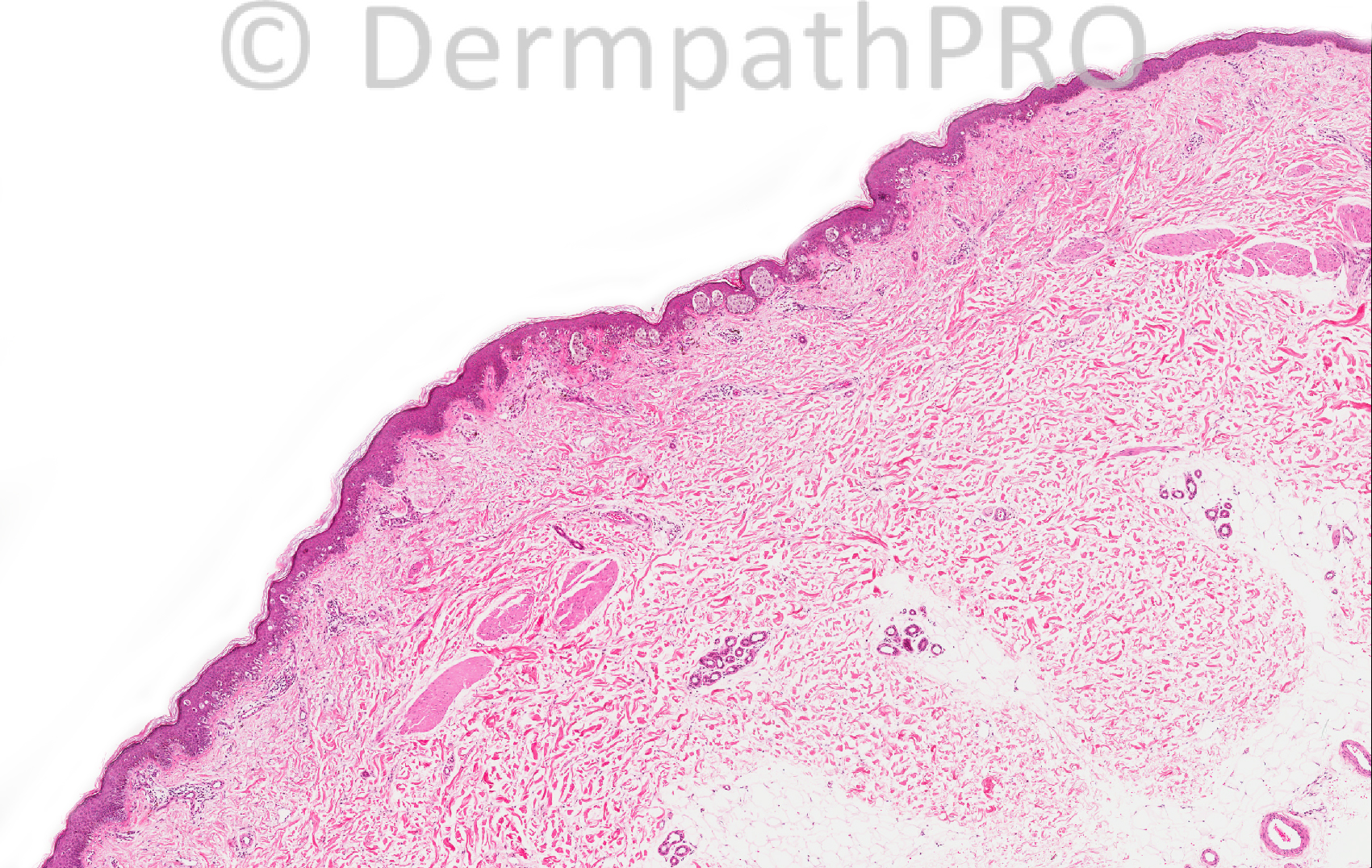
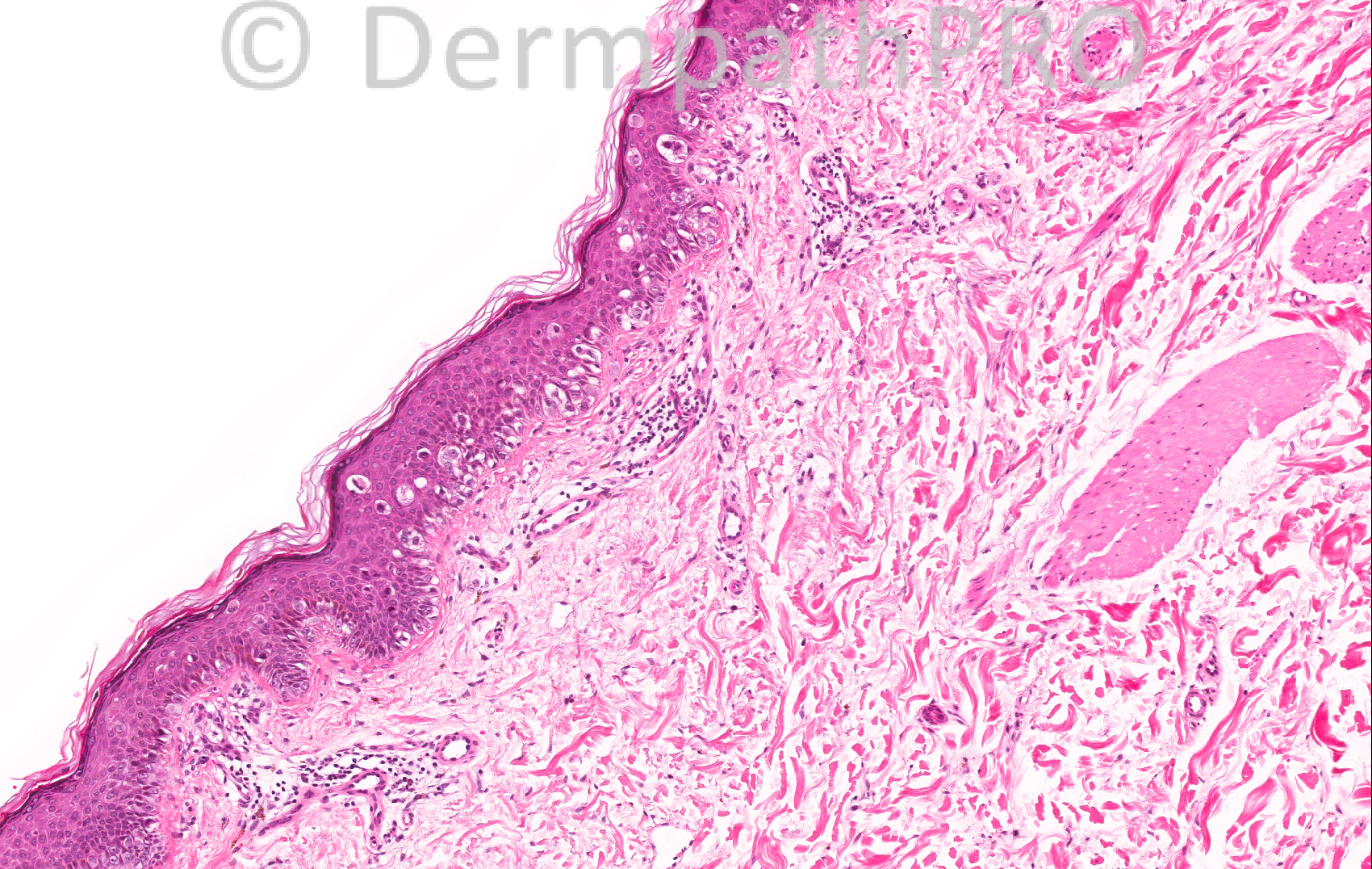
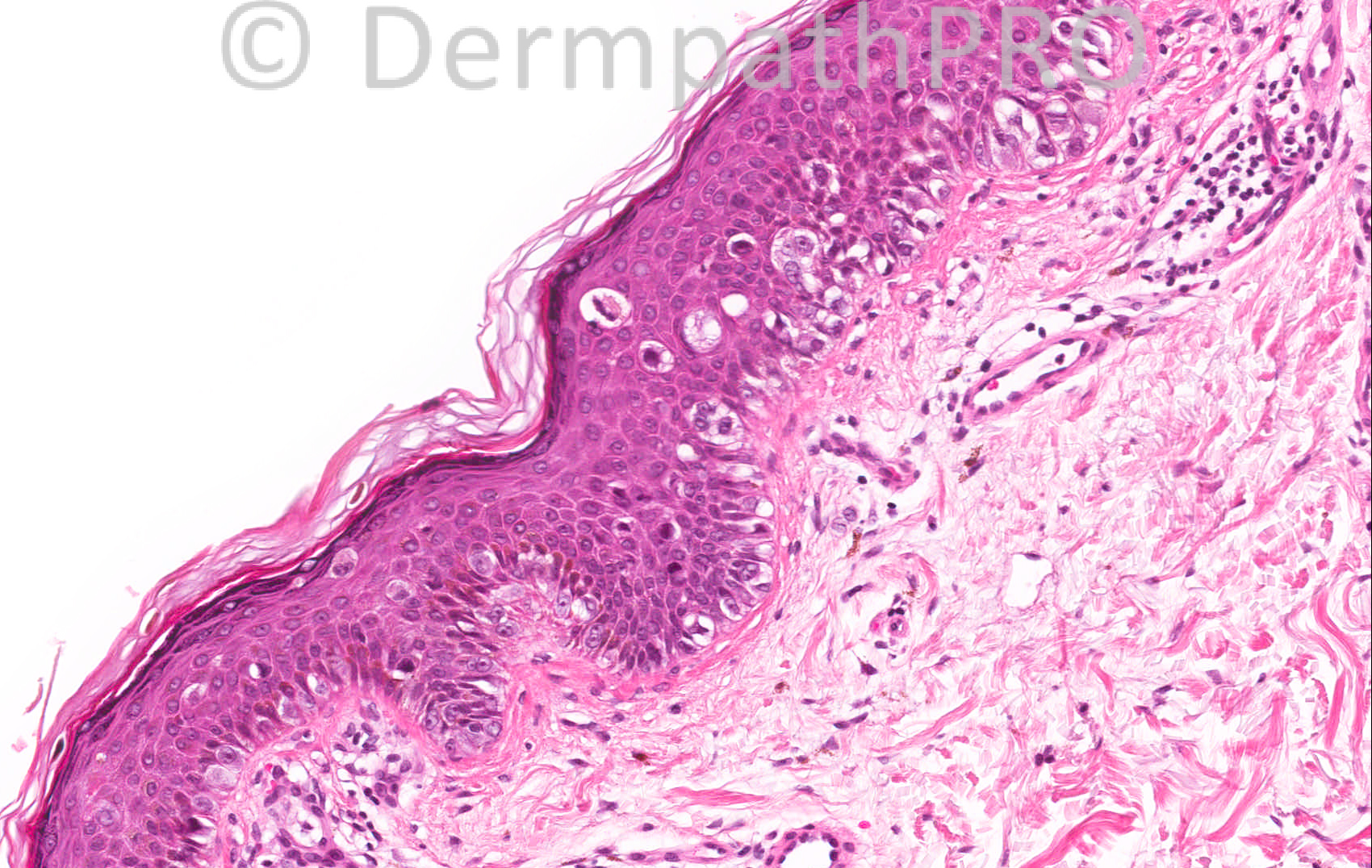
42 year old male, pigmented lesion on back


  Report Record

User Feedback


Azza Esmat

Posted

solar lentigo (in situ melanoma)

Share this comment


Link to comment
share_externally

Guest Hazem Hamed

Posted

I can not see evidence of solar damage in the dermis. Though there is a bit of fibrosis of the papillary dermis I would favor superficial spreading melanoma over a junctional dysplastic nevus.

Share this comment


Link to comment
share_externally

Guest Dr Engin Sezer

Posted

Melanoma in situ arising on dysplastic naevus.

Share this comment


Link to comment
share_externally

Arif Usmani

Posted

Melanoma in-situ vs Intraepidermal Spitz’s nevus. Favor MIS as I do not see convincing Kamino bodies. In addition there seems to be asymmetry and lack of circumscription.

Share this comment


Link to comment
share_externally

Azza Esmat

Posted

In situ superficial spreading melanoma. Agree no solar damage & site not for lentigo, but epidermis slight flat.

Share this comment


Link to comment
share_externally

Dr. Mona Abdel-Halim

Posted

SSM, in situ phase mostly arising on top of a junctional dysplastic nevus.

Share this comment


Link to comment
share_externally

In situ MM arising on dysplastic naevus

Share this comment


Link to comment
share_externally

Dr. Phillip McKee

Posted

I think that the whole lesion is [i]in situ [/i]melanoma. I believe that the nests are also part of the lesion. I do not think that there is any evidence of a pre-existent dysplastic nevus. Many thanks for your comments. [color=#FF0000][b] I wanted to remind you about the difficult case module which is free. We will be adding a couple of new ones next week but it would be great if you could submit problem cases of your own. It would help to make the site more dynamic. It does not commit you to anything and just might help you sign out a case that has been worrying you. Phillip[/b][/color]

Share this comment


Link to comment
share_externally


×
×
  • Create New...