Jump to content
In this section we have spot diagnoses posted on a daily basis since June 2010, now over 4000! You can review the archived cases and read the suggested diagnoses by users and the final comment by the contributors.
Case are uploaded each week day by 10 am UK time with the correct diagnosis will generally be posted at 8 pm UK time. Why not view the most recent spot diagnosis and proffer a diagnosis?

Case Number : Case 673 - 11 Jan Posted By: Guest

Please read the clinical history and view the images by clicking on them before you proffer your diagnosis.
Submitted Date :
   (0 reviews)

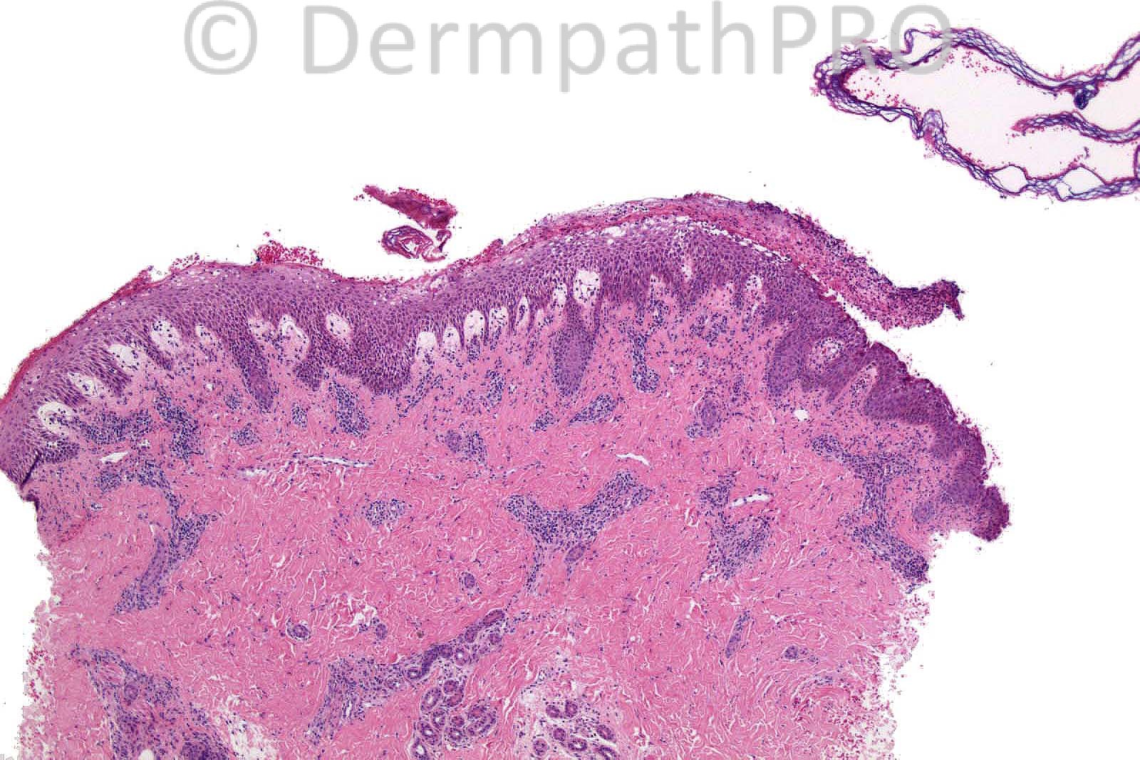
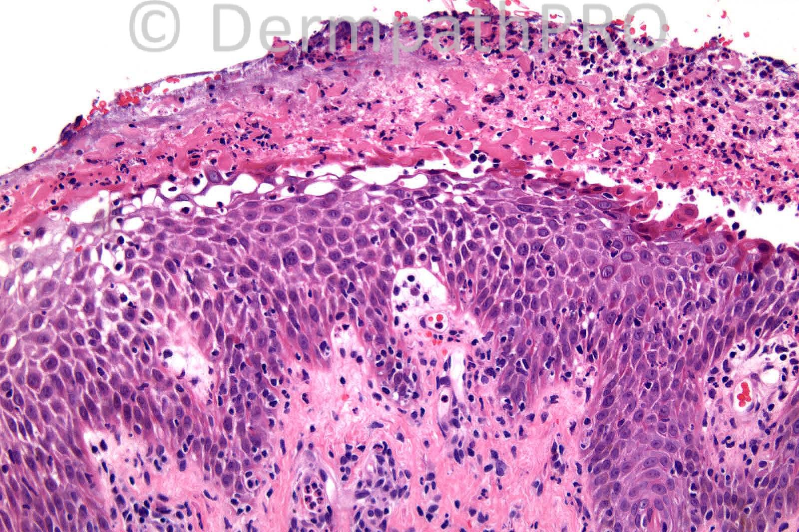
4 years-old male. Bullous lesion on leg 7/7.

Case posted by Dr. Richard Carr.


  Report Record

User Feedback


Bullous dermatosis, lesion showing a subepdermal blister containig a few neutrophils, eytrocytes and lymphocytes. The dermal papillary connective tissue remiscent of dermatotis herpetiformis. The dermis contains a perivascular inflammatory cell infiltrat. Need immunfluorescence. Bullous systemic lupus erythematosus/Chronic bullous disease of childhood/EBA?

Share this comment


Link to comment
share_externally

Dr. Mona Abdel-Halim

Posted

I think the real plan of cleavage is subcorneal, the papillary dermis shows edema. There is few acantholytic cells in the blister cavity together with lots of neutrophils. Also there is spongiosis and exocytosis of neutrophils and lymphocytes. Together with clinical correlation, this is bullous impetigo.

Share this comment


Link to comment
share_externally

Sasi Attili

Posted

[quote name='Dr. Mona Abdel Halim' timestamp='1357913357']
I think the real plan of cleavage is subcorneal, the papillary dermis shows edema. There is few acantholytic cells in the blister cavity together with lots of neutrophils. Also there is spongiosis and exocytosis of neutrophils and lymphocytes. Together with clinical correlation, this is bullous impetigo.
[/quote] Agree

Share this comment


Link to comment
share_externally

Dr. Richard Carr

Posted

Bullous impetigo is correct and Mona's description and clinical correlation
perfect!! Nice clue to the plane of cleavage is the floating
stratum corneum sign (top right). This can also be seen in superficial pemphigus.
Bacterial cocci were present on H&E (at higher power) and confirmed on Gram
(I did not want to make it too easy). Neutrophils have very potent enzymes and
can produce acantholysis in bullous impetigo which is favoured given the clinical
information (but superficial and IgA pemphigus would be in the differential
histologically).

Regards to all and best wishes for 2013.

Share this comment


Link to comment
share_externally

nice bullous impetigo. thanks

Share this comment


Link to comment
share_externally


×
×
  • Create New...