Jump to content
In this section we have spot diagnoses posted on a daily basis since June 2010, now over 4000! You can review the archived cases and read the suggested diagnoses by users and the final comment by the contributors.
Case are uploaded each week day by 10 am UK time with the correct diagnosis will generally be posted at 8 pm UK time. Why not view the most recent spot diagnosis and proffer a diagnosis?

Case Number : Case 682 - 24 Jan Posted By: Guest

Please read the clinical history and view the images by clicking on them before you proffer your diagnosis.
Submitted Date :
   (0 reviews)

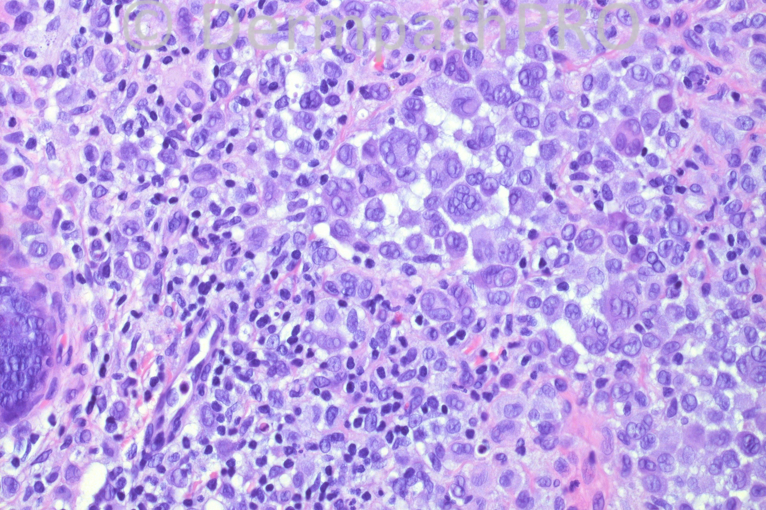
45-years-old male with a lesion on his right nose, with the clinical of nevus vs. BCC. Patient has a pending kidney biopsy performed for reasons that I am not aware of.

Case posted by Dr Hafeez Diwan


  Report Record

User Feedback


Sasi Attili

Posted

I am struggling in the absence of the physical slide to view the silhouette etc. Given the inflammatory cells and the pleomorphism, I am considering a naevoid melanoma.

Share this comment


Link to comment
share_externally

Robledo F. Rocha

Posted

A dermal melanocytic nevus infiltrated by lymphocytes and histiocytes. I think it is a halo nevus, even though there is no notice of a border of depigmentation. The giant nevocytes suggest a long-standing lesion that became a source of concern after the arrival of the inflammatory cells, or it may mean reactive cytologic atypia. I did not find any mitotic figure.

Share this comment


Link to comment
share_externally

Dr. Mona Abdel-Halim

Posted

I thought of nevoid melanoma...

Share this comment


Link to comment
share_externally

Dr. Richard Carr

Posted

Kidney may be a clue!!

Share this comment


Link to comment
share_externally

Arif Usmani

Posted

I would rule out a nevoid melanoma. I do not like the sheet-like syncytial growth pattern without apparent maturation.

Share this comment


Link to comment
share_externally

Dr. Hafeez Diwan

Posted

Obviously melanoma has to be ruled out. So I did immunos and the proliferation of larger cells is negative for melanocytic markers and is positive for CD1a. So, I interpreted this as an isolated focus of Langerhans cell histiocytosis. The kidney has been biopsied, but I don't know what it showed.

Share this comment


Link to comment
share_externally

Dr. Hafeez Diwan

Posted

And of course there is a nevus there, so I meant to say that this is a focus of LCH in association with a nevus.

Share this comment


Link to comment
share_externally

Sasi Attili

Posted

[quote name='Dr. Hafeez Diwan' timestamp='1359062242']
Obviously melanoma has to be ruled out. So I did immunos and the proliferation of larger cells is negative for melanocytic markers and is positive for CD1a. So, I interpreted this as an isolated focus of Langerhans cell histiocytosis. The kidney has been biopsied, but I don't know what it showed.
[/quote]

Dr. Diwan. Thanks for this interesting and intriguing case. Do you think this phenomenon is recruitment of the LHC by the melanocytes? DC and LCH recruitment has been described in melanoma. http://www.ncbi.nlm.nih.gov/pubmed/12754747

Share this comment


Link to comment
share_externally

Dr. Mona Abdel-Halim

Posted

Interesting :-)

Share this comment


Link to comment
share_externally

Dr. Richard Carr

Posted

Nice kidney shape nucleus(and horse shoe-kidney shapes) in this case were a clue
to do CD1a (just in case anyone missed my point). There is an eosinophil as well.

Share this comment


Link to comment
share_externally

Sasi Attili

Posted

[quote name='Dr. Richard Carr' timestamp='1359103741']
Nice kidney shape nucleus(and horse shoe-kidney shapes) in this case were a clue
to do CD1a (just in case anyone missed my point). There is an eosinophil as well.
[/quote]

In the context of a melanocytic lesion, given that melanocytes can take any shape/ form, I have to say I did not even entertain LC's. So coming to my question Richard, would you call this 'LC's associated with a naevus' or 'LCH and a nevus'. When do a group of LC's qualify to be called LCH? LCH would mean that we have to screen for systemic involvement. But are naevus associated LC's associated with systemic disease (and therefore= LCH) or are they just reactive?

Share this comment


Link to comment
share_externally

Dr. Hafeez Diwan

Posted

Very interesting comments. I run into this question often: when do LC's get to be enough to be called LCH. There can be solitary LCH; in my view, this aggregate is quite prominent and more than the usual scattered LC's. So I think this is LCH in a nevus. They could have been recruited and have formed a small nodule.

Share this comment


Link to comment
share_externally

Dr. Richard Carr

Posted

Hi Sasi,

Agree with Hafeez on this one I would call it LCH and collision of incidental naevus. Can be difficult to decide but this looks like a relatively pure nodule of LCH to me. I just can't believe this innocent presumed longstanding naevus has any relation to the LCH.

Share this comment


Link to comment
share_externally


×
×
  • Create New...