Jump to content
In this section we have spot diagnoses posted on a daily basis since June 2010, now over 4000! You can review the archived cases and read the suggested diagnoses by users and the final comment by the contributors.
Case are uploaded each week day by 10 am UK time with the correct diagnosis will generally be posted at 8 pm UK time. Why not view the most recent spot diagnosis and proffer a diagnosis?

Case Number : Case 686 - 31 Jan Posted By: Guest

Please read the clinical history and view the images by clicking on them before you proffer your diagnosis.
Submitted Date :
   (0 reviews)

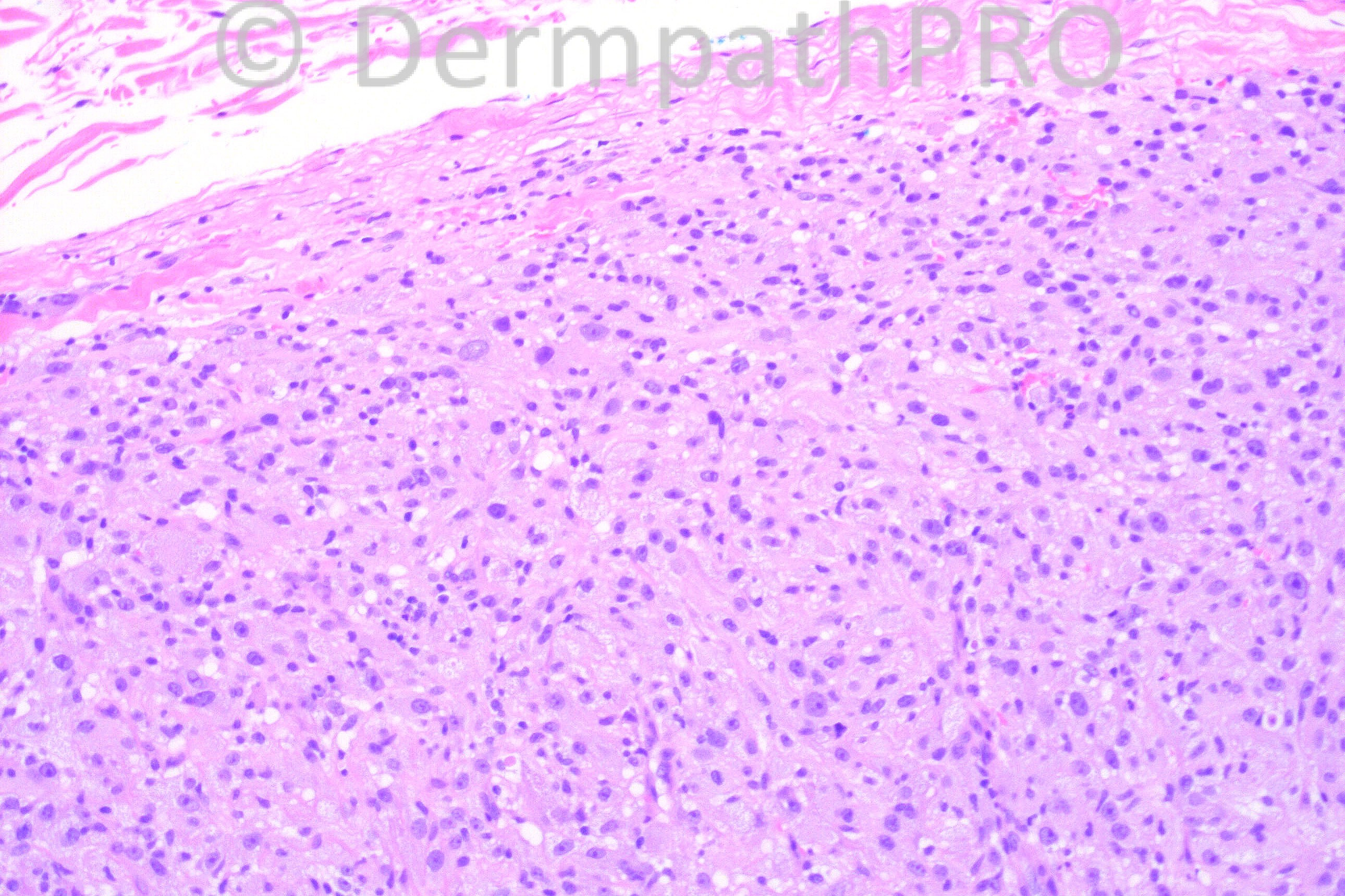
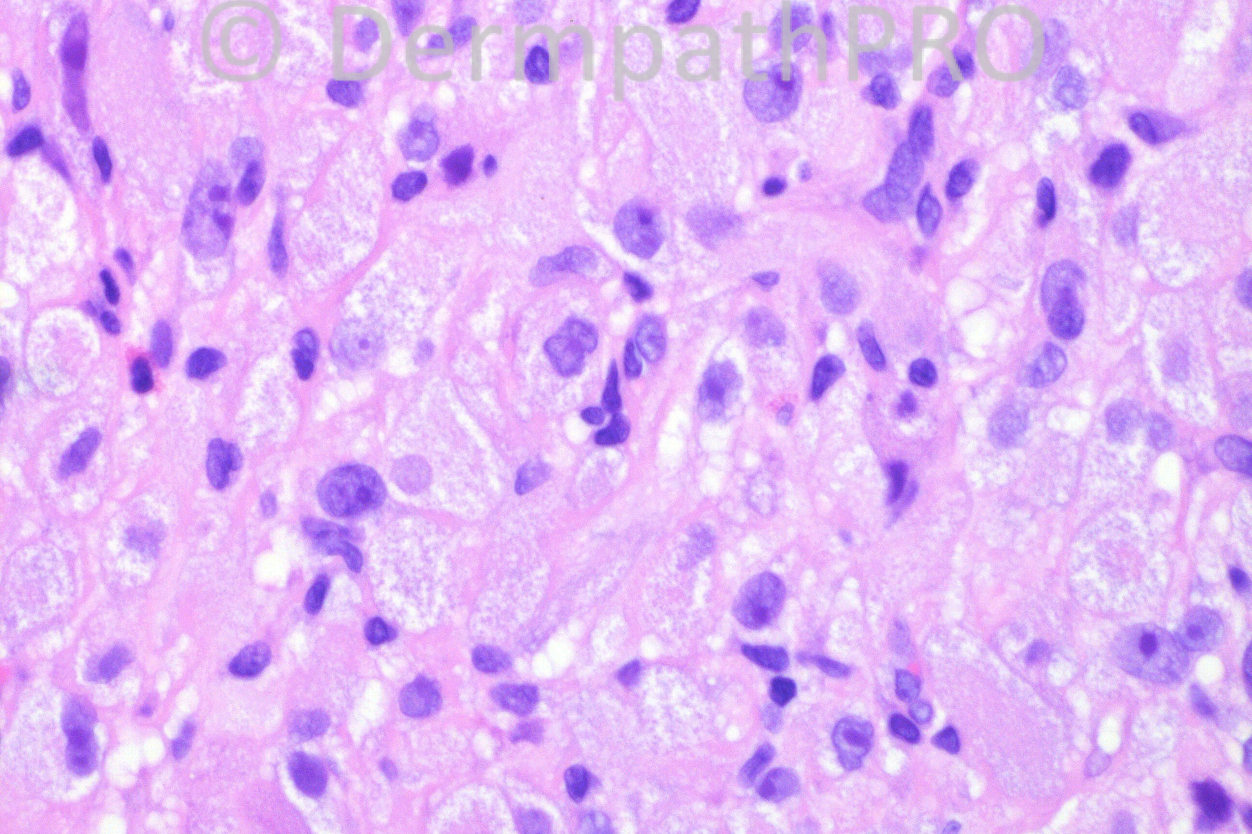
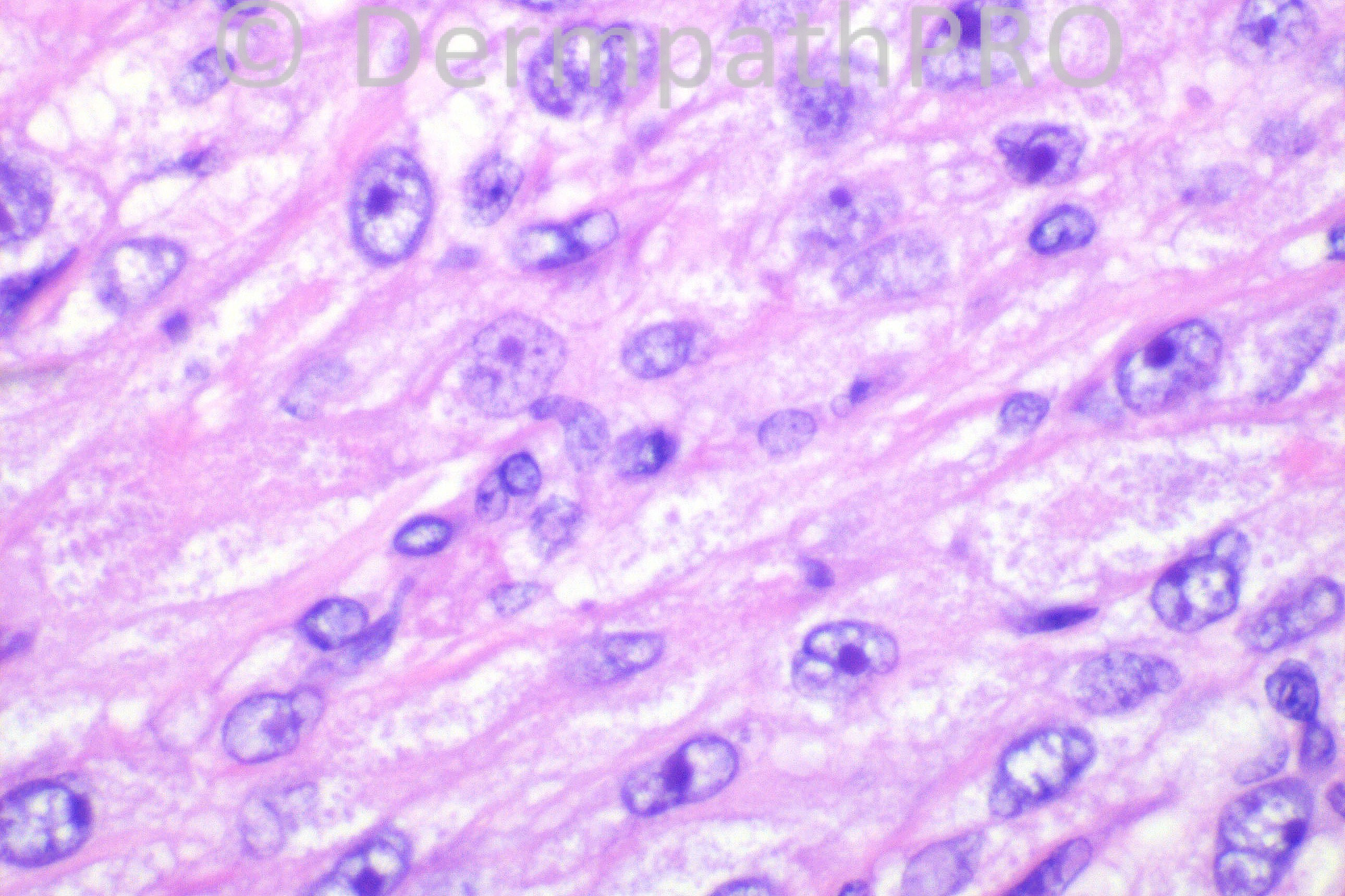
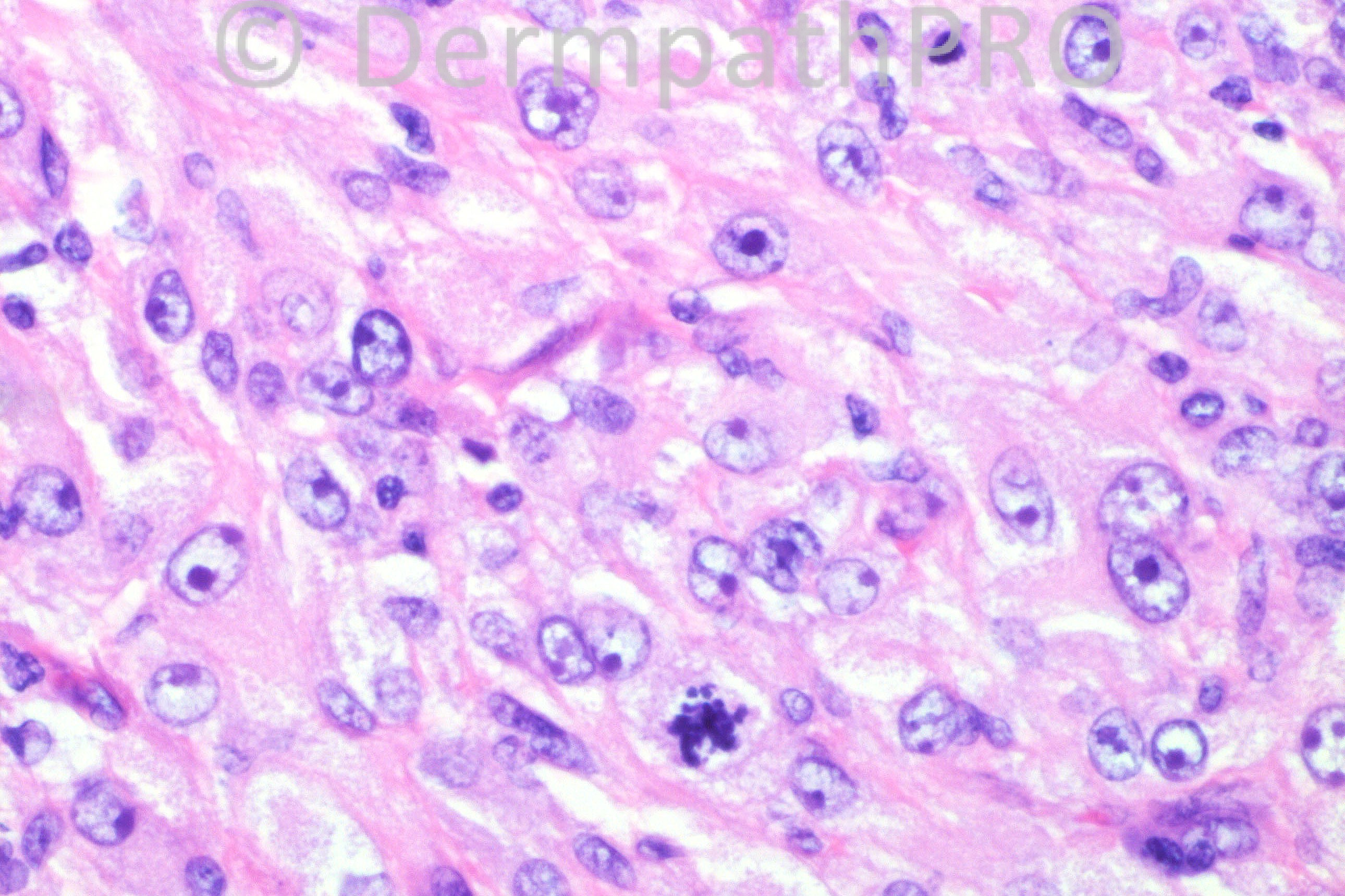
29 years-old female with a 1 cm black lesion on her left forearm. The lesion was biopsied. This tumor was negative for S-100, Mart-1, and Keratin, and was positive for CD163.

Case posted by Dr. Hafeez Diwan.


  Report Record

User Feedback


Robledo F. Rocha

Posted

Great-sized cells with abundant foamy eosinophilic cytoplasm. The nuclei are large and pleomorphic, showing oval or cleaved outlines. Nucleoli are prominent. An atypical mitotic figure can be found. I won’t hesitate calling this a malignant tumor. Immunohistochemical study suggests histiocytic sarcoma.

Share this comment


Link to comment
share_externally

Agree , malignant hisiocytosis/histiocytic sarcoma. pleomorphic foamy histiocytes with prominant nucleoli & atypical mitoses. few eosinophils.

Share this comment


Link to comment
share_externally

Arif Usmani

Posted

This appears to be a histiocytic lesion with granular cell change. I would also include infection, foreign body reaction and benign neoplasms such as granular cell dermatofibroma in the differential.

Share this comment


Link to comment
share_externally

Guest Hazem Hamed

Posted

Agree with histiocytic sarcoma. Though the cells are large, I would also consider myeloid sarcoma with monoblastic differentiation, and follicular dendritic cell sarcoma as an H&E Differential diagnosis.

Share this comment


Link to comment
share_externally

Dr. Mona Abdel-Halim

Posted

I perceived the cells as having granular cytoplasm not foamy. Thought of primitive non neural granular cell tumor ( I know it is S100 _ve and CD68 +ve), not aware of it's CD163 positivity???. It contain vesicular nuclei, small prominent nucleoli with some scattered atypia and mitoses,,, Also thought of granular cell dendrocytosis... I might however be out of the track !!!!!!!!!!!!!!

Share this comment


Link to comment
share_externally

My first thought was an S100 - granular cell tumor. I just don't see enough atypia to call this a histiocytic sarcoma. The cytoplasm is very granular, the nuclei while mildly atypical are not that pleomorphic, and the mitotic rate appears actually fairly low. I'd be surprised if this was malignant.

Share this comment


Link to comment
share_externally

Dr. Mona Abdel-Halim

Posted

For the CD163 and after some reading, I would suggest also the following DD: Granular cell variant of epithelioid cell histiocytoma...

Share this comment


Link to comment
share_externally

Dr. Hafeez Diwan

Posted

This was called non-neural (S-100 negative) malignant granular cell tumor.

Share this comment


Link to comment
share_externally

Agree with Dr Mona: Granular cell variant of epithelioid cell histiocytoma.
And also: non-neural granular cell tumor.
I wouldn't call it malignant, as those lesions can have many mitosis and prominent nucleoli.

Share this comment


Link to comment
share_externally

[color=#000000][font=arial, helvetica, clean, sans-serif][size=1]
[url="http://www.ncbi.nlm.nih.gov/pubmed/22885549#"]Am J Dermatopathol.[/url] 2012 Oct;34(7):766-9.[/size][/font][/color]
[b] [size=4]Granular[/size] [size=4]cell[/size] [size=4]variant[/size] of [size=4]epithelioid cell[/size] histiocytoma.[/b]
[color=#000000][font=arial, helvetica, clean, sans-serif][size=1]
[url="http://www.ncbi.nlm.nih.gov/pubmed?term=Rabkin%20MS%5BAuthor%5D&cauthor=true&cauthor_uid=22885549"]Rabkin MS[/url], [url="http://www.ncbi.nlm.nih.gov/pubmed?term=Vukmer%20T%5BAuthor%5D&cauthor=true&cauthor_uid=22885549"]Vukmer T[/url].[/size][/font][/color]

Share this comment


Link to comment
share_externally

Sorry Dr Diwan, I didn't see your post and that the case was closed! Thank you!

Share this comment


Link to comment
share_externally

Guest Jim Davie MD

Posted

An interesting and rare case!

The high-grade cytology is very similar to that seen in the S100+ granular cell tumor that was posted this week for expert review in the 'DIFFICULT OPINION CASES' area ([url="https://dermpathpro.com/difficult_opinion_cases_1/_/case-do013-r15"]case# DO013[/url]).

Share this comment


Link to comment
share_externally


×
×
  • Create New...