Jump to content
In this section we have spot diagnoses posted on a daily basis since June 2010, now over 4000! You can review the archived cases and read the suggested diagnoses by users and the final comment by the contributors.
Case are uploaded each week day by 10 am UK time with the correct diagnosis will generally be posted at 8 pm UK time. Why not view the most recent spot diagnosis and proffer a diagnosis?

Case Number : Case 794 - 3rd July Posted By: Guest

Please read the clinical history and view the images by clicking on them before you proffer your diagnosis.
Submitted Date :
   (0 reviews)

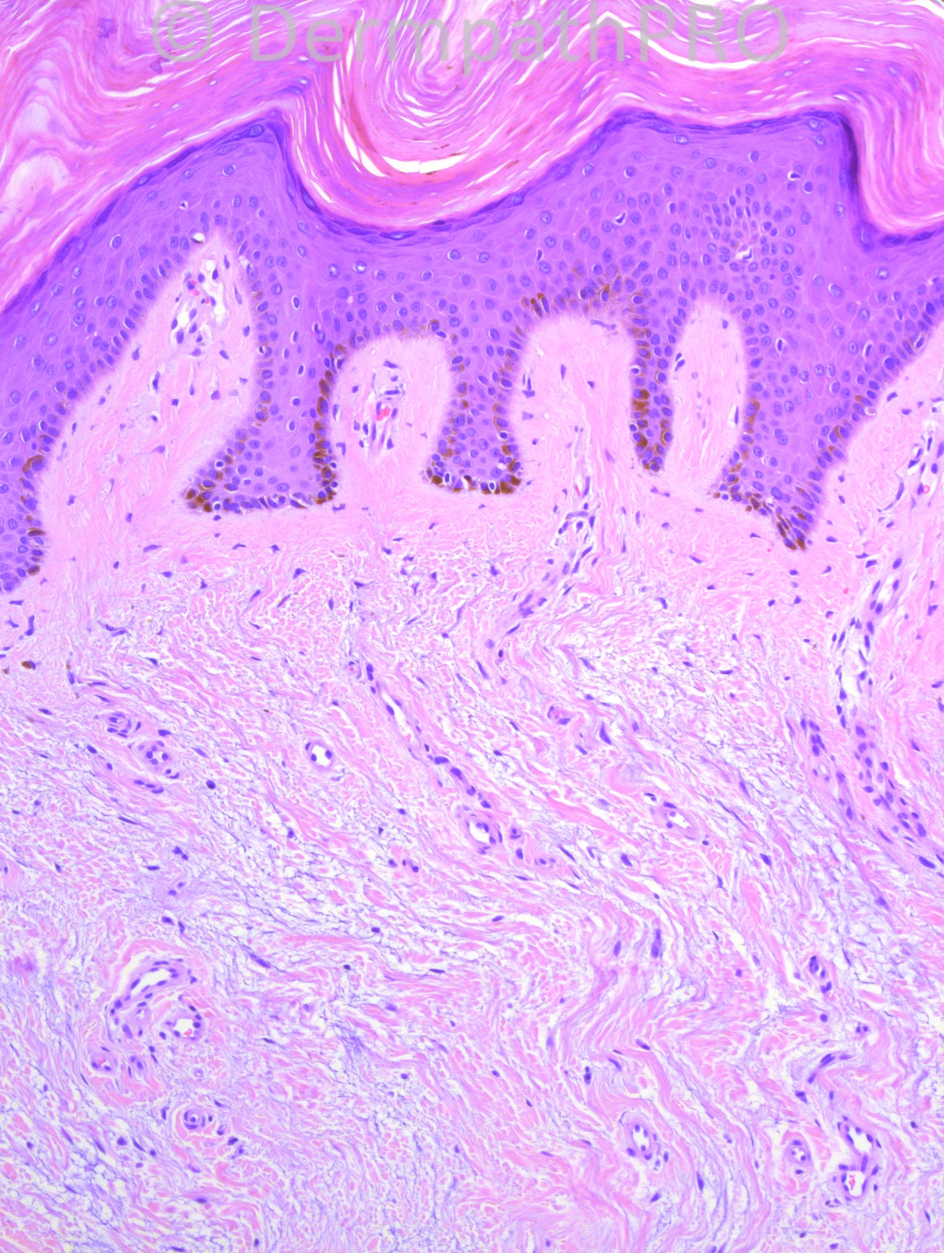
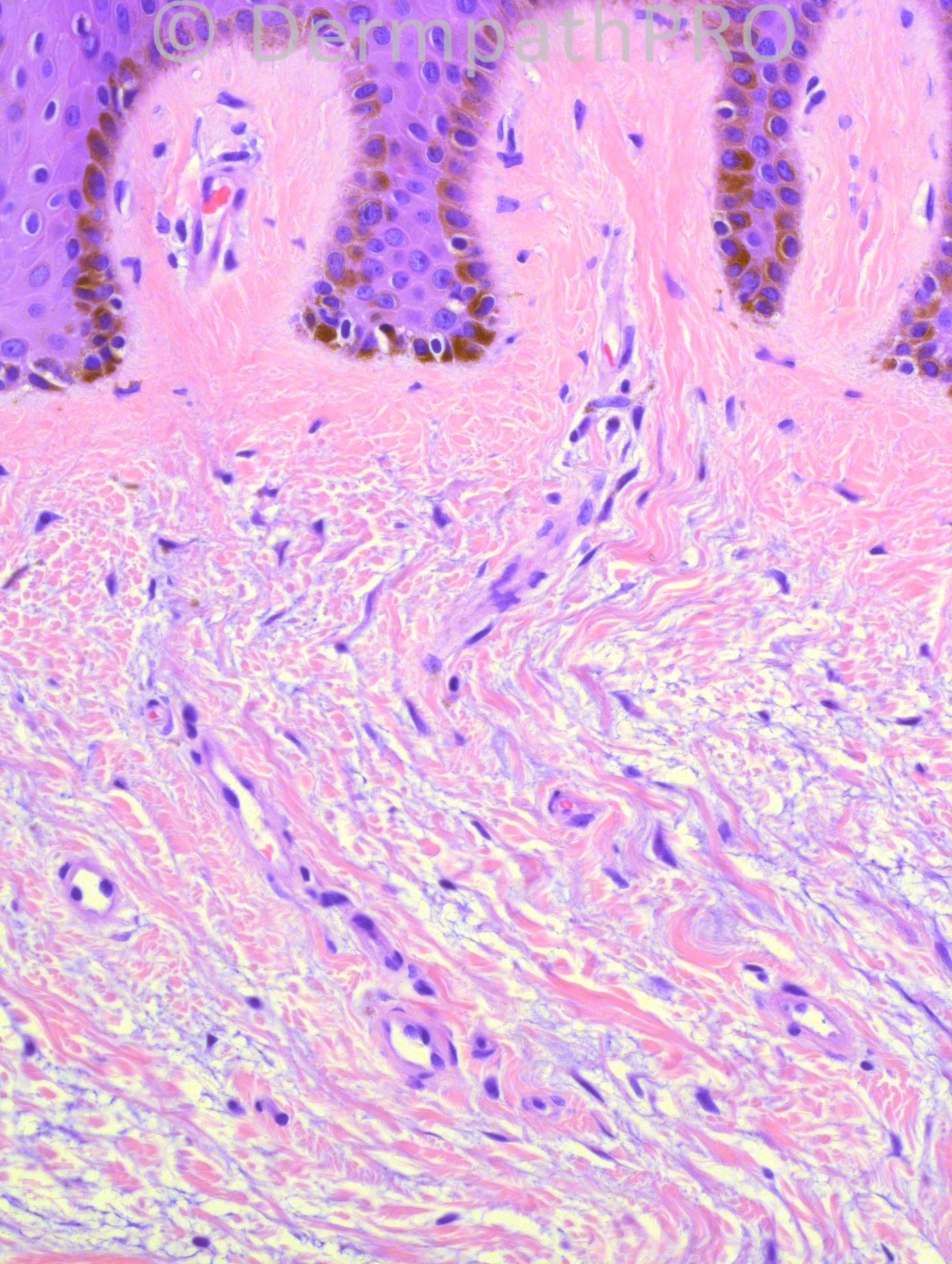
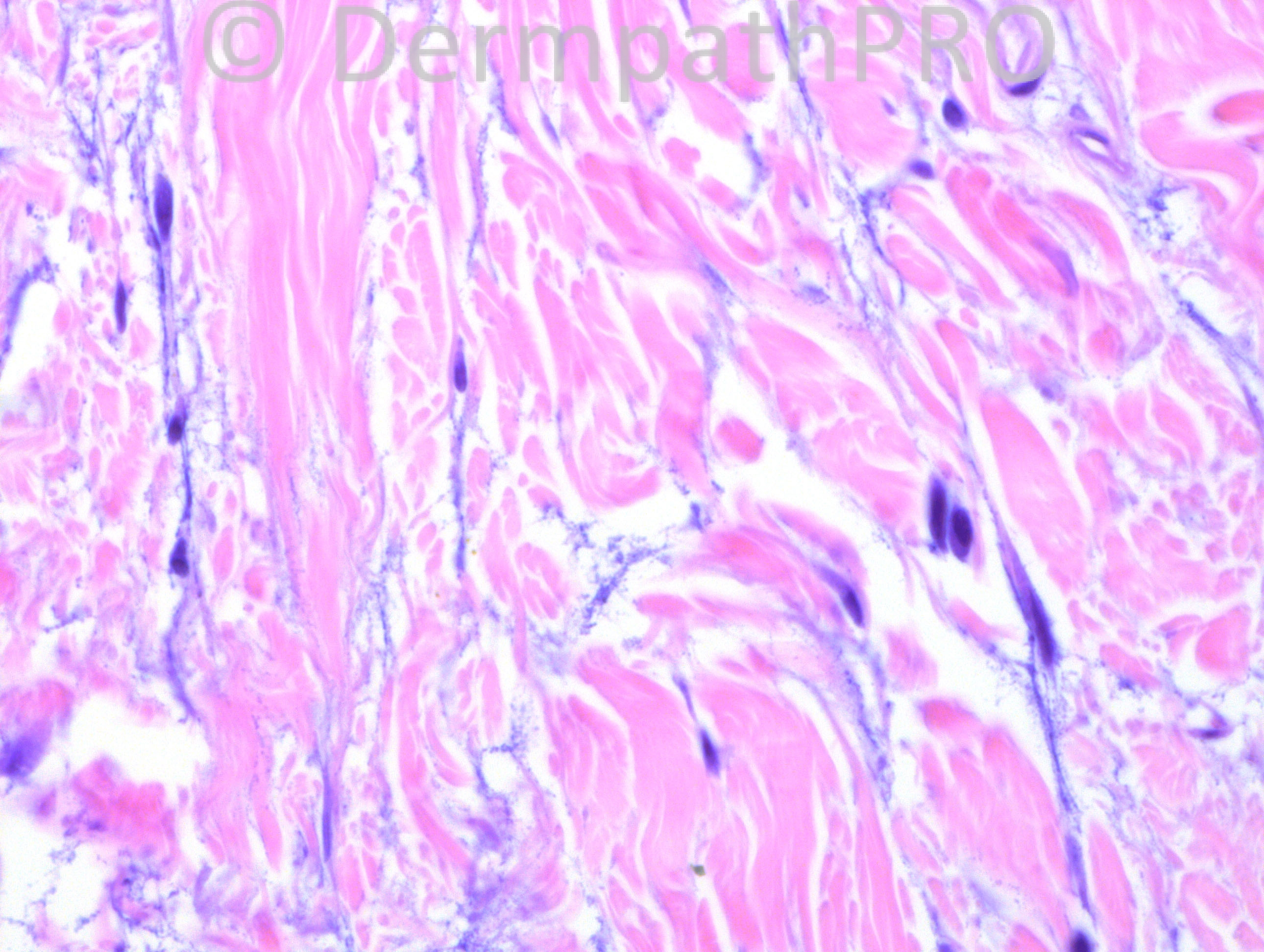
49 years-old female with raised lesions on feet and lower legs. The biopsy is from the left foot.

Case posted by Dr. Hafeez Diwan.


  Report Record

User Feedback


Robledo F. Rocha

Posted

Reticular dermis shows deposits of mucin in widened spaces between slightly increased collagen fibers accompanied by plump fibroblasts.
Lichen myxedematosus, localized form.

Share this comment


Link to comment
share_externally

Dr. Richard Carr

Posted

I shall dip my toe in! It reminded me a little of the "athlete's" or "prayer" nodules due to recurrent local friction/pressure. There may be some increased mucin here too to think about.

Share this comment


Link to comment
share_externally

Guest Maria George

Posted

I agree with Dr Rocha .The main classess of metabolic disorders with dermal deposits are
Mucinoses,porphyrias, hyalinoses, calcinoses , ochronoses and gout..The case above has mucinoses.
The main types of cutaneous mucinoses are pretibial myxedema (no or minimal increase of fibroblast), lichen myedematosus , also known as papular mucinoses (increase of fibrobblast only), and scleromyxedema (there is increase in fibroblast and fibrosis also.Patient also has monoclonal gammopathy).
Please note that there is lichen simplex-like changes in the skin.Namely, hyperkeratoses, acanthosis, and papillary dermal fibrosis.

Share this comment


Link to comment
share_externally

Guest Romualdo

Posted

I think Dr. Richard is correct. Maybe it is one of the trauma related conditions of the "athlete's" and "prayer" nodules group. This would explain the vertically oriented collagen fibers seen in dermal papillae.

Share this comment


Link to comment
share_externally

IgorSC

Posted

I think there´s a lot of mucin deposits with a little fibroblastic proliferation. Clinical information is crucial, but the location of the lesions (lower legs and feet, especially if on the dorsal aspect) favors Pretibial myxedema.

Share this comment


Link to comment
share_externally

Dr. Mona Abdel-Halim

Posted

Pretibial myxedema

Share this comment


Link to comment
share_externally

Guest Jim Davie MD

Posted

Agree with Igor: favor pretibial myxedema. History of thryoid disease would be useful.
The epidermis shows hyperkeratosis, 'dermatofibroma-like' hyperpigmentation and sharply angulated psoriasiform hyperplasia. There is absence of mucin in the papillary dermis. We are not seeing the deep reticular dermis, but there is a suggestion that the mucin content is increasing with depth. Fibroblasts are equivocally normal vs slightly increased. No significant perivascular inflammation.

Share this comment


Link to comment
share_externally

Dr. Hafeez Diwan

Posted

I called this pretibial myxedema.

Share this comment


Link to comment
share_externally

Guest Saleem Taibjee

Posted

I am also wondering about pretibial lymphoedematous mucinosis. This is usually in the context of obesity, and is distinguished from pretibial myxoedema by absence of thyroid disease and by the vertically orientated vessels.
I remembered it was presented at one of the ISDP meetings by Dr Rongioletti.
Here is a reference:
[url="http://www.ncbi.nlm.nih.gov/pubmed/19222694#"]J Cutan Pathol.[/url] 2009 Oct;36(10):1089-94. doi: 10.1111/j.1600-0560.2008.01239.x. Epub 2009 Feb 10. Obesity-associated lymphoedematous mucinosis.
[url="http://www.ncbi.nlm.nih.gov/pubmed?term=Rongioletti%20F%5BAuthor%5D&cauthor=true&cauthor_uid=19222694"]Rongioletti F[/url], [url="http://www.ncbi.nlm.nih.gov/pubmed?term=Donati%20P%5BAuthor%5D&cauthor=true&cauthor_uid=19222694"]Donati P[/url], [url="http://www.ncbi.nlm.nih.gov/pubmed?term=Amantea%20A%5BAuthor%5D&cauthor=true&cauthor_uid=19222694"]Amantea A[/url], [url="http://www.ncbi.nlm.nih.gov/pubmed?term=Ferrara%20G%5BAuthor%5D&cauthor=true&cauthor_uid=19222694"]Ferrara G[/url], [url="http://www.ncbi.nlm.nih.gov/pubmed?term=Montinari%20M%5BAuthor%5D&cauthor=true&cauthor_uid=19222694"]Montinari M[/url], [url="http://www.ncbi.nlm.nih.gov/pubmed?term=Santoro%20F%5BAuthor%5D&cauthor=true&cauthor_uid=19222694"]Santoro F[/url], [url="http://www.ncbi.nlm.nih.gov/pubmed?term=Parodi%20A%5BAuthor%5D&cauthor=true&cauthor_uid=19222694"]Parodi A[/url].

Share this comment


Link to comment
share_externally

Robledo F. Rocha

Posted

Many characteristics of obesity-associated lymphoedematous mucinosis are lacking in this case. I will list some, extracted from the referred paper:
• feet are not spared
• epidermis is not atrophic and rete ridges are not effaced
• there's no edema in upper dermis

Share this comment


Link to comment
share_externally


Join the conversation

You can post now and register later. If you have an account, sign in now to post with your account.

Guest
Add a comment...

×   Pasted as rich text.   Paste as plain text instead

  Only 75 emoji are allowed.

×   Your link has been automatically embedded.   Display as a link instead

×   Your previous content has been restored.   Clear editor

×   You cannot paste images directly. Upload or insert images from URL.

Loading...

×
×
  • Create New...