Jump to content
In this section we have spot diagnoses posted on a daily basis since June 2010, now over 4000! You can review the archived cases and read the suggested diagnoses by users and the final comment by the contributors.
Case are uploaded each week day by 10 am UK time with the correct diagnosis will generally be posted at 8 pm UK time. Why not view the most recent spot diagnosis and proffer a diagnosis?

Case Number : Case 796 - 5th July Posted By: Guest

Please read the clinical history and view the images by clicking on them before you proffer your diagnosis.
Submitted Date :
   (0 reviews)

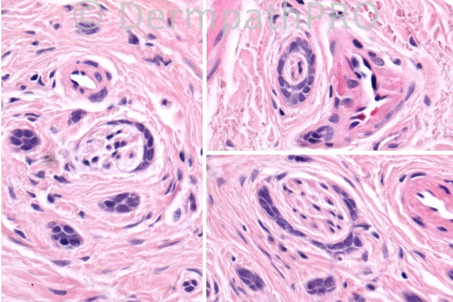
70 years-old male, lesion on the left cheek. Also had a BCC on the nose.

Case posted by Dr. Richard Carr.


  Report Record

User Feedback


Robledo F. Rocha

Posted

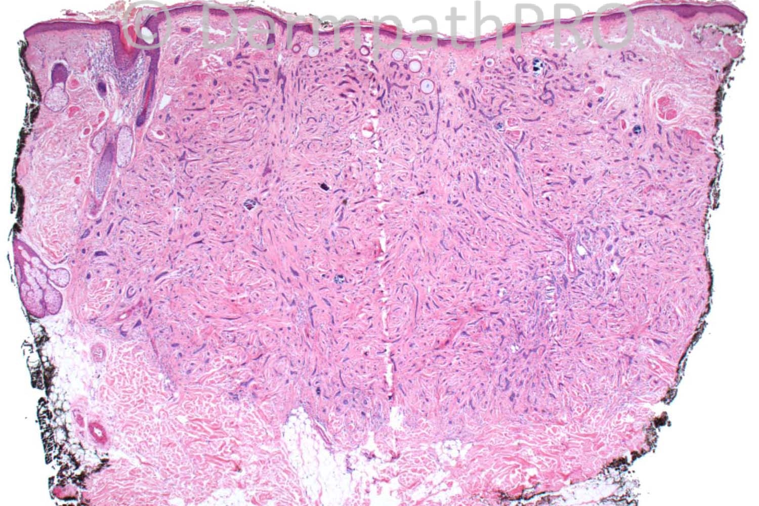
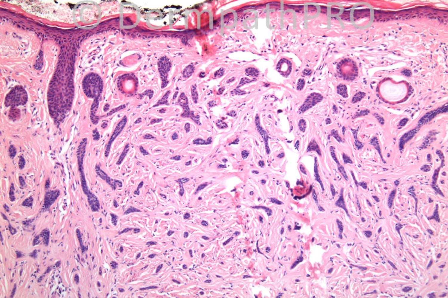
Columnar trichoblastoma (desmoplastic trichoepithelioma) showing infundibulocystic structures, a typical feature from such tumor when located on the face, and minute foci of ductal differentiation highlighted by EMA immunostaining.

Share this comment


Link to comment
share_externally

Guest Maria George

Posted

One of the dilemmas in dermpath is how to differentiate between morphea type of BCC,desmplastic TE, and MAC.
I vote for MAC in this case.

Share this comment


Link to comment
share_externally

Guest Romualdo

Posted

In favor of microcystic adnexal carcinoma is the perineural invasion shown in pictures 7 and 8. EMA highlighted perineurial cells around a small nerve in picture 8 right . Perineurial invasion is not seen in desmoplastic trichoepitheliomas.

Share this comment


Link to comment
share_externally

Sasi Attili

Posted

Funny enough, Have seen a similar case this week, with infiltration (going quite deep) and ductal differentiation, where I favoured MAC.

However this one, is not infiltrating into fat. EMA I believe is highlighting merkel cells focally (though would like to see a CK20) rather than showing definite ducts. Perineural involvement has been described in DTE (is not specific for MAC): [url="http://www.ncbi.nlm.nih.gov/pubmed/22335590"]http://www.ncbi.nlm.nih.gov/pubmed/22335590[/url]

Therefore in the absence of definite ducts or fat involvement, would favour DTE in this case.

Share this comment


Link to comment
share_externally

Guest Dr. Wilber Martínez

Posted

[color=#000000][font=arial, sans-serif][size=3]Syringoid eccrine carcinoma [/size][/font][/color][color=#000000][font=arial, sans-serif][size=3]vs Microcystic adnexal carcinoma[/size][/font][/color]

Share this comment


Link to comment
share_externally

Guest Hazem Hamed

Posted

Agree with Sasi. EMA highlights Merkel cells colonizing the tumour cell strands which are negative for EMA. Taken together with morphology this would be consistent with DTE.

Share this comment


Link to comment
share_externally

Robledo F. Rocha

Posted

Let me explain in more details why I favor columnar trichoblastoma over microcystic adnexal carcinoma:
• lesion is located on the cheek rather than upper lip or nasolabial fold
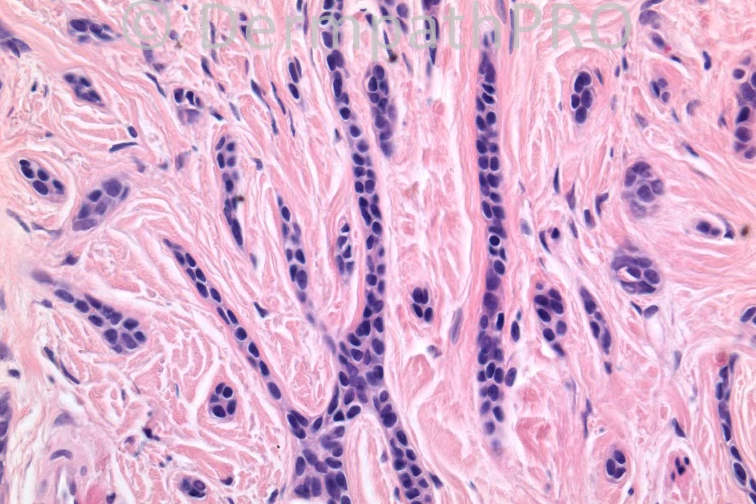
• in the lower part of the tumor there aren't ductal structures but only narrow linear or slightly branching cords of compact basaloid cells
• tumor displays sharply defined lateral margins, best seen at scanning power magnification (picture #1)
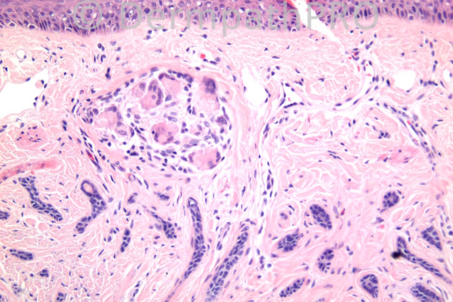
• foreign body giant cell reaction to keratin (picture #3) is more frequent in columnar trichoblastoma than in microcystic adnexal carcinoma
• tadpole-like configuration (picture #5) can be found in columnar trichoblastoma
• as pointed above, perineural infiltration (picture #7) can ocurr, albeit rare, in columnar trichoblastoma

Share this comment


Link to comment
share_externally

Guest Graham Reilly

Posted

I am favouring MAC over desmoplastic TE in view of the extensive perineural involvement and the ductal formation.I can`t see any papillary mesenchymal bodies. Looking forward to Richards comments.

Share this comment


Link to comment
share_externally

Robledo F. Rocha

Posted

[quote name='Mark A. Hurt MD' timestamp='1373026776']
In the book on "tumors with apocrine differentiation" by Requena et al, in 1998, they described lesions of MAC in contrast with syringomatous carcinoma. I have always thought syringomatous carcinoma should be part of the spectrum of MAC. Who agrees or disagrees, and why?
[/quote]
Agree. I think syringomatous carcinoma (or syringoid eccrine carcinoma, as Dr. Martínez called it in his comment above) is a microcystic adnexal carcinoma composed almost exclusively of ductal elements.

Share this comment


Link to comment
share_externally

Guest Maria George

Posted

As I mentioned before it is well known dilemma.However , IHC, may help.
[color=#000000][font=arial, helvetica, clean, sans-serif][size=3]BerEP4 differentiates between MAC (-ve) and morpheaform BCC (+ve) but not between MAC and DTE whereas PHLDA1 differentiates between DTE (+ve) and morpheaform BCC (-ve) but shows variable staining in MAC.Epithelial part of DTE will be +ve for CK20 and stromal component will be +ve for CD34. CK15 and CK19 are helpful adjuncts in the differential diagnosis of sclerosing [/size][/font][/color][color=#000000][font=arial, helvetica, clean, sans-serif][size=3]adnexal[/size][/font][/color][color=#000000][font=arial, helvetica, clean, sans-serif][size=3] neoplasms but are second in line to BerEP4 and PHLDA1.[/size][/font][/color]

[url="http://www.ncbi.nlm.nih.gov/pubmed?term=Sellheyer%20K%5BAuthor%5D&cauthor=true&cauthor_uid=23723527"]Sellheyer K[/url][color=#000000][font=arial, helvetica, clean, sans-serif][size=3], [/size][/font][/color][url="http://www.ncbi.nlm.nih.gov/pubmed?term=Nelson%20P%5BAuthor%5D&cauthor=true&cauthor_uid=23723527"]Nelson P[/url][color=#000000][font=arial, helvetica, clean, sans-serif][size=3], [/size][/font][/color][url="http://www.ncbi.nlm.nih.gov/pubmed?term=Kutzner%20H%5BAuthor%5D&cauthor=true&cauthor_uid=23723527"]Kutzner H[/url][color=#000000][font=arial, helvetica, clean, sans-serif][size=3], [/size][/font][/color][url="http://www.ncbi.nlm.nih.gov/pubmed?term=Patel%20RM%5BAuthor%5D&cauthor=true&cauthor_uid=23723527"]Patel RM[/url][color=#000000][font=arial, helvetica, clean, sans-serif][size=3].[/size][/font][/color]
[b] [url="http://www.ncbi.nlm.nih.gov/pubmed/23398472"]The immunohistochemical differential diagnosis of microcystic adnexal carcinoma, desmoplastic trichoepithelioma and morpheaform basal cellcarcinoma using BerEP4 and stem cell markers.[/url][size=2][url="http://www.ncbi.nlm.nih.gov/pubmed#"]J Cutan Pathol.[/url][/size][size=2] 2013 Apr;40(4):363-70.[/size][/b]

As I mentioned before, the images shown contains many clues for MAC.

Share this comment


Link to comment
share_externally

Guest Maria George

Posted

Sorry for too much postings .But for Mark's query, if there are epithelial components linked to both hair follicles and sweat glands you use the term ''adenexal'' carcinoma.But when there is no follicular differentiation , for example keratin microcysts, like the case shown above you call it sweat gland carcinoma or malignant syringomas .But some people may not go to that depth and prefer to lump it as MAC.

Share this comment


Link to comment
share_externally

Dr. Richard Carr

Posted

Great discussion - well well well!!! This is a classic desmoplastic trichoepithelioma (DTE) in every single aspect (exept for the rare occurrence of perineural invaison) although I agree extending to the full thickness of the dermis it does not extend into subcutis (MAC is almost always deeper at first diagnosis but is a perilous problem in superficial biopsies). Sasi is absolutely right I am showing you Merkel cells on the EMA (this is a very old case and I did not do a CK20) and the perineurium of the small nerve. There are no ducts sorry! It took me a long time searching the sections to find the perineural invasion and it was all within the confines of the border of the lesion. Thank you also to Sasi for the helpful reference. Trevor Beer also illustrated an example at the London Dermpath symposium and I believe publishsed two cases. BerEP4 does highlight focal ductal staining (only) in MAC and in my experience is variable to widespread in DTE's though of not diffuse strong as in infiltrative BCC. I have rarely if ever seen papillary mesenchymal bodies in DTE but the presence of Merkel cells is very helpful in distinguishing the tumour from MAC or infiltrative BCC. I must draw your attention to the beutiful curvilinear pattern here so characteristic of DTE and complete absence of larger or irregular and bizarre shapes (although none of you went for infiltrative BCC) not to mention the perfect stroma - really a joy to behold!.

Share this comment


Link to comment
share_externally

Dr. Richard Carr

Posted

In my haste to get home last night I forgot to admit that when I reported this case about 10 years ago I was not so confident of ruling out a basal cell carcinoma - because of the perineural invasion I mistakenly (in my current view of the case) favoured a diagnosis of an indolent infiltrative BCC and even published the case on that basis in our cutaneous "basaloid" tumour review papers in [i]Diagnostic Histopathology. That said those papers are still mostly accurate, fairly comprehensive up to the literature in 2005/6 and I hope useful. If anyone would like PDF copies I can invite you to a drop box folder that also includes a lecture on the topic epithelial tumours and immunohistochemistry. Send your e-mail requesting access to r[email="richard.carr@swh.nhs.uk"]ichard.carr@swh.nhs.uk[/email] [/i]

Share this comment


Link to comment
share_externally


Join the conversation

You can post now and register later. If you have an account, sign in now to post with your account.

Guest
Add a comment...

×   Pasted as rich text.   Paste as plain text instead

  Only 75 emoji are allowed.

×   Your link has been automatically embedded.   Display as a link instead

×   Your previous content has been restored.   Clear editor

×   You cannot paste images directly. Upload or insert images from URL.

Loading...

×
×
  • Create New...