Jump to content
In this section we have spot diagnoses posted on a daily basis since June 2010, now over 4000! You can review the archived cases and read the suggested diagnoses by users and the final comment by the contributors.
Case are uploaded each week day by 10 am UK time with the correct diagnosis will generally be posted at 8 pm UK time. Why not view the most recent spot diagnosis and proffer a diagnosis?

Case Number : Case 799 - 10th July Posted By: Guest

Please read the clinical history and view the images by clicking on them before you proffer your diagnosis.
Submitted Date :
   (0 reviews)

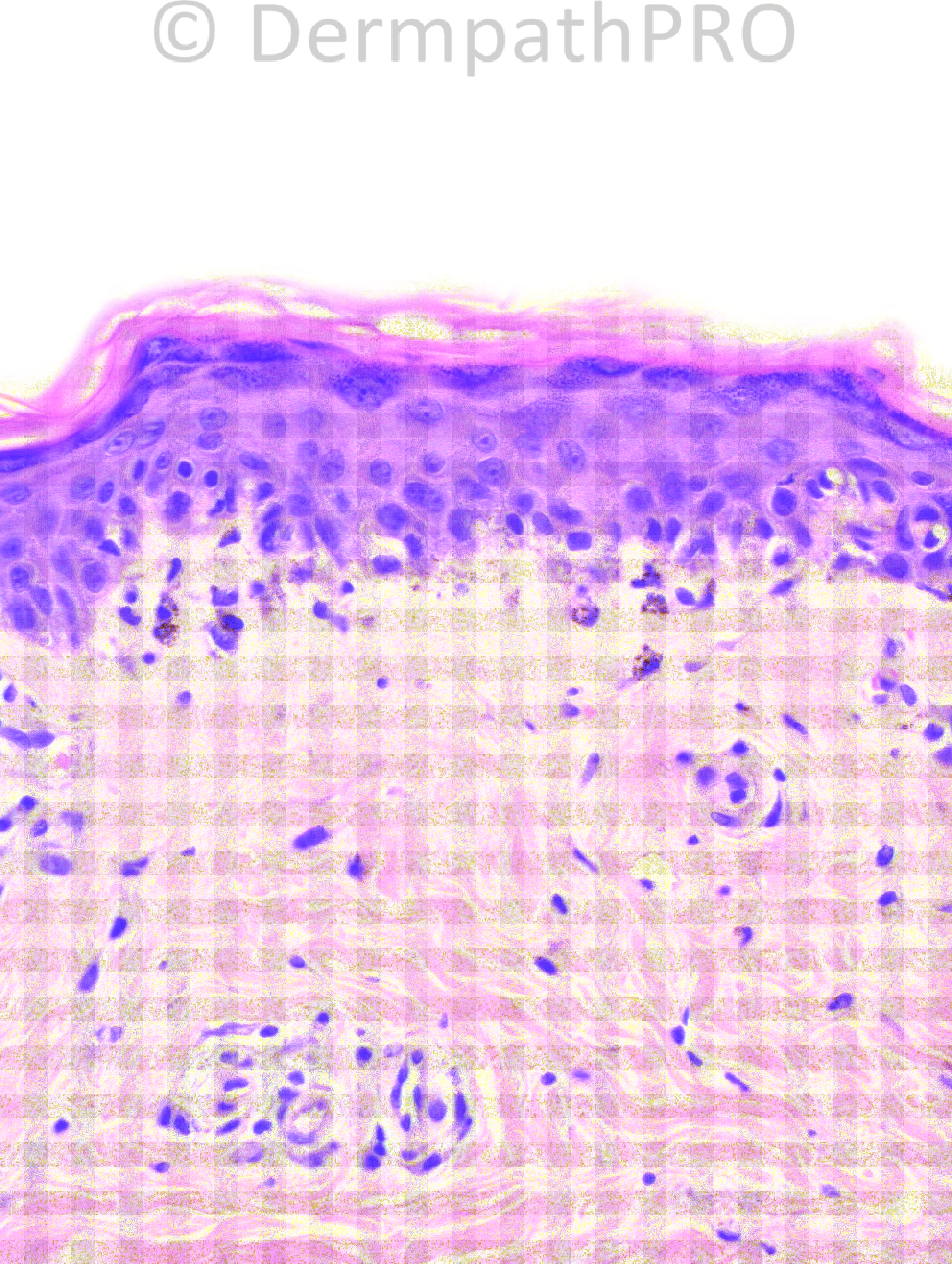
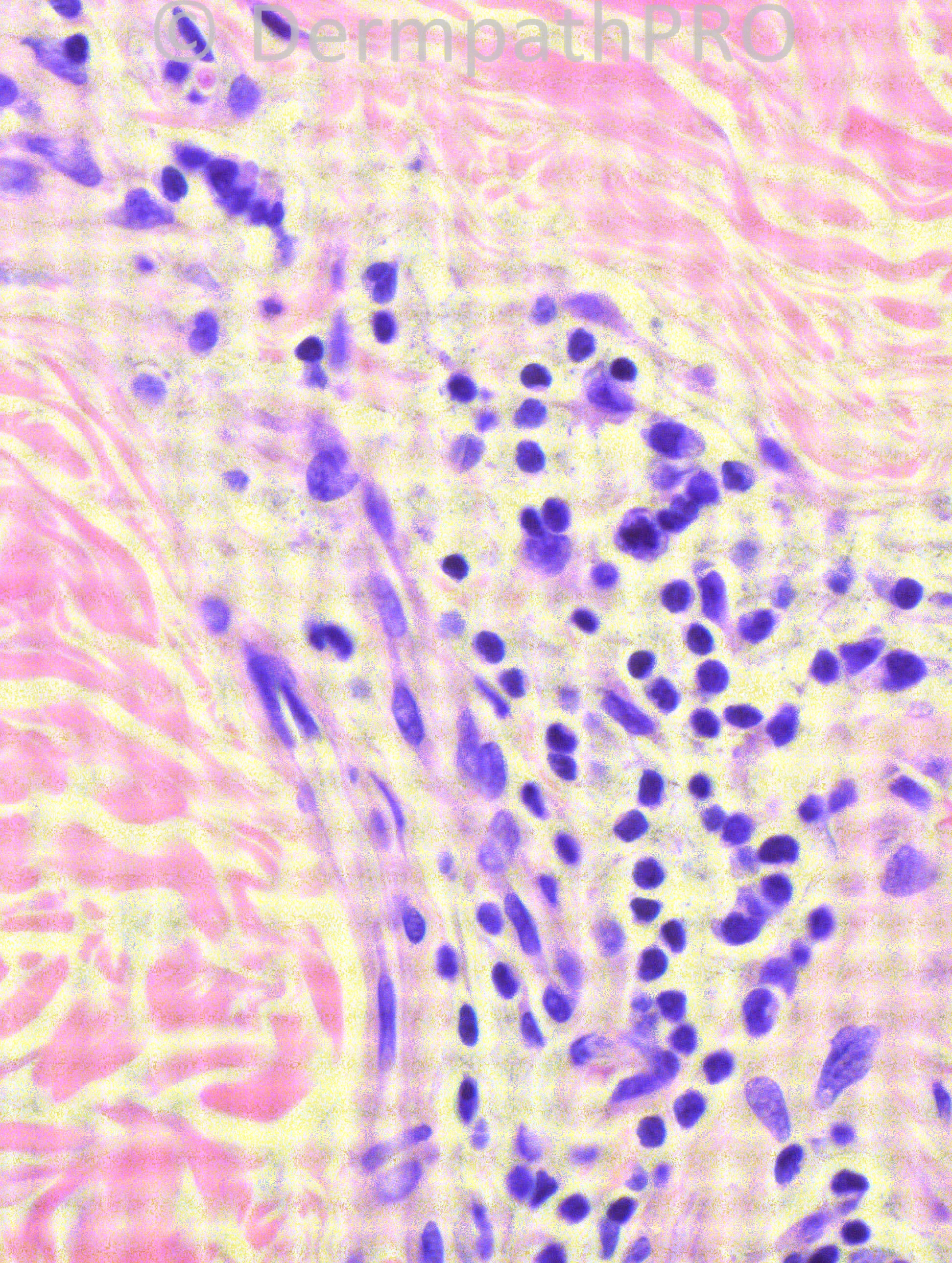
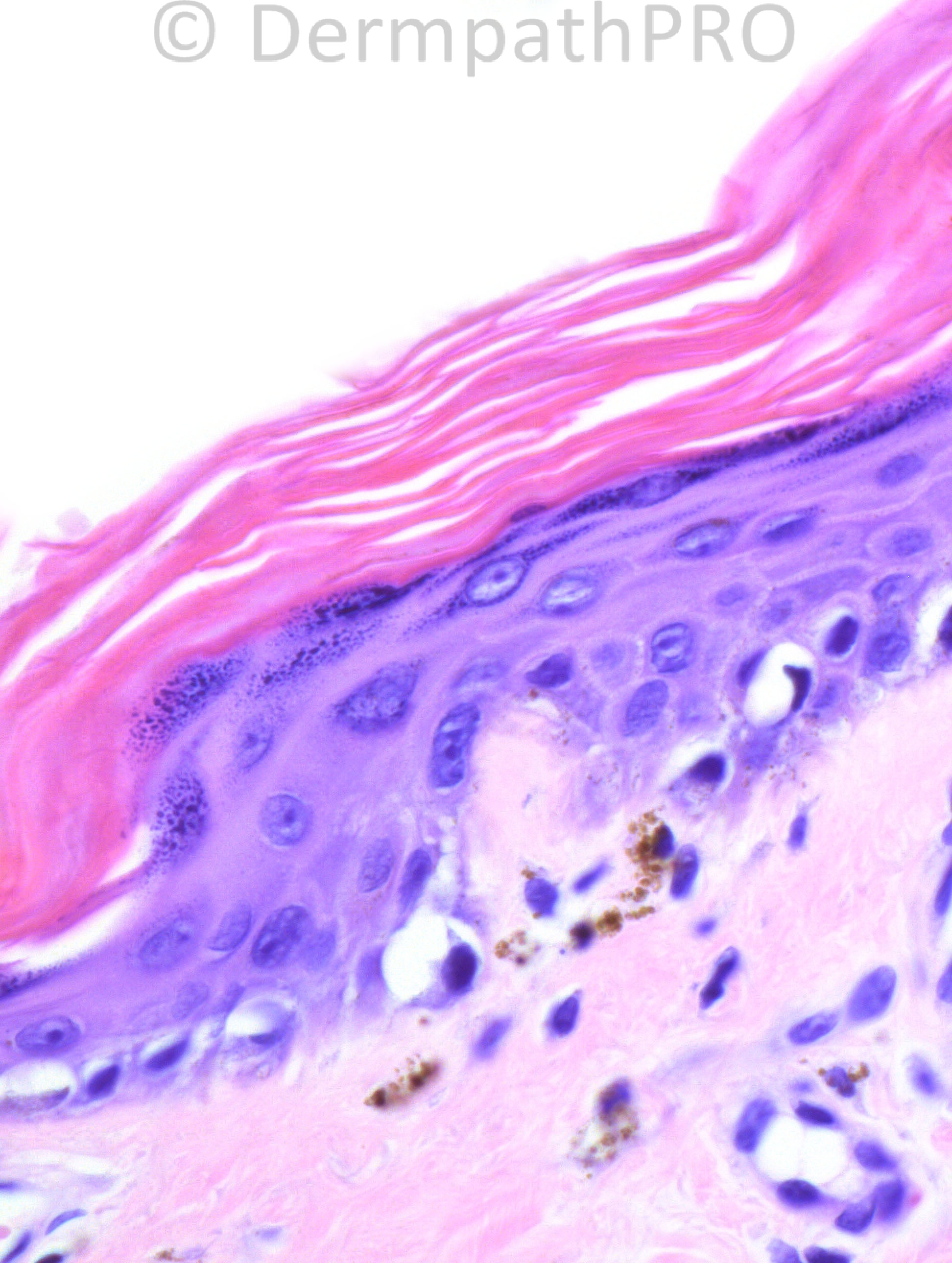
54 years-old female with inflammatory myopathy, on high dose prednisone. A skin biopsy from the right thigh was taken.

Case posted by Dr. Hafeez Diwan.

  Report Record

User Feedback


Guest Romualdo

Posted

Drug induced lupus erythematosus.

Share this comment


Link to comment
share_externally

Guest Romualdo

Posted

Yes, [url="https://dermpathpro.com/user/149-i-abdul-kadir/"][color="#151b54"]I. Abdul-kadir[/color][/url] is probably correct! I did not remember dermatomyositis. Although epidermal alterations are almost identical in the two conditions the meager inflammatory infiltrate and the clinical information of myopathy are in keeping with dermatomyositis.

Share this comment


Link to comment
share_externally

Dermatomyositis. The biopsy findings are the same (lupus and dermatomyositis).

Share this comment


Link to comment
share_externally

Dr. Mona Abdel-Halim

Posted

DM vs LE. I think mucin is seen in the third image, this will favor LE.

Share this comment


Link to comment
share_externally

Robledo F. Rocha

Posted

[size="4"][size=4]On microscopic pictures alone, I favor dermatomyositis over lupus erythematous just because lymphocytic infiltrate is sparse, but this finding is not enough to rule out lupus erythematous once both diseases share many microscopic features, including increased dermal mucin. [/size][/size]
[size="4"][size=4]On the other hand, history case of inflammatory myopathy strengthens the hypothesis of dermatomyositis. Furthermore, in dermatomyositis it’s not uncommon a mismatched response to systemic corticosteroid between muscle disease and cutaneous manifestations.[/size][/size]

Share this comment


Link to comment
share_externally

Guest Jim Davie MD

Posted

[size=4]Systemic lupus erythematosus, vs. dermatomyositis.

- Clinical inflammatory myopathy can be seen in both diseases.
- I agree the histology is too close for differentiation. (Steroids may cause a sparse inflammatory infiltrate, otherwise a sparse inflammatory infiltrate might give a clue for slightly favoring DM, as Robledo mentioned.)

Direct immunofluorescence studies (negative in DM, sometimes positive in systemic LE) and ANA serologies may be useful for this case, in the absence of additional clinical clues.[/size]

Share this comment


Link to comment
share_externally

Dr. Hafeez Diwan

Posted

I called this dermatomyositis. A muscle biopsy showed marked myositis.

Share this comment


Link to comment
share_externally


Join the conversation

You can post now and register later. If you have an account, sign in now to post with your account.

Guest
Add a comment...

×   Pasted as rich text.   Paste as plain text instead

  Only 75 emoji are allowed.

×   Your link has been automatically embedded.   Display as a link instead

×   Your previous content has been restored.   Clear editor

×   You cannot paste images directly. Upload or insert images from URL.

Loading...

×
×
  • Create New...