Jump to content
In this section we have spot diagnoses posted on a daily basis since June 2010, now over 4000! You can review the archived cases and read the suggested diagnoses by users and the final comment by the contributors.
Case are uploaded each week day by 10 am UK time with the correct diagnosis will generally be posted at 8 pm UK time. Why not view the most recent spot diagnosis and proffer a diagnosis?

Case Number : Case 800 - 11th July Posted By: Guest

Please read the clinical history and view the images by clicking on them before you proffer your diagnosis.
Submitted Date :
   (0 reviews)

58-year-old female with hyperpigmented, thickened lesions on arms. A prior biopsy from the shoulder was read by someone as “morphea.” The present biopsy is from the right arm.

Case posted by Dr Hafeez Diwan.


  Report Record

User Feedback


Guest Saleem Taibjee

Posted

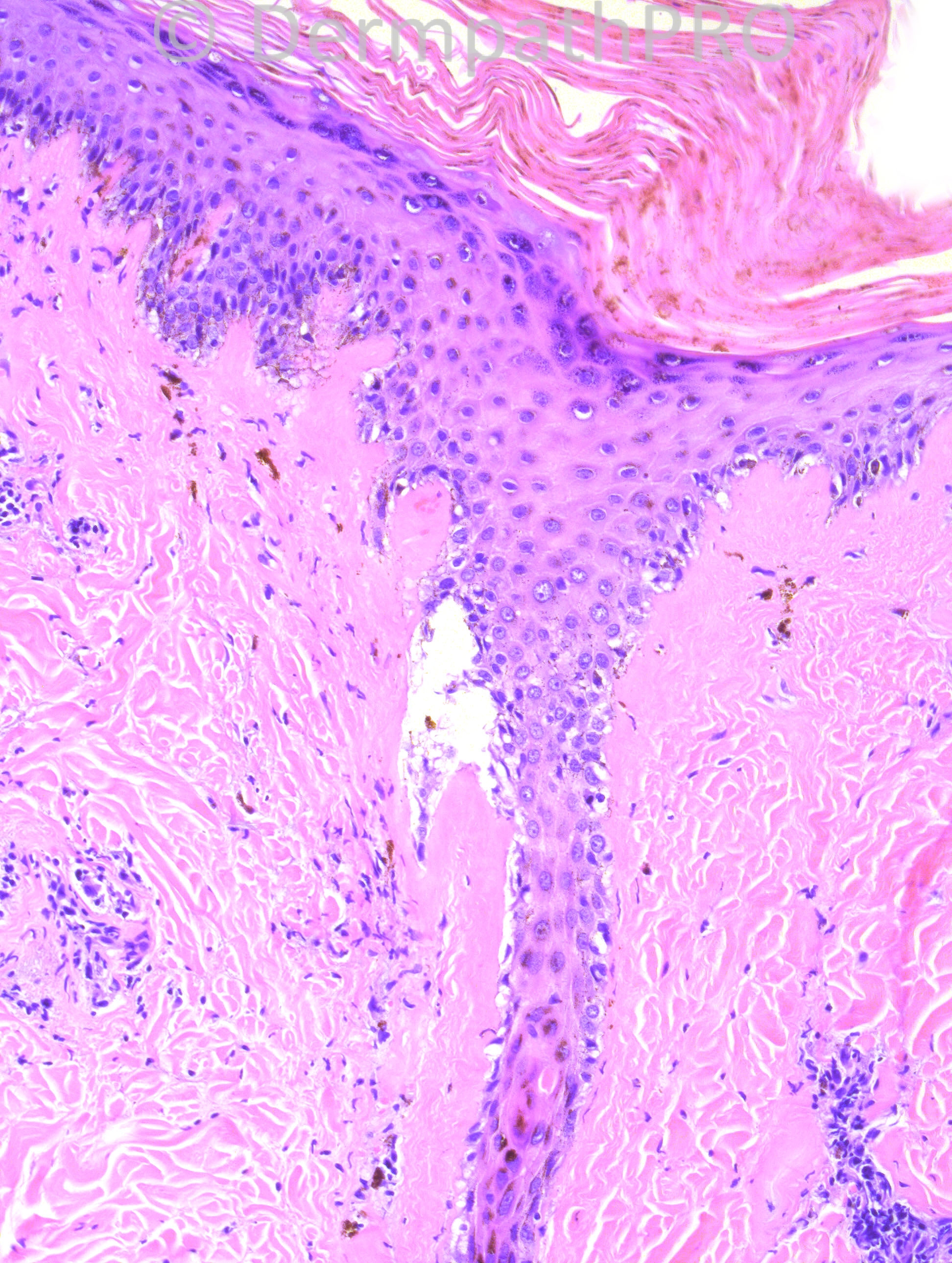
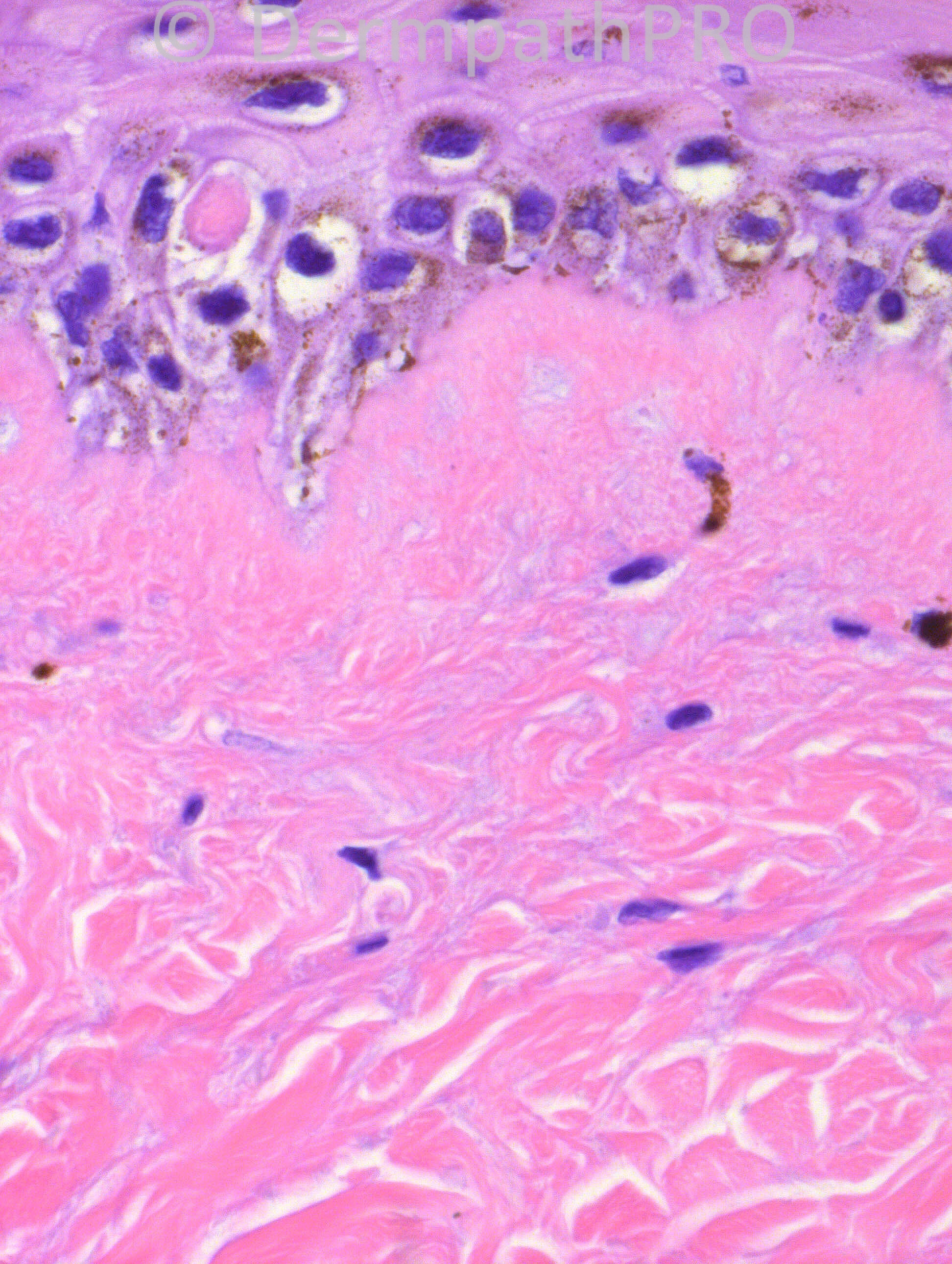
The additional interface element made me consider the following histological differential:
Sclerodermoid variant of dermatomyositis
Sclerodermoid graft-versus-host
Lichen sclerosus-morphoea overlap (I think there may be homogenisation of the superficial dermis here perhaps favouring the latter?).

Share this comment


Link to comment
share_externally

Sasi Attili

Posted

I agree with Saleem. The vacuolar interface and sclerosis made me wonder about sclerodermatoid GVHD. LS/ Morphoea overlap is a good differential but CPC required.

Share this comment


Link to comment
share_externally

Guest Rodrigo Restrepo

Posted

Agree Abdul Chronic lupus erythematosus.

Share this comment


Link to comment
share_externally

I think that serologic information is essencial, but the differential diagnosis could be LS/morphea overlap or even a Lupus erythematosus and morphea overlap. The clinical information would be mostly important for this last one.

Share this comment


Link to comment
share_externally

Guest Dr. Engin Sezer

Posted

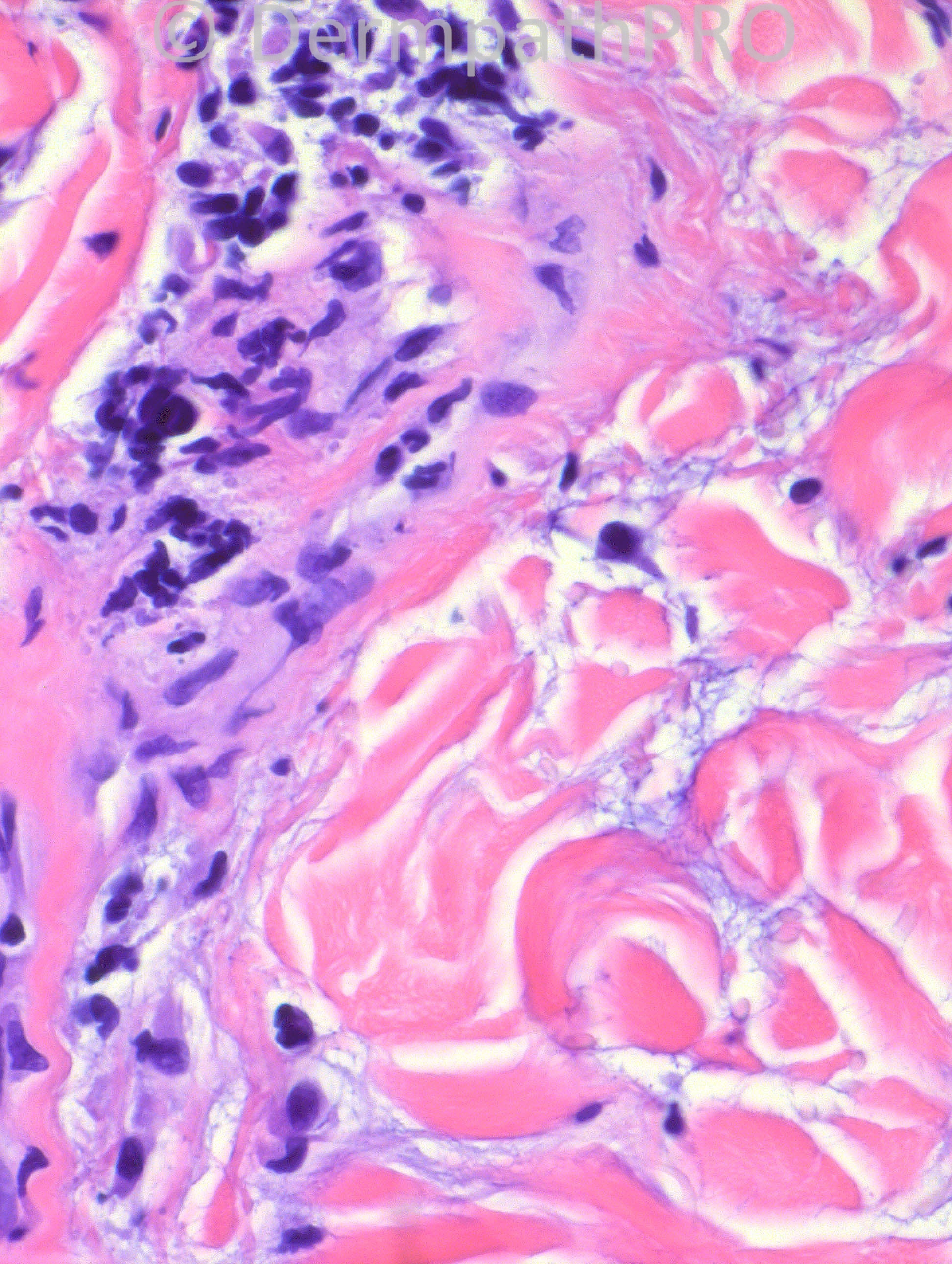
Band of hyalinized collagen bundles in the upper dermis with epidermal interphase changes combined with sclerotic, thickened collagen with mucin between the bundles, consistent with LS/morphea overlap

Share this comment


Link to comment
share_externally

Robledo F. Rocha

Posted

[size=4][size=4]I think this is a scarring chronic lupus erythematosus. Microscopic pictures show an interface dermatitis with lymphocytic infiltrate around both superficial and deep vascular plexuses, and increased mucin between collagen fibers in reticular dermis.[/size][/size]
[size=4][size=4]An atrophic hair follicle can be found. It features vacuolar damage of the infundibular basal cells and an underlying fibrous tract. These findings, in addition to the presence of melanophages and sclerosis in upper dermis, explain the clinical characteristics of thickened and hyperpigmented lesions that probably also display alopecia.[/size][/size]

Share this comment


Link to comment
share_externally

Guest Dr. Francisco Vílchez

Posted

Too much mucin and interface dermatitis...I think chronic lupus.

Share this comment


Link to comment
share_externally

Dr. Hafeez Diwan

Posted

Agreed. This is lupus. The patient has a positive ANA.

Share this comment


Link to comment
share_externally


Join the conversation

You can post now and register later. If you have an account, sign in now to post with your account.

Guest
Add a comment...

×   Pasted as rich text.   Paste as plain text instead

  Only 75 emoji are allowed.

×   Your link has been automatically embedded.   Display as a link instead

×   Your previous content has been restored.   Clear editor

×   You cannot paste images directly. Upload or insert images from URL.

Loading...

×
×
  • Create New...