Jump to content
In this section we have spot diagnoses posted on a daily basis since June 2010, now over 4000! You can review the archived cases and read the suggested diagnoses by users and the final comment by the contributors.
Case are uploaded each week day by 10 am UK time with the correct diagnosis will generally be posted at 8 pm UK time. Why not view the most recent spot diagnosis and proffer a diagnosis?

Case Number : Case 803 - 16th July Posted By: Guest

Please read the clinical history and view the images by clicking on them before you proffer your diagnosis.
Submitted Date :
   (0 reviews)

No history available.

Case posted by Dr. Mark Hurt.


  Report Record

User Feedback


Guest Romualdo

Posted

In situ superficial spreading melanoma.

Share this comment


Link to comment
share_externally

Robledo F. Rocha

Posted

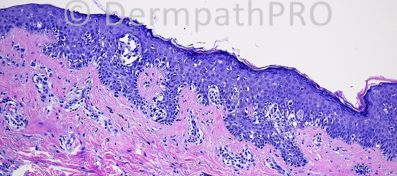
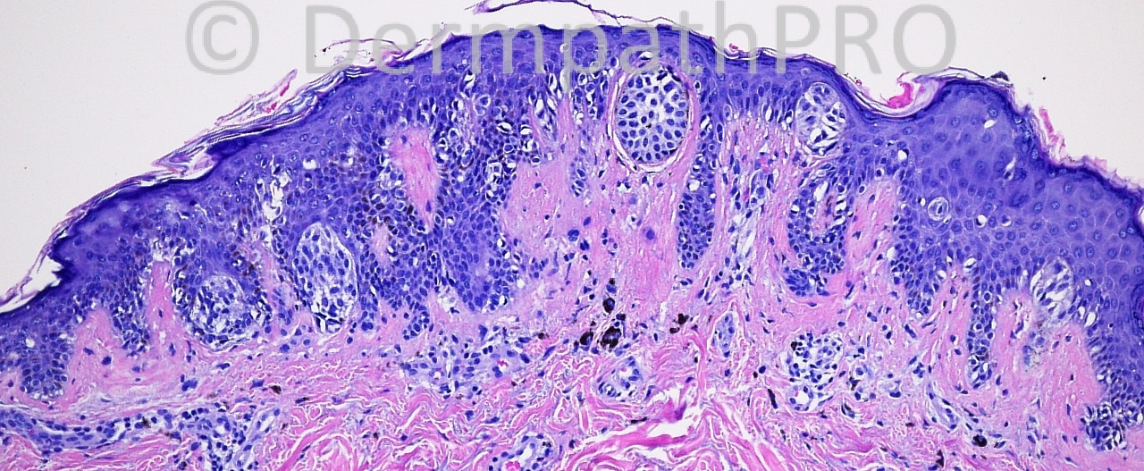
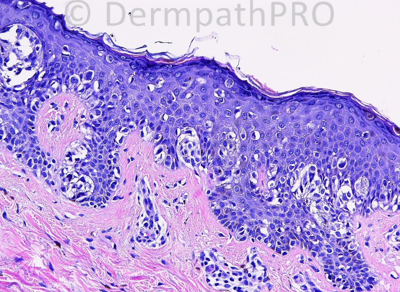
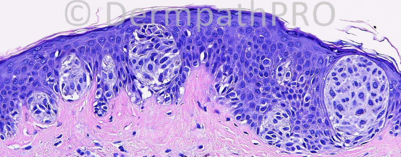
Assymetric junctional and pagetoid proliferation of atypical epithelioid melanocytes with nested and lentiginous growth patterns. I would call this superficial spreading melanoma, in-situ stage.
Is the site a man's back or a woman's leg?

Share this comment


Link to comment
share_externally

Guest Rodrigo Restrepo

Posted

Agree, superficial spreading melanoma.

Share this comment


Link to comment
share_externally

[color=#1C2837][font=arial, verdana, tahoma, sans-serif][size=4]Superficial spreading melanoma.[/size][/font][/color]

Share this comment


Link to comment
share_externally

Asymmetric junctional nests and atypical lentiginous proliferation. Also pagetoid extension at the center and at the borders of the lesion. No doubt Superficial Spreading Melanoma, in situ.

Share this comment


Link to comment
share_externally

Guest Dr. Francisco Vílchez

Posted

Agree, superficial spreading melanoma.

Share this comment


Link to comment
share_externally

Guest DR. Engin Sezer

Posted

Nested melanoma [i]in situ[/i]

Share this comment


Link to comment
share_externally

Sasi Attili

Posted

For some reason I am not so malignant on this one. The cells have a epithelioid morphology with nesting and single cell ascent in the center of the lesion. However the cells seems to be getting smaller with ascent.

Atypical special site naevus (depending on CPC) or an epidermal Spitz, would be what I would favour. I would however what to see the lesion excised completely.

Share this comment


Link to comment
share_externally

Guest Marcia

Posted

Melanoma in situ, superficial spreading type.

Share this comment


Link to comment
share_externally

Dr. Richard Carr

Posted

This lesion is relatively well nested, predominantly junctional (definitely a few cells in the papillary dermis (middle left). Some spitzoid morphology and focal pagetoid spread. I agree with Sasi we might accept many of these features in some special site naevi. Clinical correlation is obviously important but assuming these images are representative (i.e. a small lesion of around 4mm diameter) I would label as an atypical / odd small predominantly junctional and pagetoid Spitzoid lesion without overtly worrying features for outright melanoma and provided it is completely excised no further management required. It falls into a grey area of small superficial atypical lesions that are hard to classify / predict their biology and given the arbitrary features I often don't label them - i.e. I admit defeat but take a practical approach as provided they have been examined carefully (with additional levels to rule out a proper "invasive" melanoma) and removed in their entirety there is no chance of any adverse outcome to the patient for this lesion! There is an inverse relationship between size and difficulty of diagnosis in such cases.

Share this comment


Link to comment
share_externally

Dr. Mona Abdel-Halim

Posted

I am also not with malignant here, more with atypical site nevus

Share this comment


Link to comment
share_externally

Guest Jim Davie MD

Posted

In the absence of clinical context, I agree this would favor melanoma in situ, with the differential including a special site (likely vulvar/genital) nevus.
Features favoring malignant: lentiginous predominance, pagetoid downwards spread, asymmetric pagetoid scatter over non-nested lentiginous junctional zones, asymmetric pigment distribution, pleomorphism of nest size and shape, high-grade atypia with dusty grey cytoplasm, variable nuclear hyperchromatism, randomly prominent nucleoli, thick nuclear membranes. Overall asymmetry, with rete peg irregularity in size/position.

Share this comment


Link to comment
share_externally

Sasi Attili

Posted

This is a tricky case for which there would be a wide range of opinions. I suspect there might be a East/ West divide as to what this lesion is called. Most people in the UK I think might not go as far as calling this a melanoma. Wolter Mooi gave an excellent lecture at the recently concluded BAD meeting in Liverpool, talking exactly on this issue. He favoured the term 'Uncertain Malignant Potential' for these tumours which are thin and the certainty of malignancy is difficult to assess, in view of the large number of cases that would be necessary to achieve statistical significance!

Obviously this is a debatable topic and I am not sure we will ever find an answer to this question. [b]However if you call this malignant, you can never be wrong [/b][i](apart from colleagues who might disagree)[/i][b]!! [/b]So it is a safe bet to err on the side of caution and this is the approach most pathologists might take.

On a slightly different note, Wolter Mooi has a melanocyte slide set that is accessible for free online and I would recommend checking out: [url="http://melanocytepathology.com/"]http://melanocytepathology.com/[/url].

Share this comment


Link to comment
share_externally

Guest Romualdo

Posted

Thanks Dr. Sasi for divulgating this excellent site!

Share this comment


Link to comment
share_externally

Sasi Attili

Posted


If one thinks about it, even a normal cell has the 'potential' to become malignant. A melanoma cell has to after all arise from a normal cell. In my opinion it is the statistical chance that the potential can manifest, that is important. One can be certain about the malignant potential of even a normal cell i.e. it has the potential. It is the chance of the potential manifesting, that is debatable. This assumes clinical significance in the case so called early melanoma's. I think the diagnosis of an early melanoma has too many variables and subjective criteria, for it to be called a defined 'diagnosis'. That is where all the problem arises. Your statement 'assuming the criteria are legitimate' aptly sums it up.

Share this comment


Link to comment
share_externally


Join the conversation

You can post now and register later. If you have an account, sign in now to post with your account.

Guest
Add a comment...

×   Pasted as rich text.   Paste as plain text instead

  Only 75 emoji are allowed.

×   Your link has been automatically embedded.   Display as a link instead

×   Your previous content has been restored.   Clear editor

×   You cannot paste images directly. Upload or insert images from URL.

Loading...

×
×
  • Create New...