Jump to content
In this section we have spot diagnoses posted on a daily basis since June 2010, now over 4000! You can review the archived cases and read the suggested diagnoses by users and the final comment by the contributors.
Case are uploaded each week day by 10 am UK time with the correct diagnosis will generally be posted at 8 pm UK time. Why not view the most recent spot diagnosis and proffer a diagnosis?

Case Number : Case 805 - 18th July Posted By: Guest

Please read the clinical history and view the images by clicking on them before you proffer your diagnosis.
Submitted Date :
   (0 reviews)

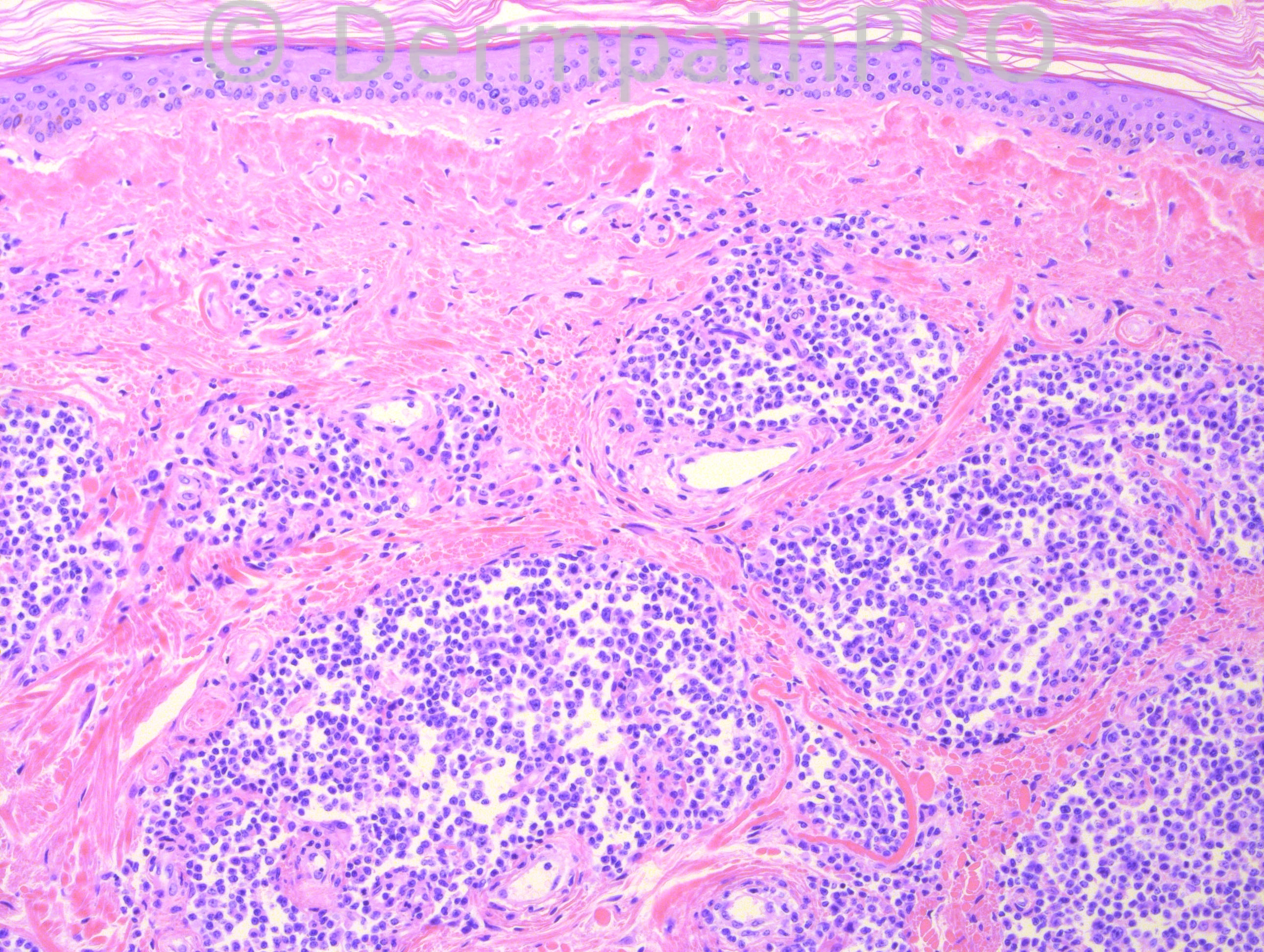
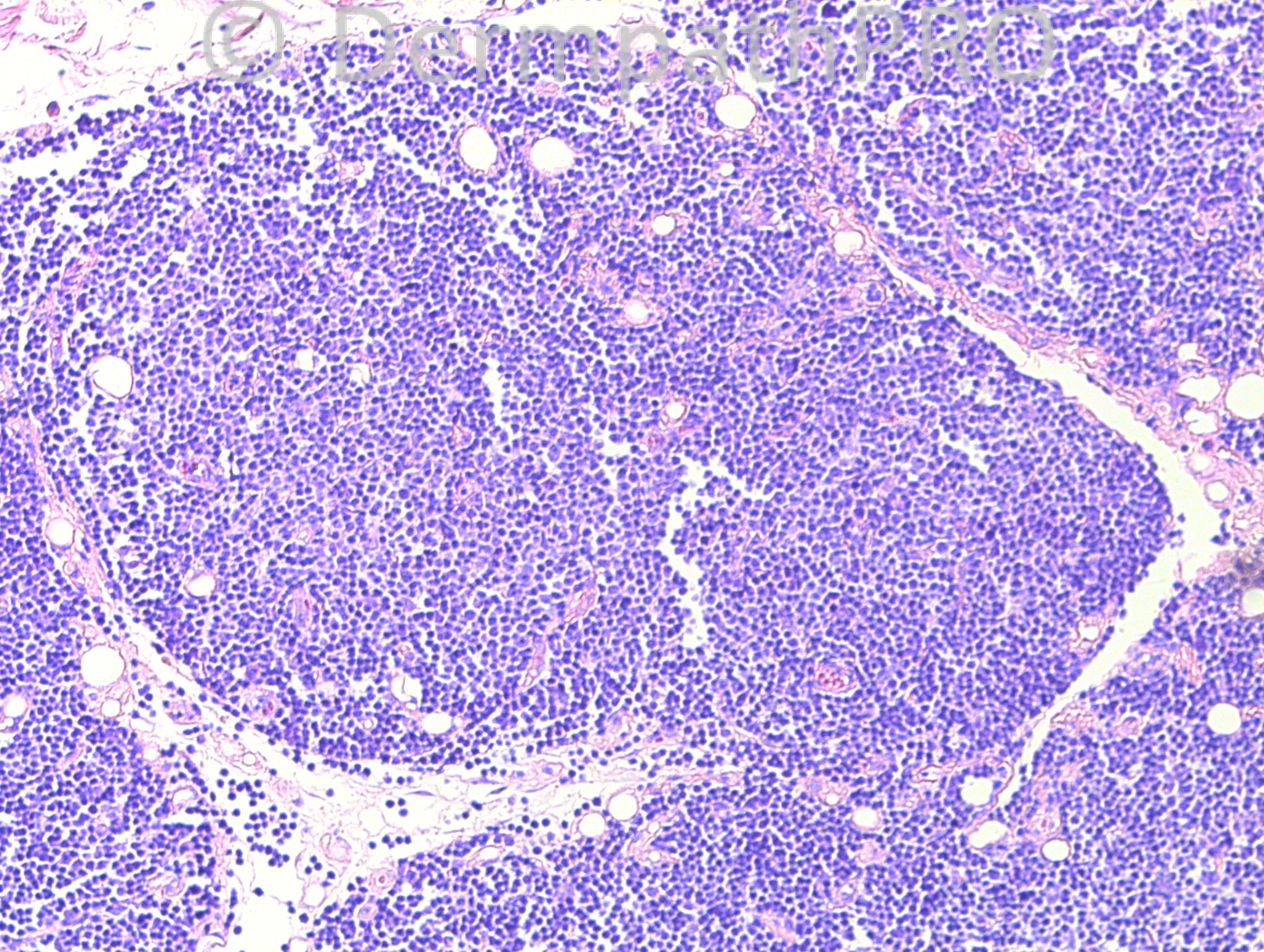
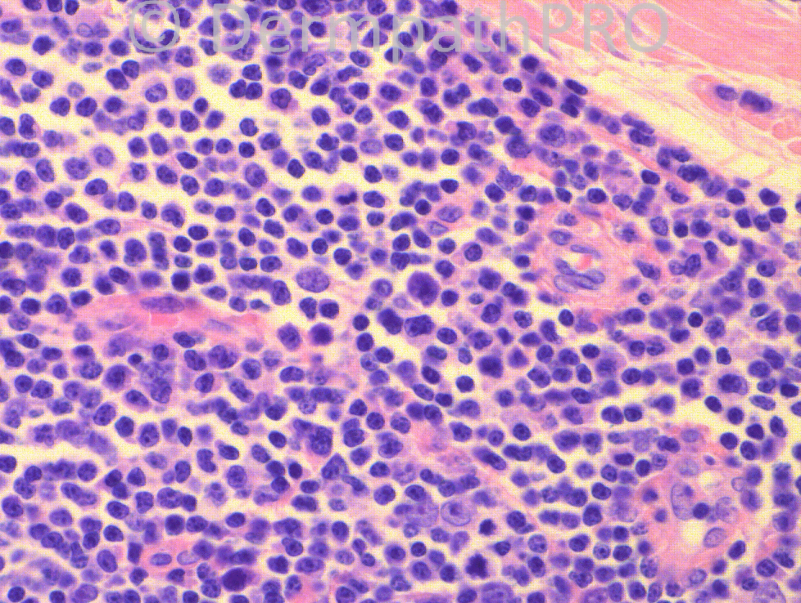
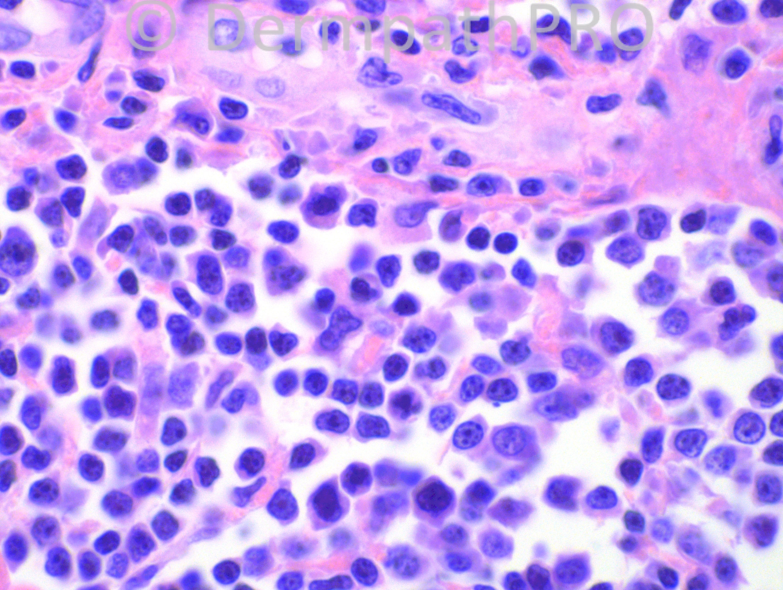
85 years-old female with a lesion on the neck.

Case posted by Dr. Hafeez Diwan


  Report Record

User Feedback


Agree, Marginal zone lymphoma. The infiltrate extends to the subcutaneous tissue and is composed of lymphocytes, plasmacytoid cells and plasma cells. There´s an amyloid-like substance at the superficial dermis.
The age and localization, neck, also favors MZL.

Share this comment


Link to comment
share_externally

Guest Rodrigo Restrepo

Posted

Marginal zone lymphoma

Share this comment


Link to comment
share_externally

Robledo F. Rocha

Posted

Dermal and subcutaneous nodular infiltrate composed of medium-sized lymphocytes showing cleaved nucleus and pale cytoplasm, admixed with plasma cells and lymphoplasmacytoid cells. Despite sparing the epidermis, a lymphoepithelial lesion involving an eccrine duct is shown on picture #4.
Agree with my colleagues' diagnosis of primary cutaneous marginal-zone B-cell lymphoma.
Amyloid deposition in this cutaneous lymphoma was reported and reviewed by a [url="http://www.ncbi.nlm.nih.gov/pubmed/20507739"]Chinese paper[/url].

Share this comment


Link to comment
share_externally

Guest Jim Davie MD

Posted

[size=4]I agree, MZL is the most likely diagnosis (although this case appears less infiltrative than other cases I have seen). Mixed population as described, with expected absence of tingible-body macrophages in these images. No reactive or neoplastic follicular centers.

For the sake of expanding our differential diagnosis, other considerations:
1. Diffuse-pattern variant of follicular center lymphoma (FCL)...either primary or secondary to skin... may be virtually indistinguishable from MZL. Both may show prominent lymphoplasmacytic differentiation and poorly formed or absent follicles. Staining may be similar in tough cases, especially if numerous reactive T-cells obscure interpretation.
2. Pseudolymphoma.

Stains may help. Top on my list should be Ig light-chain stains to determine lambda/kappa light chain ratio on the plasmacytoid component.[/size]

Share this comment


Link to comment
share_externally

Guest Maria George

Posted

Just to say something different what about angilymphoid hyperplasis with eosinophilia vs Kimura.

Share this comment


Link to comment
share_externally

I would also add plasmacytoma to the differential.

Share this comment


Link to comment
share_externally

Dr. Hafeez Diwan

Posted

Marginal zone lymphoma. There was kappa predominance by immunohistochemistry.

Share this comment


Link to comment
share_externally


Join the conversation

You can post now and register later. If you have an account, sign in now to post with your account.

Guest
Add a comment...

×   Pasted as rich text.   Paste as plain text instead

  Only 75 emoji are allowed.

×   Your link has been automatically embedded.   Display as a link instead

×   Your previous content has been restored.   Clear editor

×   You cannot paste images directly. Upload or insert images from URL.

Loading...

×
×
  • Create New...