Jump to content
In this section we have spot diagnoses posted on a daily basis since June 2010, now over 4000! You can review the archived cases and read the suggested diagnoses by users and the final comment by the contributors.
Case are uploaded each week day by 10 am UK time with the correct diagnosis will generally be posted at 8 pm UK time. Why not view the most recent spot diagnosis and proffer a diagnosis?

Case Number : Case 806 - 19th July Posted By: Guest

Please read the clinical history and view the images by clicking on them before you proffer your diagnosis.
Submitted Date :
   (0 reviews)

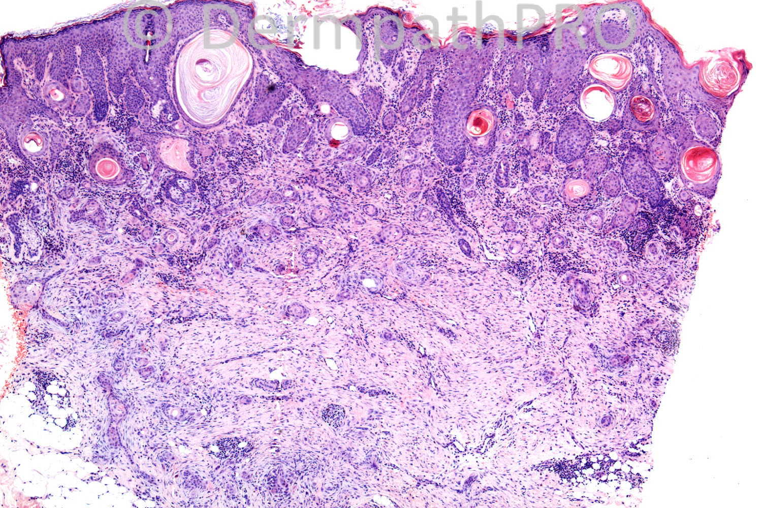
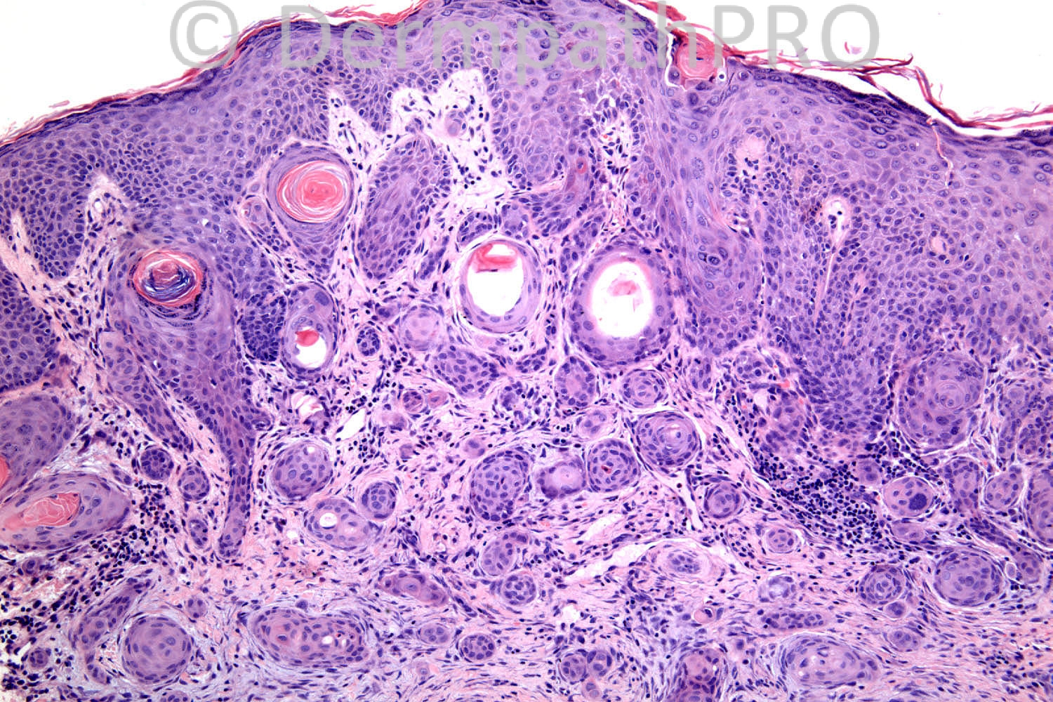
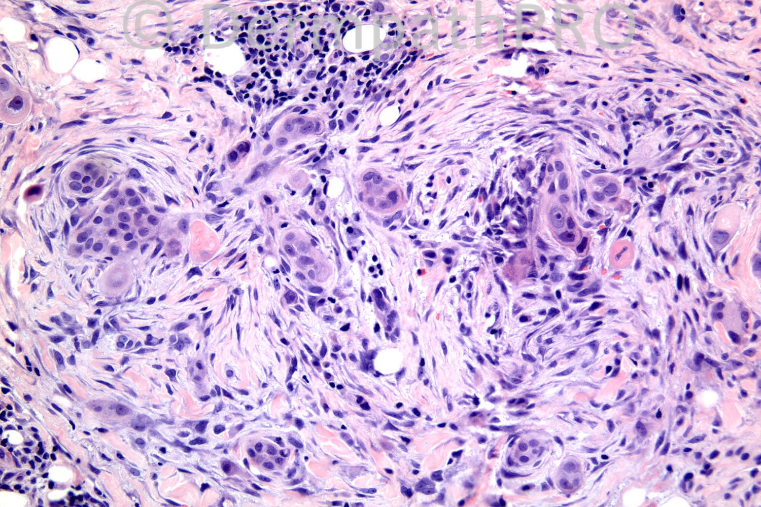
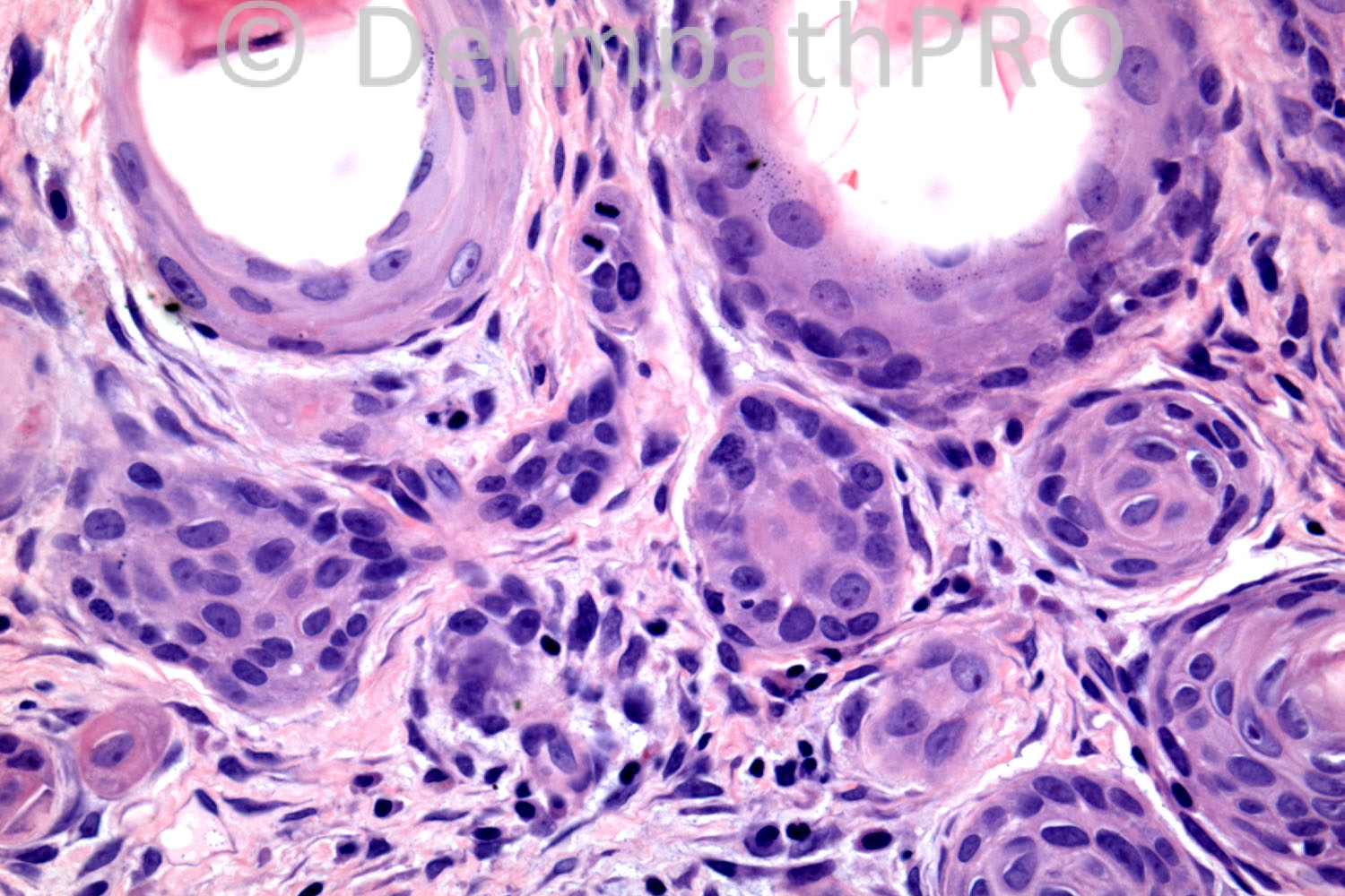
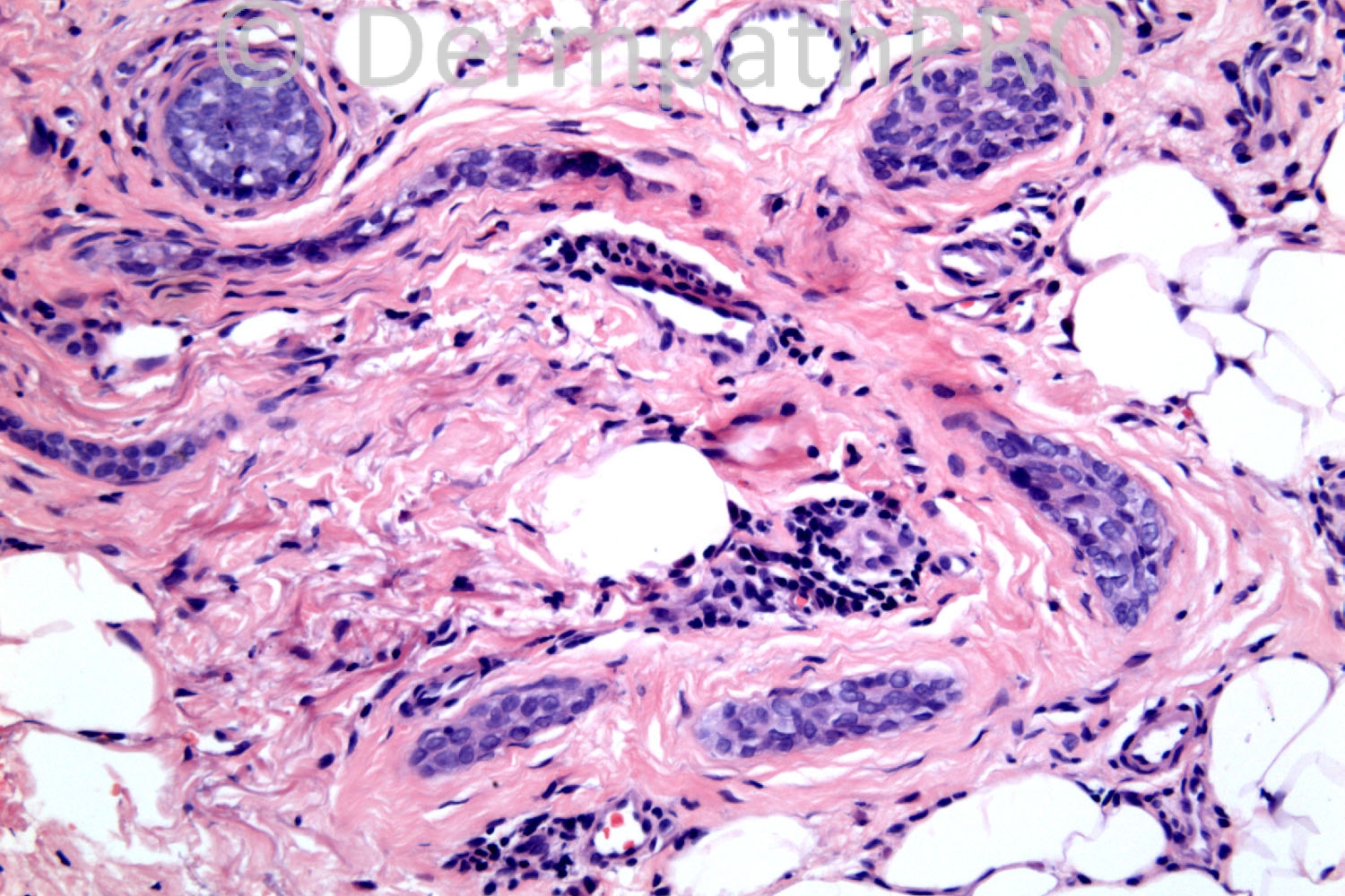
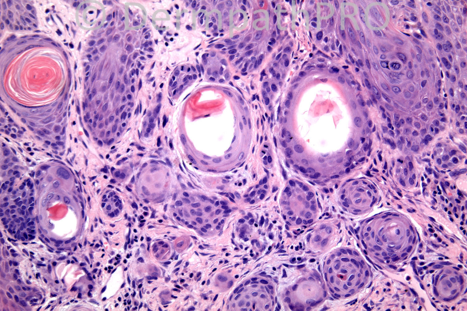
65 years-old male. Lesion behind ear; several prior NMSC's.

Case posted by Dr. Richard Carr.

  Report Record

User Feedback


Guest Romualdo

Posted

Microcystic adnexal carcinoma, despite the presence of focal atypia.

Share this comment


Link to comment
share_externally

Guest Maria George

Posted

invasive squamous cell carcinoma versus carcinosacoma.

Share this comment


Link to comment
share_externally

Dr. Mona Abdel-Halim

Posted

I will diagnose this as invasive follicular SCC with focal spindle cell changes.

Share this comment


Link to comment
share_externally

Robledo F. Rocha

Posted

There’s a sly stratification that gives to this tumor the appearance of a microcystic adnexal carcinoma, but the wide connection to the epidermis, the obvious nuclear anaplasia, and the focal spindle-cell component compel me to consider infundibulocystic squamous cell carcinoma as the most likely diagnosis.

Share this comment


Link to comment
share_externally

Guest Rodrigo Restrepo

Posted

Microcystic adnexal carcinoma

Share this comment


Link to comment
share_externally

Sasi Attili

Posted

My DD includes MAC and Squamoid Eccrine ductal Carcinoma (essentially a low grade locally invasive epidermal tumour), favour the latter.

Share this comment


Link to comment
share_externally

Dr. Richard Carr

Posted

Hey Phillip - what an honour. Great responses today - well done on a difficult case - I don't think it can be over-emphasied how difficult this diagnosis can be in real life in small biopsies. I called this MAC (the keratocysts in this case are typical - and can easily cause confusion with follicular tumours - some believe MAC is a mixed follicular - ductal lesion in any case). I agree a report along the lines of a low-grade infiltrative carcinoma is appropriate with advice that the patient may possibly have extensive disease and margin control surgery should be considered. The ductal differentiation (as in this case) can be exceedingly focal especially in superficial biopsies. Interestingly the patient had a "second lesion" nearby that an experienced colleague brought to me as ?syringoma. In fact it was all one process and the patient ended up having a large excision at Moh's surgery. The cellular atypia present in this case is not typical but the lesion lacked appreciable mitotic activity of a more high grade (adenosquamous) carcinoma. I do agree the "reactive" stroma is rather cellular / prominent fibroblastic in this case (and that was a feature of the original case series as were the basaloid cords).

Regards to all and enjoy your weekeds - I will be enjoying the Golf, Cricket and Tour de France, we are having an amazing drought / heat wave in UK that is set to last another month or two.

Share this comment


Link to comment
share_externally


Join the conversation

You can post now and register later. If you have an account, sign in now to post with your account.

Guest
Add a comment...

×   Pasted as rich text.   Paste as plain text instead

  Only 75 emoji are allowed.

×   Your link has been automatically embedded.   Display as a link instead

×   Your previous content has been restored.   Clear editor

×   You cannot paste images directly. Upload or insert images from URL.

Loading...

×
×
  • Create New...