Jump to content
In this section we have spot diagnoses posted on a daily basis since June 2010, now over 4000! You can review the archived cases and read the suggested diagnoses by users and the final comment by the contributors.
Case are uploaded each week day by 10 am UK time with the correct diagnosis will generally be posted at 8 pm UK time. Why not view the most recent spot diagnosis and proffer a diagnosis?

Case Number : Case 808 - 23rd July Posted By: Admin_Dermpath

Please read the clinical history and view the images by clicking on them before you proffer your diagnosis.
Submitted Date :
   (0 reviews)

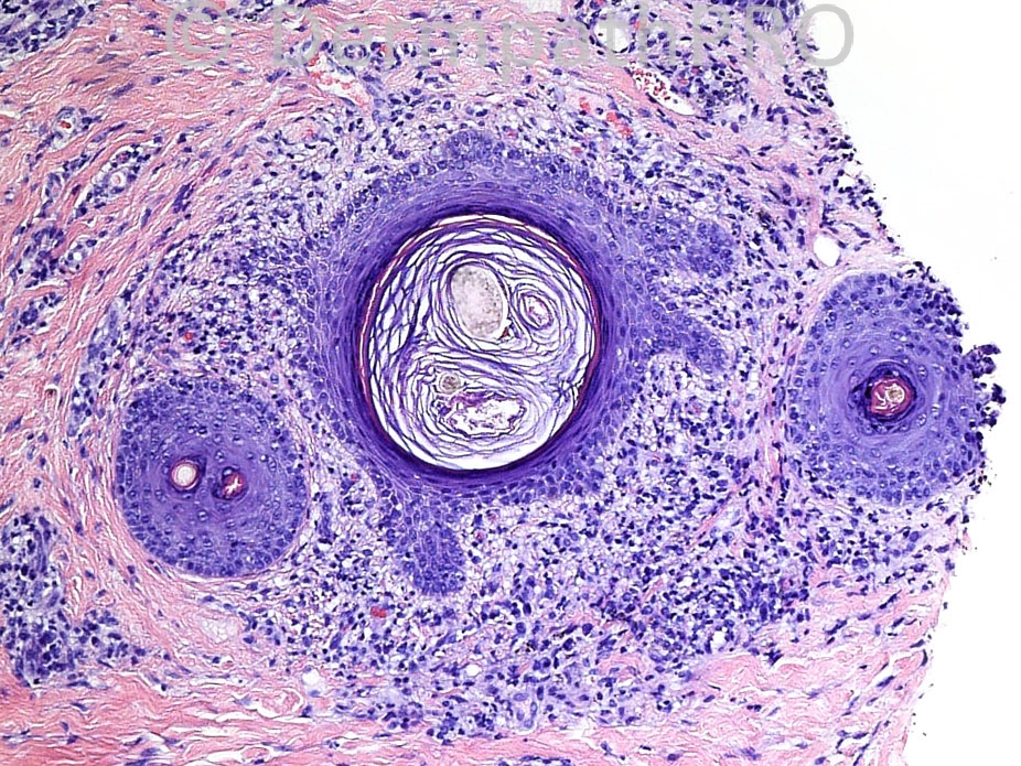
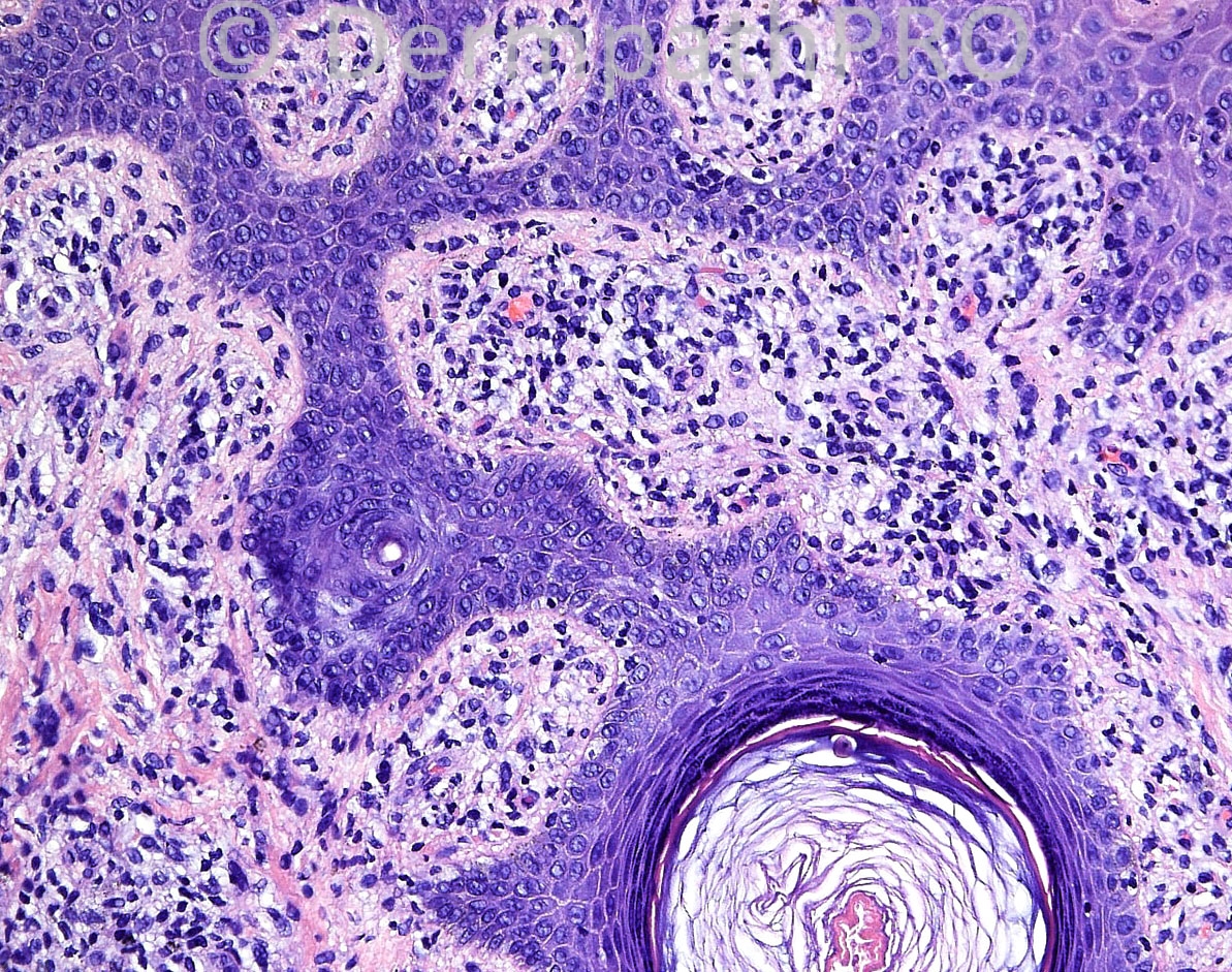
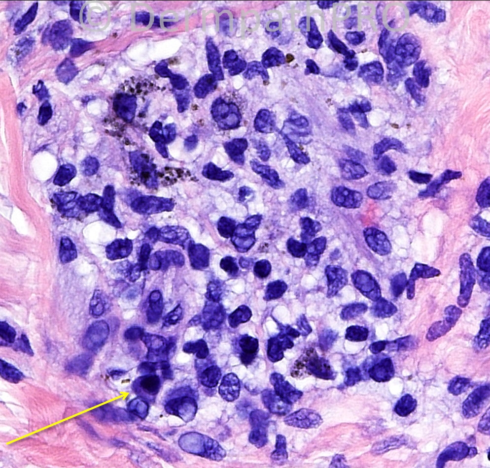
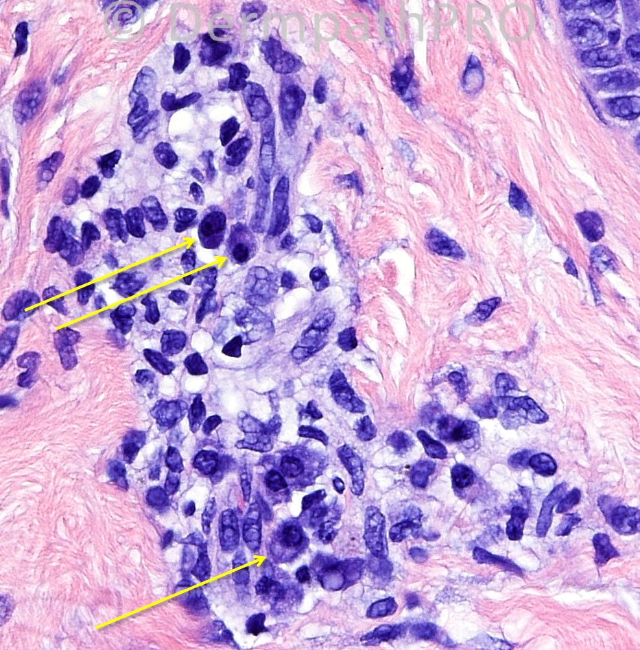
21 years-old white male, DS, Occiput biopsy.


Case posted by Dr Mark Hurt.


  Report Record

User Feedback


Robledo F. Rocha

Posted

[color="#000000"][font="Palatino Linotype, serif"][size="4"]Acne keloidalis nuchae. Classic microscopic features are present and include lymphoplasmacytic infiltrate surrounding dilated hair follicles that show thinning of the infundibular epithelium and polytrichia, loss of sebaceous glands, and interfollicular dermal scarring with a fuzzy lamellar appearance. Site, age and gender are also typical.[/size][/font][/color]

Share this comment


Link to comment
share_externally

I agree that there are many things in which make me think about Folliculitis Keloidalis Nuchae, but the histological findings are not specific and this disease is more commom in African-american people. In my opinion, the case is probably a late stage of Folliculitis decalvans in which neutrophils could not be find, however plasma cells are quite common.

Share this comment


Link to comment
share_externally

Guest Engin Sezer

Posted

Two pack compond follicles other then 6 pack suggest a lymhocyte-mediated cicatricial alopecia such as keloidalis nuchae

([url="http://www.ncbi.nlm.nih.gov/pubmed/21083676"]The amount counts: distinguishing neutrophil-mediated and lymphocyte-mediated cicatricial [b]alopecia[/b] by compound follicles.[/url] Pincus LB, Price VH, McCalmont TH.

[b]J Cutan Pathol[/b]. 2011 Jan;38(1)

Share this comment


Link to comment
share_externally

Sasi Attili

Posted

Though the presence of plasma cells should make one exclude syphilis, these are not very specific in the context of a lymphocytic scarring alopecia.

Share this comment


Link to comment
share_externally

Sasi Attili

Posted

discoid lupus would be my favoured diagnosis, of the clinical differentials listed. However, would like to see vertical sections too!

Share this comment


Link to comment
share_externally

The clinical impression of Seborrheic dermatitis doesn´t fit, so I would expect the lesion has crusting and erythema. The paper Engin Sezer showed is really helpful and is a required reading for all dermpath-lovers, but at the same time it favors LMCA, the interfollicular dermal fibrosis and the plasma cells in this case favors a NMCA (neutrophilic-mediated cicatricial alopecia) at a late stage. Amazing case.

Share this comment


Link to comment
share_externally

Guest Jim Davie MD

Posted

[i][before spirochete stains were posted ....][/i]

Late-stage lichen planopilaris might be a consideration, in addition to lupus and the above entities.
- LPP clinically presents with erythema and scaling at the follicles, so makes sense with the clinical differential diagnosis that includes seb derm and lupus.
- This is a scarring non-suppurative alopecia with interface component and follicular plugging. Although vacuolar change is not prominent, this is a high-level horizontal section with targeted perifollicular and epidermal interface inflammation, and spiky follicular epithelial cross sections with hypergranulosis. The presence of melanophages and slight basement membrane thickening is congruent with an interface component.

Share this comment


Link to comment
share_externally

Dr. Richard Carr

Posted

I was going to suggest syphilitc alopecia on the H&E (I thought M21 might be a clue). This has no features of scarring to me and is not nuchal!

Share this comment


Link to comment
share_externally

Dr. Richard Carr

Posted

If it is syphilis - thanks Mark I have never seen a case of syphilitic alopecia.

Share this comment


Link to comment
share_externally

Robledo F. Rocha

Posted

Look at all those spirochetes highlighted by immunostain!!! Dra. Maria George is right, it's syphilitic alopecia.

Share this comment


Link to comment
share_externally

Dr. Mona Abdel-Halim

Posted

Lovely, wounderful...

Share this comment


Link to comment
share_externally

Difficult case. I did not think of Sypihils for two reasons: first, I never saw a case beatiful like this; second, clinically, Syphilitic alopecia has a moth-eaten patern of hair loss and resembles more non-cicatricial alopecia, mostly alopecia areata. Great case.

Share this comment


Link to comment
share_externally

Guest Romualdo

Posted

[quote name='Guest' timestamp='1374575261']
Syphilitic alopecia
.
[/quote]

Well done Dr. Maria George!!

Share this comment


Link to comment
share_externally


Join the conversation

You can post now and register later. If you have an account, sign in now to post with your account.

Guest
Add a comment...

×   Pasted as rich text.   Paste as plain text instead

  Only 75 emoji are allowed.

×   Your link has been automatically embedded.   Display as a link instead

×   Your previous content has been restored.   Clear editor

×   You cannot paste images directly. Upload or insert images from URL.

Loading...

×
×
  • Create New...