Jump to content
In this section we have spot diagnoses posted on a daily basis since June 2010, now over 4000! You can review the archived cases and read the suggested diagnoses by users and the final comment by the contributors.
Case are uploaded each week day by 10 am UK time with the correct diagnosis will generally be posted at 8 pm UK time. Why not view the most recent spot diagnosis and proffer a diagnosis?

Case Number : Case 810 - 25th July Posted By: Guest

Please read the clinical history and view the images by clicking on them before you proffer your diagnosis.
Submitted Date :
   (0 reviews)

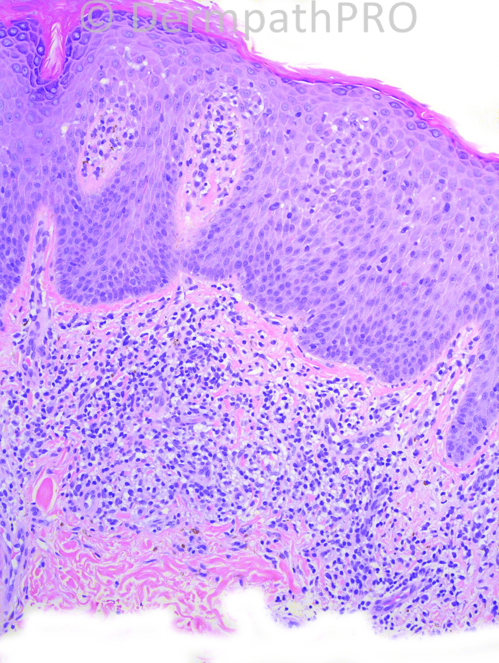
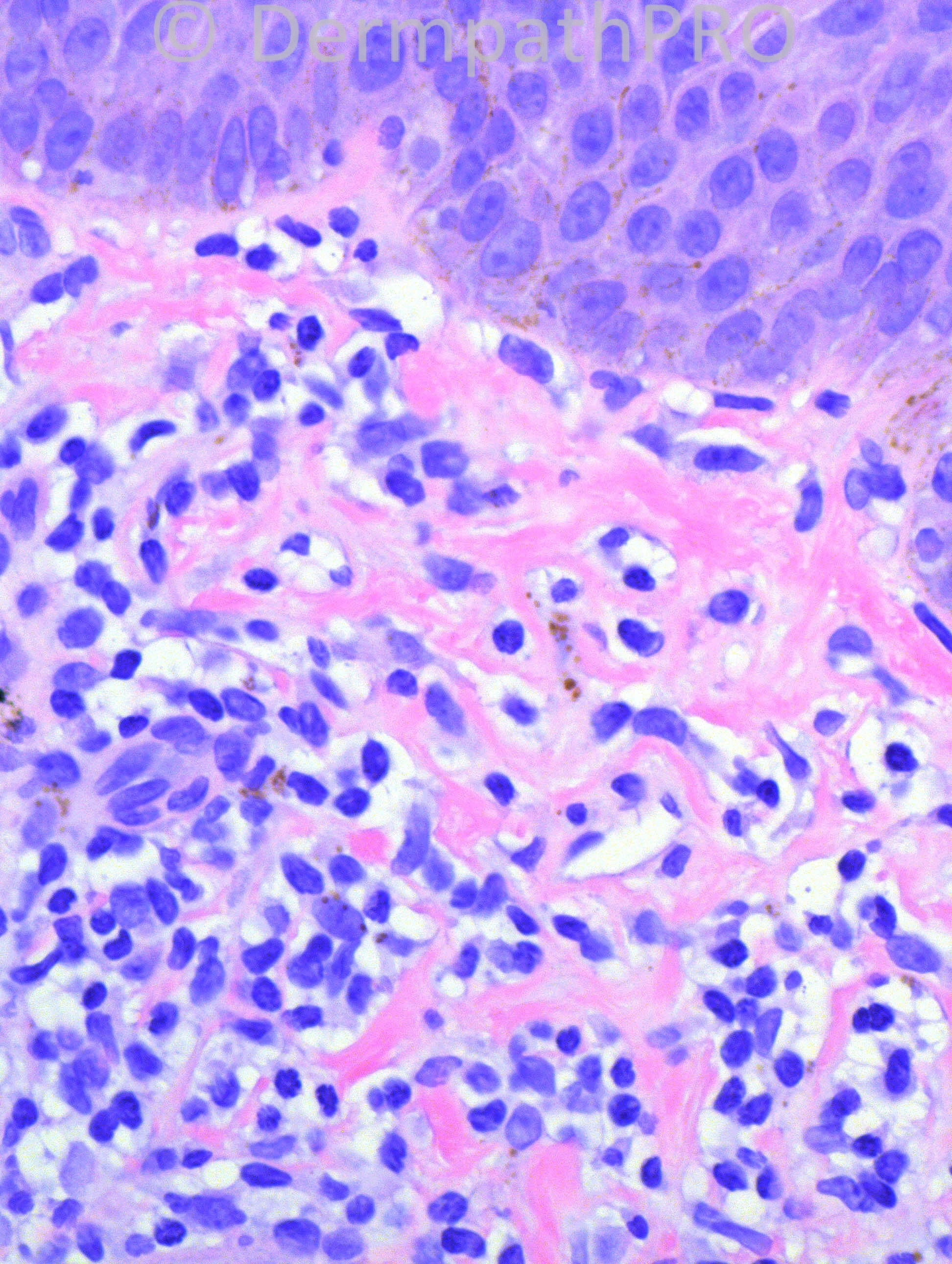
46-year-old male with right thigh biopsy.


Case posted by Dr. Hafeez Diwan.


  Report Record

User Feedback


Guest Saleem Taibjee

Posted

Some lymphoid atypia? CTCL? Obviously needs clinical correlation

Share this comment


Link to comment
share_externally

Superficial lymphoid infiltrate, acanthosis with focal parakeratosis and mild spongiosis. The pigments under the inflammatory infiltrate looks like hemossiderin. Pigmented purpuric dermatosis (despite the gender)?

Share this comment


Link to comment
share_externally

Guest immediately labrmp

Posted

Lichen aureus based on haemosiderin deposition, extravasation of RBCs and band-like lymphoid infiltration. Dx: PPD-like MF (unlikely)

Share this comment


Link to comment
share_externally

Guest Romualdo

Posted

I think it is necessary to perform Perls stain to confirm the nature of the pigment present in the dermis. I am not so convinced but If it turns out to be hemosiderin, then the possibilities of pigmented purpuric dermatosis or mycosis fungoides developing from a pigmented purpuric dermatosis must be considered (some atypical lymphocytes with large cerebriform nuclei are seen in the epidermis and coarse collagen bundles are apparently present in the dermis). If there is no hemosiderin then I think the diagnosis will be mycosis fungoides. Immunostains and clinicopathological correlation are necessary. MF-like drug eruptions may look very similar.

Share this comment


Link to comment
share_externally

Robledo F. Rocha

Posted

A superficial vacuolar lymphocytic infiltrate with epidermal hyperplasia, mild spongiosis, mound of parakeratosis, and extravasated erythrocytes in the papillary dermis.
My first hypothesis was pityriasis rosea, but it´s hard to sustain this after a case history of an isolated lesion on thigh, unless an inverted herald patch is taken into account.
I interpret the brown pigment as melanin incontinence, so pigmented purpuric dermatitis is not my choice.
Exocytosis of large lymphocytes strikes me about the possibility of mycosis fungoides (so-called small-plaque parapsoriasis), but in this case I would expect those large lymphocytes surrounded by a clear halo and aligned along the basal layer of the epidermis.
By elimination, I favor erythema annulare centrifugum.

Share this comment


Link to comment
share_externally

Sasi Attili

Posted

Quite tough without knowing the history, but would plump for PPD with the differential of CTCl based on the slightly atypical lymphocytes

Share this comment


Link to comment
share_externally

Guest Jim Davie MD

Posted

I will toss in with the lichenoid pigmented purpura group on this one. Subacute eczematous dermatitis would be another consideration. Clinical is key.
MF cannot be excluded as it can masquerade as just about any inflammatory dermatosis, but I agree with Robledo that the junction shows too little involvement by lymphocytes to put MF on the top of the differential Dx list.
[size=4] A spongiotic psoriasiform dermatitis with lichenoid infiltrate, lymphocytic and rare non-specific RBC extravasation and exocytosis, with focal parakeratosis. There is equivocal and non-specific rare satellite necrosis. I think there is mixed hemosiderin and melanin in the dermis. [/size][size=4]The superimposed effects of chronic irritation (LSC/prurigo) complicates the interpretation, with addition of non-specific periretal and dermal 'wiry fibrosis' that would hint at consideration of MF (although more impressive fibrosis can be seen in some prurigo cases). [/size]

Share this comment


Link to comment
share_externally

Mycosis fungoides. Methinks there's disproportionate epidermotropism.

Share this comment


Link to comment
share_externally

Dr. Hafeez Diwan

Posted

I called this mycosis fungoides. There was marked CD4 predominance. Clinically, this was MF as well.

Share this comment


Link to comment
share_externally


Join the conversation

You can post now and register later. If you have an account, sign in now to post with your account.

Guest
Add a comment...

×   Pasted as rich text.   Paste as plain text instead

  Only 75 emoji are allowed.

×   Your link has been automatically embedded.   Display as a link instead

×   Your previous content has been restored.   Clear editor

×   You cannot paste images directly. Upload or insert images from URL.

Loading...

×
×
  • Create New...