Jump to content
In this section we have spot diagnoses posted on a daily basis since June 2010, now over 4000! You can review the archived cases and read the suggested diagnoses by users and the final comment by the contributors.
Case are uploaded each week day by 10 am UK time with the correct diagnosis will generally be posted at 8 pm UK time. Why not view the most recent spot diagnosis and proffer a diagnosis?

Case Number : Case 813 - 30th July Posted By: Guest

Please read the clinical history and view the images by clicking on them before you proffer your diagnosis.
Submitted Date :
   (0 reviews)

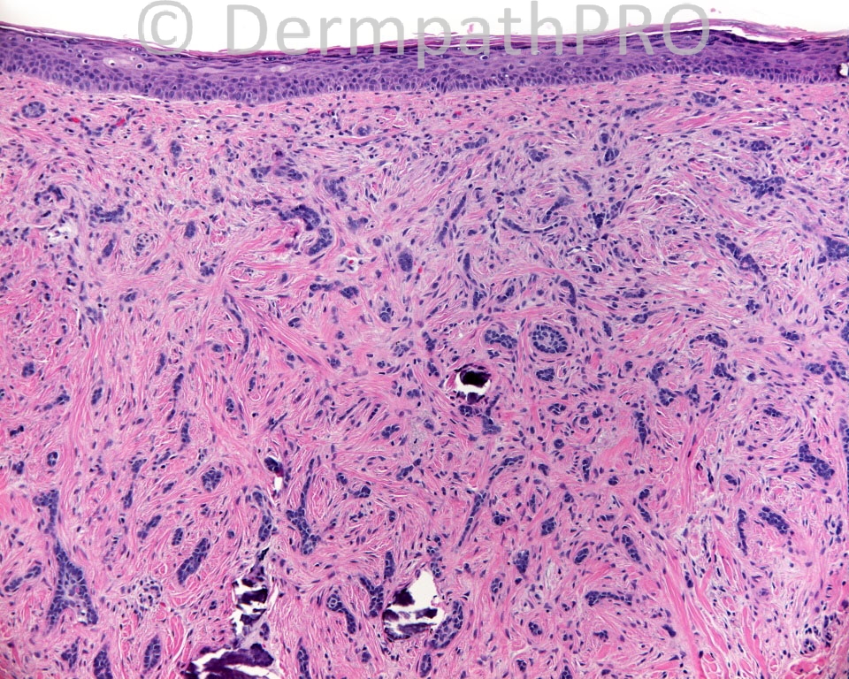
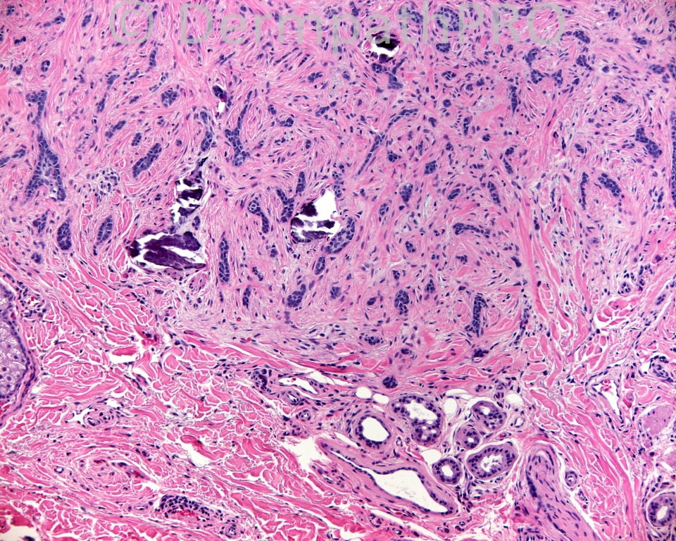
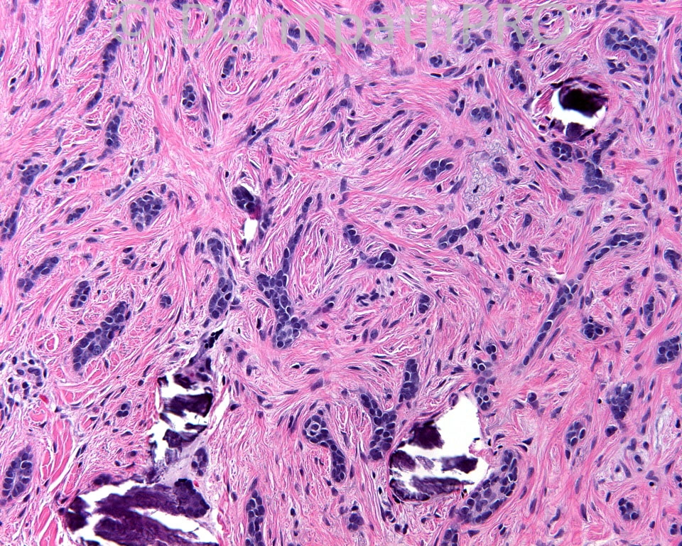
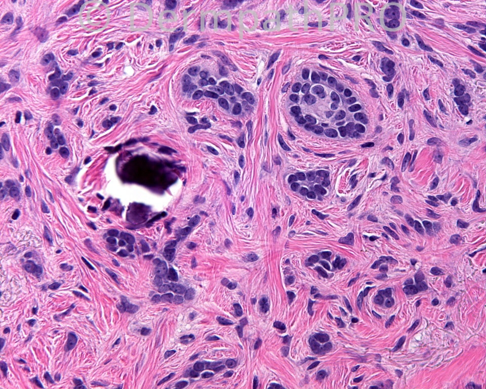
The patient is a 51 year-old white man with a lesion from the right cheek of the face.

Case posted by Dr Mark Hurt.


  Report Record

User Feedback


Guest Maria George

Posted

Desmoplastic Trichoepithelioma.

Share this comment


Link to comment
share_externally

Guest Dr. Francisco Vílchez

Posted

Nice desmoplastic trichoepithelioma.

Share this comment


Link to comment
share_externally

Guest Romualdo

Posted

In the complete absence of horn cysts and scarcity of elongated epithelial strands I go with a morpheaform basal cell carcinoma.

Share this comment


Link to comment
share_externally

Guest Graham Reilly

Posted

Desmoplastic trichoepithelioma - central depression seen in first slide.

Share this comment


Link to comment
share_externally

Sasi Attili

Posted

[color=#1C2837][font=arial, verdana, tahoma, sans-serif][size=4]Desmoplastic Trichoepithelioma[/size][/font][/color]

Share this comment


Link to comment
share_externally

Guest Rodrigo Restrepo

Posted

Morpheaform basal cell carcinoma.

Share this comment


Link to comment
share_externally

Robledo F. Rocha

Posted

[color=#000000][font=Palatino Linotype, serif][size=4]I favor columnar trichoblastoma (desmoplastic trichoepithelioma) over morpheaform basal cell carcinoma:[/size][/font][/color][list]
[*][color=#000000][font=Palatino Linotype, serif][size=4]on scanning magnification, it appears as a symmetric lesion with depressed center;[/size][/font][/color]
[*][color=#000000][font=Palatino Linotype, serif][size=4]cellular fibrous stroma clearly demarcated from surrounding reticular dermis;[/size][/font][/color]
[*][color=#000000][font=Palatino Linotype, serif][size=4]no discernible mitotic figures can be found;[/size][/font][/color]
[*][color=#000000][font=Palatino Linotype, serif][size=4]calcification foci with rounded contours, probably glossing over keratinous microcysts;[/size][/font][/color]
[*][color=#000000][font=Palatino Linotype, serif][size=4]a transversally cutted papillary mesenchymal body can be seen [size=4][color=#000000]on the upper right quadrant of the picture #6[/color][/size].[/size][/font][/color]
[/list]

Share this comment


Link to comment
share_externally

Guest Romualdo

Posted

If possible, I ask Dr. Mark Hurt to answer two questions, when closing the case: 1.Is there a mitotic figure in picture 7? 2. Is there a papillary mesenchymal body on the upper right quadrant of picture 6, as Robledo said?

Share this comment


Link to comment
share_externally

Mark A. Hurt MD

Posted

My diagnosis was columnar trichoblastoma (desmoplastic trichoepithelioma).

The lesion is small, circumscribed, and with a dell of depression on the surface. There are epithelial columns, cords, and strands enmeshed within a stroma that encircles the epithelial components. The stroma contains clefts, yet it is closely apposed to the germinative components.

To Romualdo's questions about a papillary mesenchymal body on figure 6, no, I don't think it is one, at least not in the classical sense of a "ball in claw" relationship of the epithelium to the stroma. Additionally, I don't think that there is mitosis in figure 7; I think it is an illusion.

For those of you with the adnexal tumor book by Kazakov, Michal, Kacerovska, & McKee, take a look at Figure 7.28 on page 218; it is identical to the case above. Additionally look at the book by Ackerman, Reddy, & Soyer from 2001. Their figures 22-98 (page 596) through 22-110 (page 609) illustrate also the principal features for the diagnosis and the main foil (morpheiform bcc).

Share this comment


Link to comment
share_externally

I would like to ask you all if you would make this diagnosis straight on the H&E or would use a IHQ?
Thanks.

Share this comment


Link to comment
share_externally

Mark A. Hurt MD

Posted

I would likely make the diagnosis on this case from the H&E alone (in fact, I did). In cases in which there is only a partial biopsy, it becomes a problem, in my experience. I attempted for years to find some kind of marker that really helped solve the dilemma in some cases, but haven't had much luck. For instance, CK20 is said to help sort out the differential of DTE (positive Merkel cells) vs MBCC (no Merkel cells), but I have had mixed results. Recently, I came across the [url="http://www.ncbi.nlm.nih.gov/pubmed/?term=20711172"]article[/url] by Abbas, Richards & Mahalingam, which seems promising; it employs fibroblast-activation protein, which is positive in the stroma of MBCC and negative in the stroma of DTE. If any are willing to elaborate on what they do in their practices, we will all appreciate it.

Share this comment


Link to comment
share_externally

Thank you, Dr. Mark Hurt. I do not get good and clear results with CK20 too and I made only once the Androgen receptor that helped me in a superficial biopsy. I will read this article, I didn´t know about this fibroblast-activation protein.
Thank you again!

Share this comment


Link to comment
share_externally

Dr. Richard Carr

Posted

It is an H&E diagnosis (but Dr Akerman does state it can be one of the most difficult diagnoses in dermatopathology - I keep a spreadsheet wtih his 26 differentiating criteria!!). I find CK20 useful in small biopsies (almost always >5 MC in the basaloid cords, even in small biopsies), never seen any MC in an DTE-like infiltrative BCC. I have only collected about 20 or 30 DTE in my immuno database though (they are quite uncommon in my practice) but would be happy to collaborate on a bigger series. I have never seen prominent papillary mesenchymal bodies in DTE other than associated with trapped hair follicles (often numerous vellus follicles in cases of DTE that almost always occur on the central face).

Share this comment


Link to comment
share_externally

Mark A. Hurt MD

Posted

Thank you, Richard. Do you have a sense of how to characterize the stroma of DTE vs MBCC? I think they are quite different; DTE's seem to have a much more stereotyped and organized stroma, but MBCC's usually have a less stereotyped stroma. What are your thoughts on this?

Share this comment


Link to comment
share_externally

Mark A. Hurt MD

Posted

One more question, Richard. How many of the 26 criteria do you think are essential in differentiating between the two lesions? That is, which do you think are the most important? My sense of criteria, as such, is that usually no more than 4 or 5 are essential.

Share this comment


Link to comment
share_externally

Dr. Richard Carr

Posted

Thanks Mark,
Here goes - you will tell something about my personality from this response!

Akerman uses the terms: Rims of compact collagen (encircling nests in DTE).
I like the curvilinear cords in DTE (but I have not recorded if that is distinguishing but looks very typical in your case).

Akerman's criteria are as follows (and I have been scoring cases prospectively):
1. Dell - not so useful in small biospies and not always present in DTE
2. Papillated epidermis (often in DTE, Flat in BCC) - not so useful
3. Symmetry (DTE yes; BCC no) - very useful in excision bx, obviously not helpful in small biopsies
4. Aggregations of larger size (DTE: No) - very useful
5. Variation in size and shapes (DTE: little variation) - very useful
6. Bizarre shapes (DTE: No) - very useful
7. Foci of typical (nodular) BCC (DTE: No) - very useful when present
8. Retraction artefact - not so helpful as absent usually in infiltrative BCC
9. Rims of compact collagen: DTE present around cords; not usually present in BCC - very useful
10. Squamous differentiation: DTE present in some aggregations (not in BCC) - not overly helpful
11. Follicular differentiation (hair bulbs, papillae, trichohyaline granules): (present in DTE) - not helpful
12. Aberrant follicles connected to epideris: (DTE may be present; absent in BCC) - not helpful
13. Aberrant hair shafts in nests: - not helpful
14. Epithelial keratin filled cysts: (numerous in DTE): very helpful
15. Shadow cells in cysts: not helpful
16. Granulomas around ruptured cysts: quite helpful
17. Calcification assoc. with keratin cysts: Very helpful
18. Sebaceous differnetiation: not helpful
19. Sebaceous cells around infundibular epithelium: not helpful
20. Mitotic figures (Rare in DTE): very helpul (Ki67 can be useful usually <5% in DTE often ~10% or greater in BCC)
21. Single cell necrosis / apoptosis: (absent in DTE): yes absent in DTE usually but can be quite low in infiltrative BCC
22. Melanocytic naevus (often present in DTE): only in one or two of our cases so not very helfpul!
23. Nerve and muscle (DTE not invaded): Bewared you can see perineural invasion within the confines of a DTE occasionally (I suspect this is under-recognised or has resulted in wrong classification as I did see previous Spot Diagnosis case).
24. Solar elastosis: Above stroma of DTE (admixed with tumour in BCC): My database indicates this criterion is useless!
25. Stromal fibroblasts (slight increase in DTE only, markedly increased in BCC): Quite useful
26. Lymphocytic infiltration (slight or absent in DTE): All DTE seem to lack inflammatory cell infiltrate in my database but I am not always sure inflammatory cells are prominent in BCC - could be useful if inflammation is prominent though.
Other comments: The DTE have not invaded subcutis but may be full thickness dermal (deepest in my series 2.5mm). Despite all these criteria a few cases in small biopsies I could not decide and in one or two of those sparse MC were noted. I usually advise complete excision with conservative margins (or margin control) for such (uncertain) cases. I also have two or three examples of collision tumours with DTE and infiltrative BCC - again MC with CK20 useful to illustrate in those cases. Hope this helps!

Share this comment


Link to comment
share_externally

Mark A. Hurt MD

Posted

Wow! This is excellent, Richard. I think you are right about the rims of compact collagen and symmetry as being very useful.

Share this comment


Link to comment
share_externally

Robledo F. Rocha

Posted

On the difference in the perception of the same object, here depicted by the presence or not of a papillary mesenchymal body on picture #6, please read this [url="https://dermpathpro.com/blog/3/entry-88-the-form-of-perception-and-the-object-of-perception/"]entry[/url] in Dr. Mark Hurt's Blog.

Share this comment


Link to comment
share_externally

Mark A. Hurt MD

Posted

Thanks, Robledo, for the "plug" about the blog, The "form-object distinction" is a very important philosophical principle, and it has wide implications in dermatopathology. By the way, I did not identify that distinction, but I do know the source, if anyone is interested.

Share this comment


Link to comment
share_externally


Join the conversation

You can post now and register later. If you have an account, sign in now to post with your account.

Guest
Add a comment...

×   Pasted as rich text.   Paste as plain text instead

  Only 75 emoji are allowed.

×   Your link has been automatically embedded.   Display as a link instead

×   Your previous content has been restored.   Clear editor

×   You cannot paste images directly. Upload or insert images from URL.

Loading...

×
×
  • Create New...