Jump to content
In this section we have spot diagnoses posted on a daily basis since June 2010, now over 4000! You can review the archived cases and read the suggested diagnoses by users and the final comment by the contributors.
Case are uploaded each week day by 10 am UK time with the correct diagnosis will generally be posted at 8 pm UK time. Why not view the most recent spot diagnosis and proffer a diagnosis?

Case Number : Case 775 - 6th June Posted By: Guest

Please read the clinical history and view the images by clicking on them before you proffer your diagnosis.
Submitted Date :
   (0 reviews)

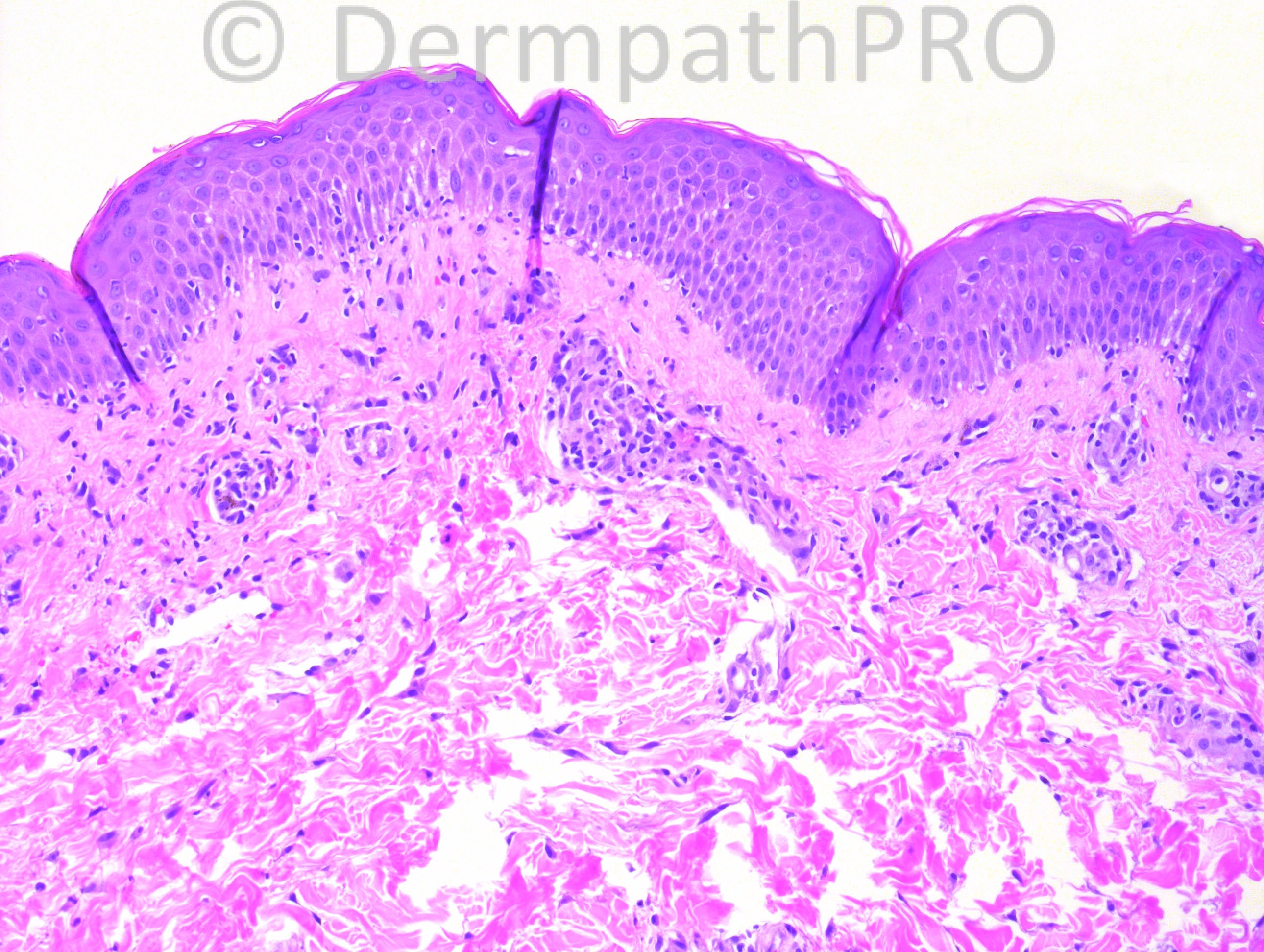
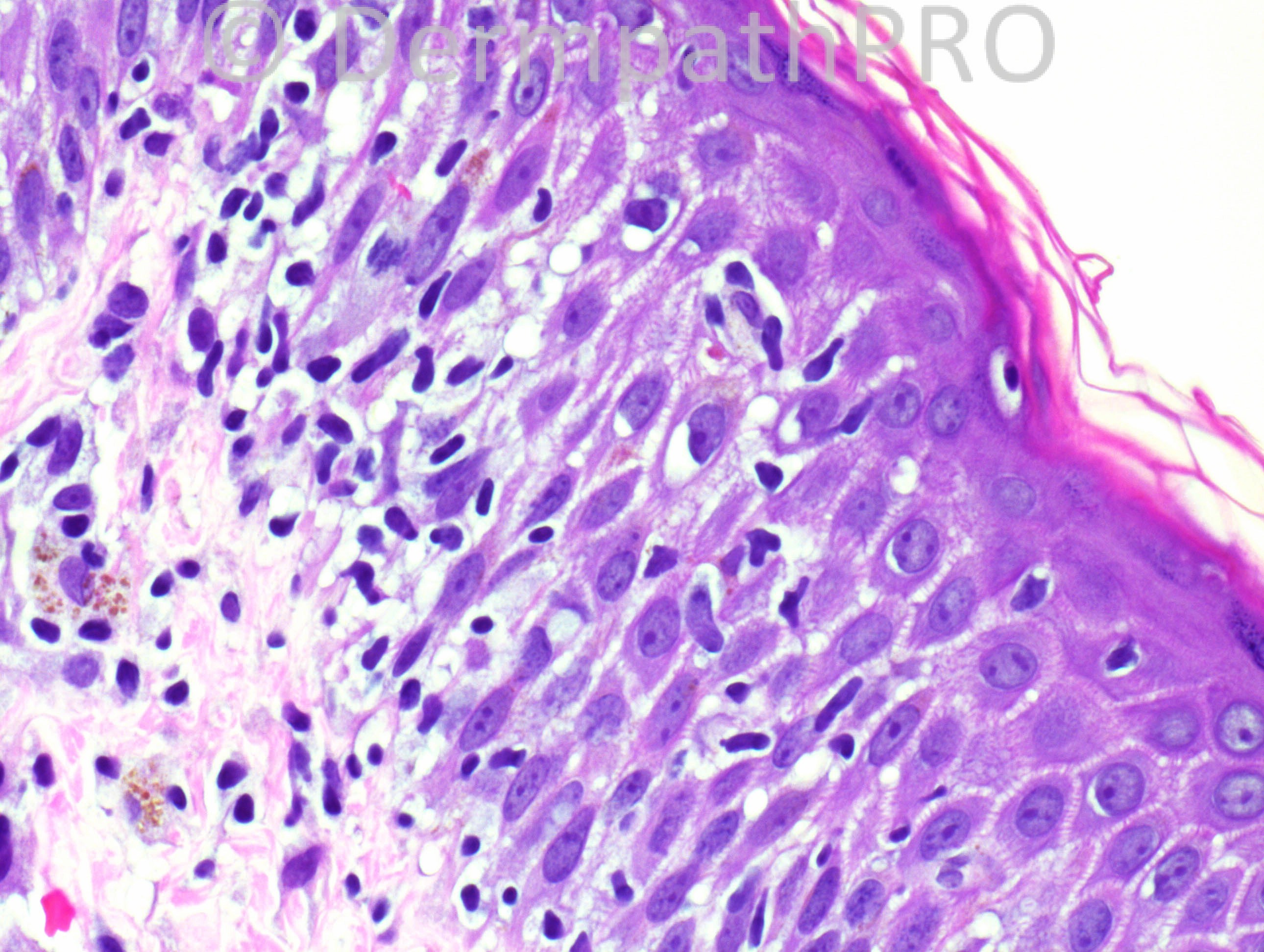
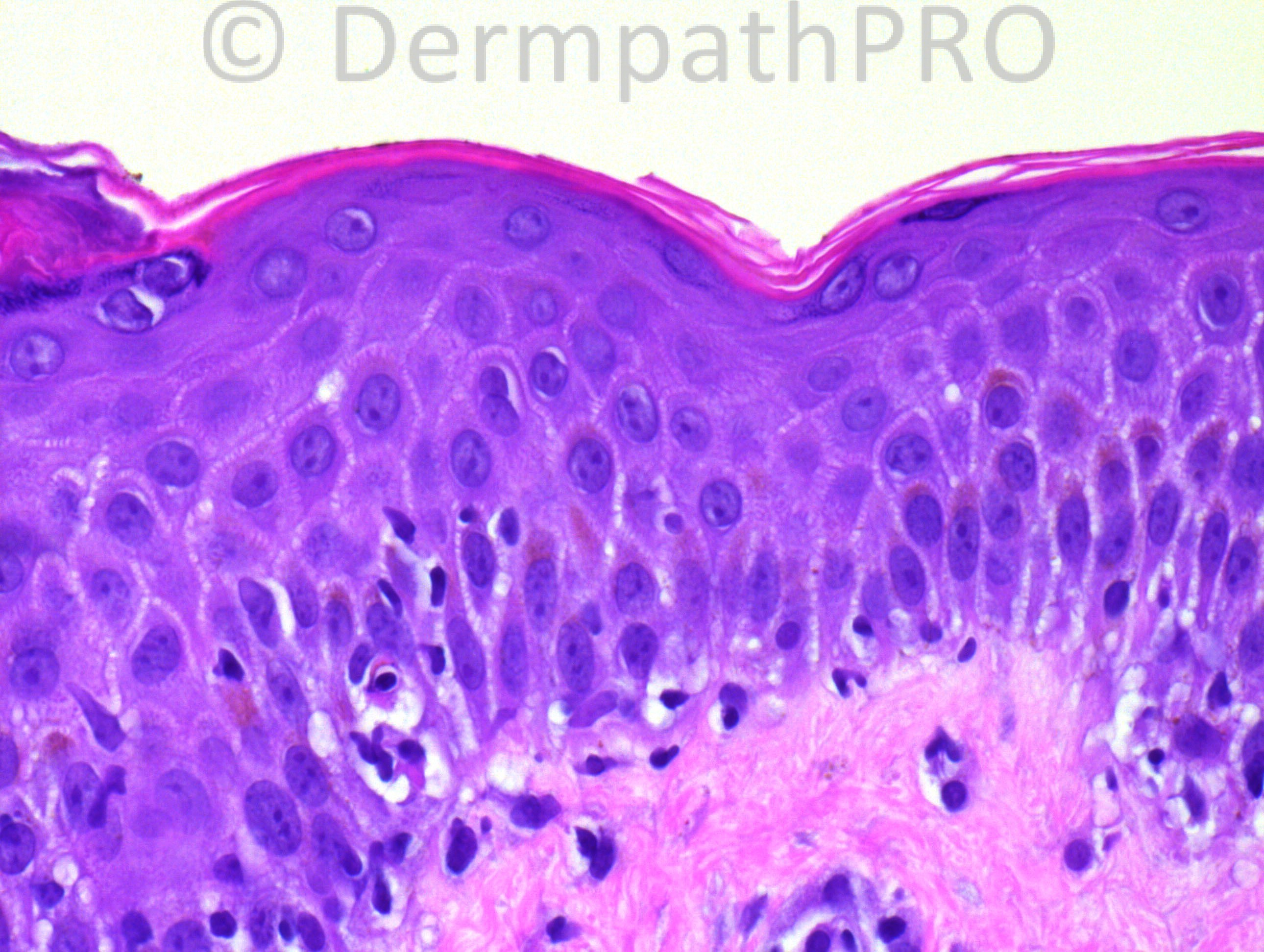
19 years-old Hispanic female with history of refractory Hodgkin lymphoma (nodular sclerosing type), status-post MUD bone marrow transplant (day 121 post-transplant), with a maculopapular rash.

Case posted by Dr. Hafeez Diwan.


  Report Record

User Feedback


Guest Dr Engin Sezer

Posted

GVHD

Share this comment


Link to comment
share_externally

Guest Romualdo

Posted

The combined presence of GVHD features and marked spongiosis suggests the eczematoid variant of GVHD.

Share this comment


Link to comment
share_externally

Robledo F. Rocha

Posted

Microscopic features are typical of acute graft-versus-host disease in the proper setting of the given case history, but the late onset of the cutaneous rash is not. I go with late manifestation of acute graft-versus-host disease.
It could be graded as 2 since there are both basal vacuolization and dyskeratotic keratinocytes.
Lymphocytic infiltrate is sparse, as might be expected after bone marrow transplantation.

Share this comment


Link to comment
share_externally

Dr. Richard Carr

Posted

MUD???

Share this comment


Link to comment
share_externally

Robledo F. Rocha

Posted

MUD = matched unrelated donor.

Share this comment


Link to comment
share_externally

Guest Jaime Mejía MD

Posted

GVHD

Share this comment


Link to comment
share_externally

Dr. Mona Abdel-Halim

Posted

GVHD

Share this comment


Link to comment
share_externally

Guest Maria George

Posted

Necrotic keratinocyte surrounded by cytotoxic lymphocyte (satellite cell necrosis).
Goofd for GVHD.

Share this comment


Link to comment
share_externally

Guest Jim Davie MD

Posted

It looks like acute GVHD by histology, agree grade 2. Agree that onset is very late for acute, as this would be chronic GVHD by history, if rigorously following date definitions of acute-vs-chronic (>100 days post-transplant = chronic). Would doubt a drug etiology with the smoking gun we have.

Chronic GVHD can have variable clinical presentations, including eczematous or maculopapular process similar to those of acute GVHD. ref: Abstract [url="http://www.ncbi.nlm.nih.gov/pubmed/20868623"]J Cutan Med Surg. 2010 Sep-Oct;14(5):249-53.[/url]

We don't have clinical history as to whether this is a first presentation or a persistent process. There is spongiosis, satellite cell necrosis, lymphocyte exocytosis, and subtle vacuolar change. There is modest accumulation of melanophages that suggest a subacute process, but no sclerotic changes or lichen-planus like epidermal changes that would be expected in a classic chronic GVHD.

Share this comment


Link to comment
share_externally

Dr. Hafeez Diwan

Posted

GVHD.

Share this comment


Link to comment
share_externally


×
×
  • Create New...