Jump to content
In this section we have spot diagnoses posted on a daily basis since June 2010, now over 4000! You can review the archived cases and read the suggested diagnoses by users and the final comment by the contributors.
Case are uploaded each week day by 10 am UK time with the correct diagnosis will generally be posted at 8 pm UK time. Why not view the most recent spot diagnosis and proffer a diagnosis?

Case Number : Case 777 - 10th June Posted By: Guest

Please read the clinical history and view the images by clicking on them before you proffer your diagnosis.
Submitted Date :
   (0 reviews)

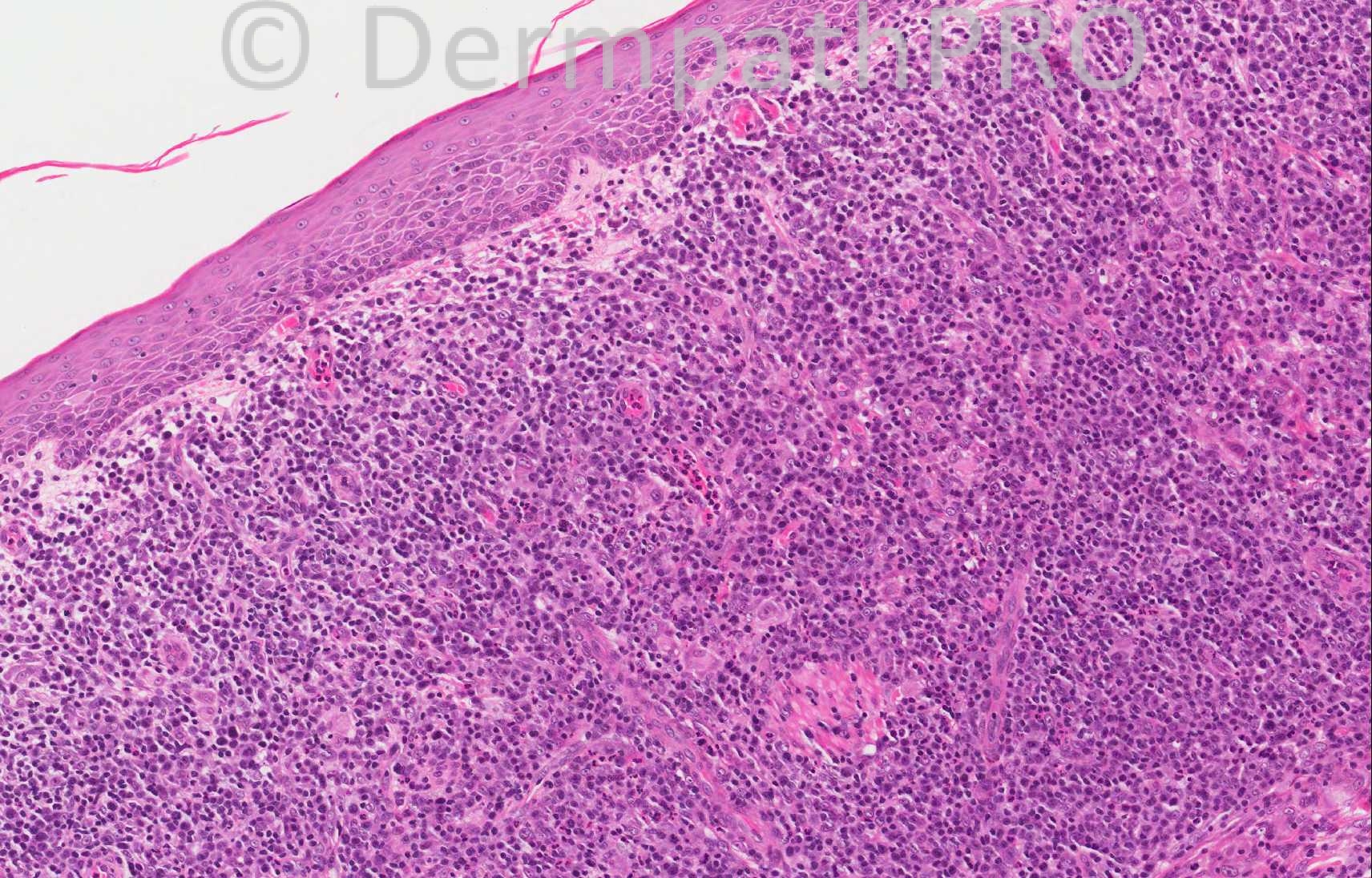
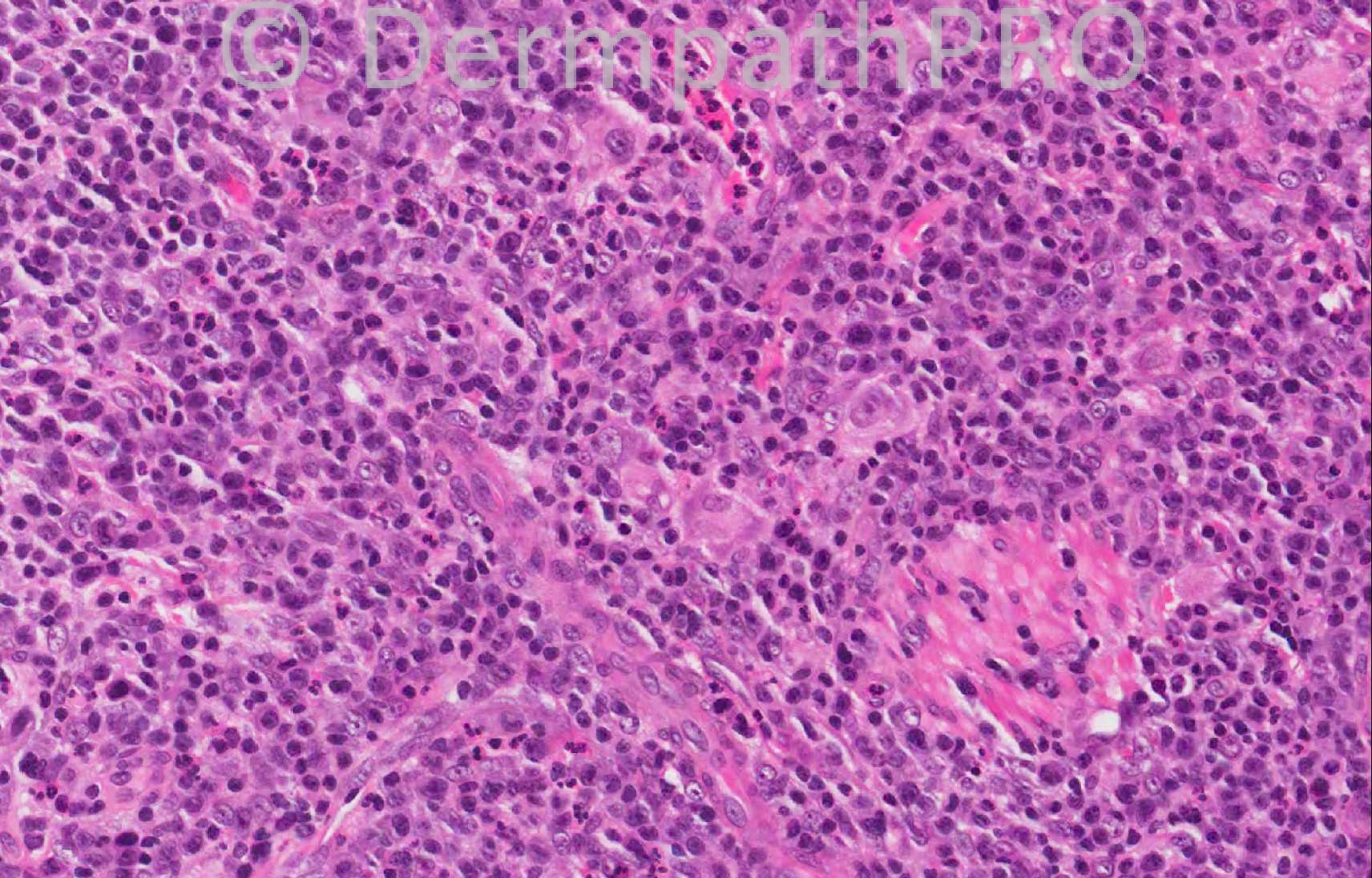
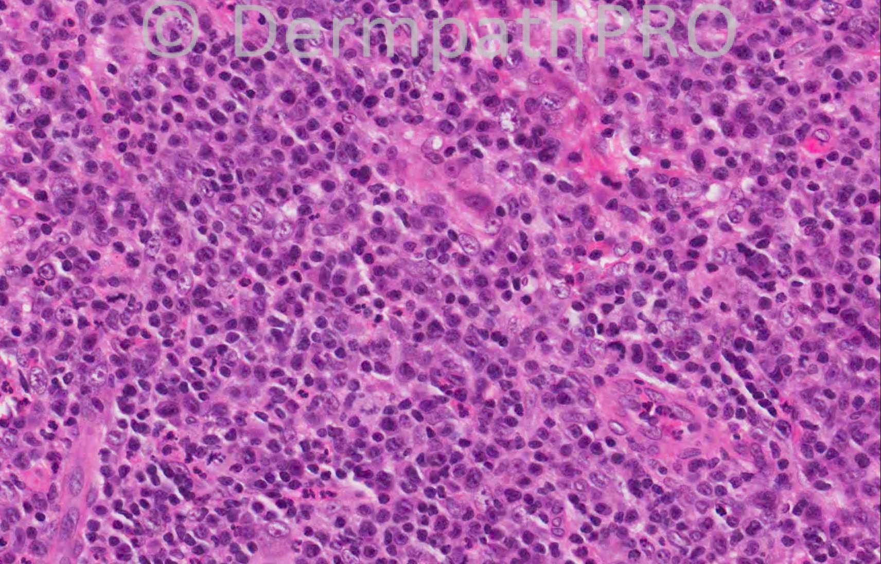
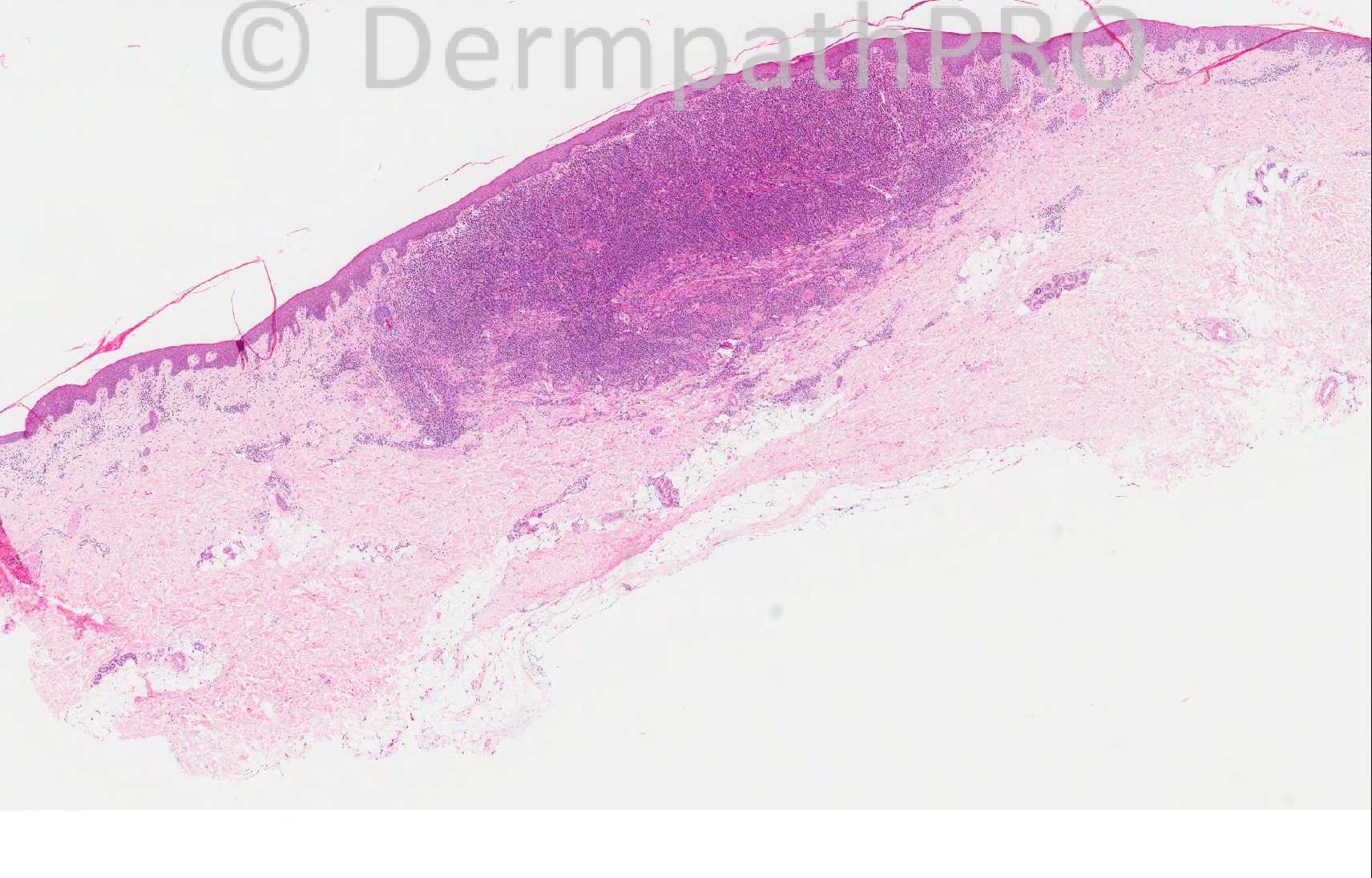
84 years-old male. Lesion left arm ?lymphoma ?nodular prurigo, recurrent nodules.

Thank you to Dr. Leena Joseph for providing this case, she is a Consultant Skin Pathologist at the Whythenshawe Hospital.

  Report Record

User Feedback


Guest Maria George

Posted

[color=#444444][font=arial, sans-serif][size=1][right]Localized dense lymphohistiocytic infiltrate with Grenz area below epidermis.D.D may include pseudolymphoma, lymphoma, plasmacytoma, and Langerhans cell histiocytosis .IHC is recommended .Initial panel may include,CD3, CD4, CD5, CD7,CD8,CD20,CD1a,,CD68,kappa and lambda light chains, Bcl2,Bcl6, and [/right][/size][/font][/color][color=#444444][font=arial, sans-serif][size=1][right]CD138 (Syndecan-1).[/right][/size][/font][/color]

Share this comment


Link to comment
share_externally

Guest Dr Engin Sezer

Posted

My first impression is anaplastic large cell lymhoma vs. diffuse large B-cell lymhoma

Share this comment


Link to comment
share_externally

Dr. Mona Abdel-Halim

Posted

Lymphoma vs. pseudolymphoma for work up

Share this comment


Link to comment
share_externally

Guest Romualdo

Posted

I think this is a lymphoma. My favorite is anaplastic large cell lymphoma.

Share this comment


Link to comment
share_externally

Robledo F. Rocha

Posted

[color=#000000][color=#000000]A dense mixed infiltrate with predominance of small lymphocytes and plasma cells, but with the remarkable presence of eosinophils, worrisome large cells and some mitotic figures.[/color][/color]
[color=#000000][color=#000000]My first impression is pseudolymphoma due to many features that suggest a benign condition:[/color][/color][list]
[*][color=#000000][color=#000000]this [/color][color=#000000]non-ulcerated[/color][color=#000000] lesion is [/color][color=#000000]symmetric and well-circumscribed;[/color][/color]
[*][color=#000000][color=#000000]it shows a [/color][color=#000000]top-heavy / wedge-shaped arrangement of the infiltrate;[/color][/color]
[*][color=#000000][color=#000000]there is also Grenz zone, spared epidermis, and prominent blood vessels with plump endothelial cells.[/color][/color]
[/list]
[color=#000000][color=#000000]I favor lymphomatoid drug eruption, maybe associated with [url="http://www.ncbi.nlm.nih.gov/pubmed/7632065"]antidepressant therapy[/url].[/color][/color]

Share this comment


Link to comment
share_externally

Guest Jim Davie MD

Posted

Agree with the above of lymphoma vs. pseudolymphoma. Recurrent nodules in the history would favor a benign reactive process. Stain panel similar to Maria's with light chain would be worthwhile to begin a workup. There is no reactive follicular architecture present. If malignant, a MZL or less likely FCL (diffuse pattern) would be in my differential given the persence of plasma cells and lymphoplasmacytoid cells.

Share this comment


Link to comment
share_externally

Dr. Richard Carr

Posted

My first impression was nodular scabies (nodular infiltrate heavy in plasma cells and sparse eosinophils). Lots of plasma cells so easy enough to do a Kappa and Lambda to r/o lymphoma.

Share this comment


Link to comment
share_externally

Iskander H. Chaudhry

Posted

The diagnosis for this was lymphoproliferative disorder - lymphomatoid papulosis.

Share this comment


Link to comment
share_externally

Guest Maria George

Posted

Thank you [color=#1C2837][font=arial, verdana, tahoma, sans-serif][size=4][right]Iskander .But from A-E ,which type you gave it to this one?[/right][/size][/font][/color]

Share this comment


Link to comment
share_externally

Robledo F. Rocha

Posted

Lymphomatous cutaneous infiltrates are really a jack-in-the-box!
I would classify this as a type A (histiocytic type) lymphomatoid papulosis. Do the panel agree?

Share this comment


Link to comment
share_externally

Guest Shanesmith

Posted

[i]Lymphoepithelial carcinoma[/i]

Share this comment


Link to comment
share_externally

Sasi Attili

Posted

A bit late. My first impression was Lyp

Share this comment


Link to comment
share_externally

Guest Dr.Ebtisam FAIDA

Posted

Pseudo lymphoma vs. lymphoma for immunostudy

Share this comment


Link to comment
share_externally


×
×
  • Create New...