Jump to content
In this section we have spot diagnoses posted on a daily basis since June 2010, now over 4000! You can review the archived cases and read the suggested diagnoses by users and the final comment by the contributors.
Case are uploaded each week day by 10 am UK time with the correct diagnosis will generally be posted at 8 pm UK time. Why not view the most recent spot diagnosis and proffer a diagnosis?

Case Number : Case 779 - 12th June Posted By: Guest

Please read the clinical history and view the images by clicking on them before you proffer your diagnosis.
Submitted Date :
   (0 reviews)

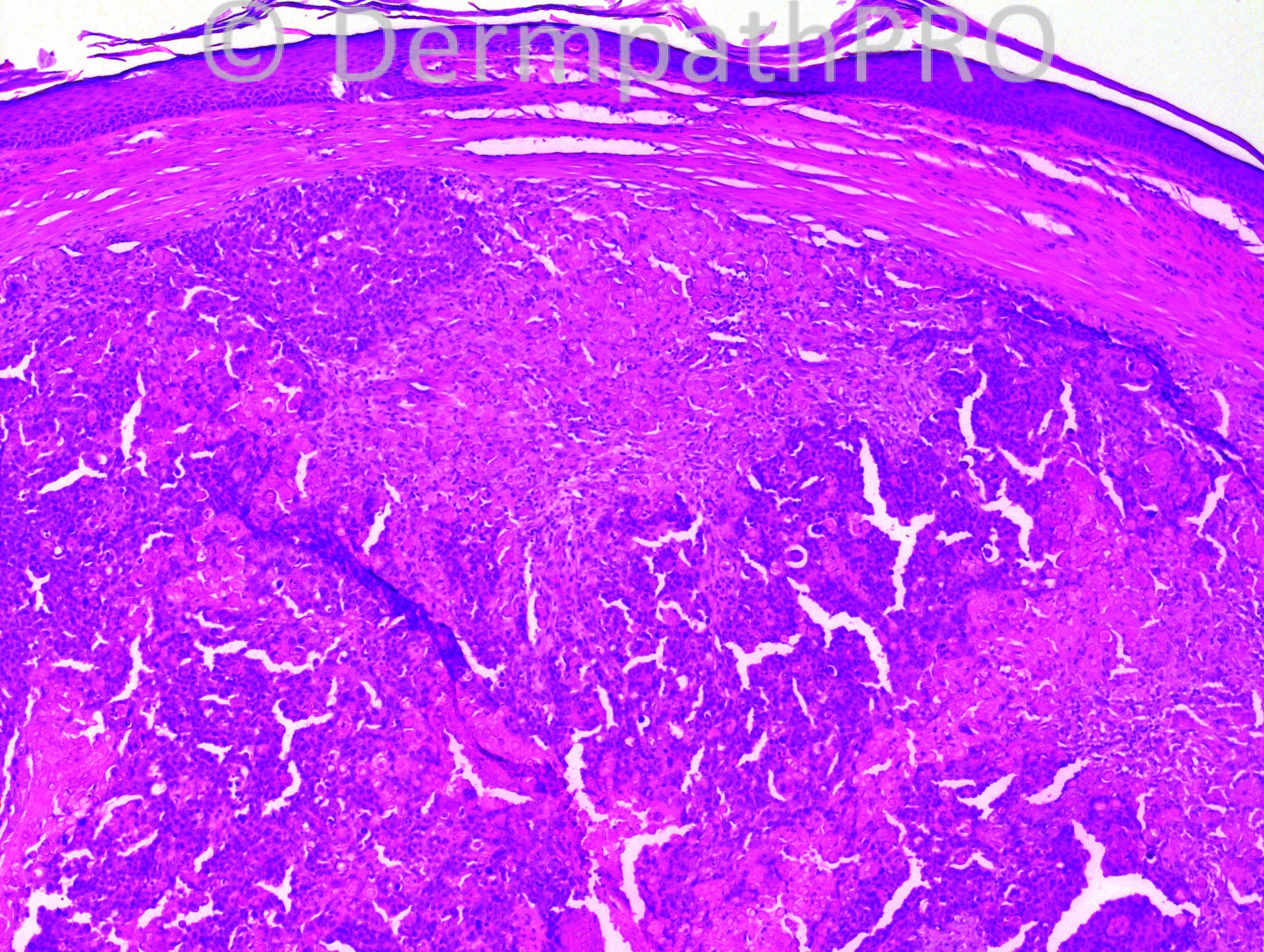
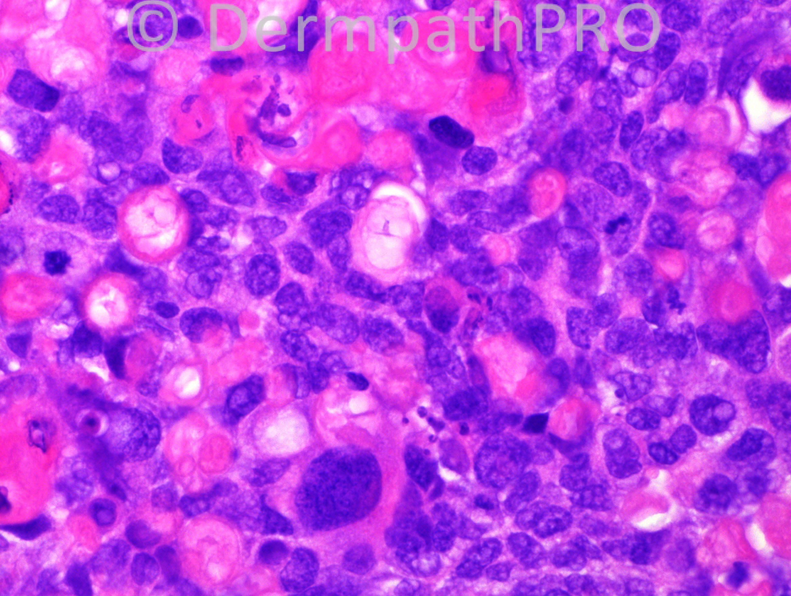
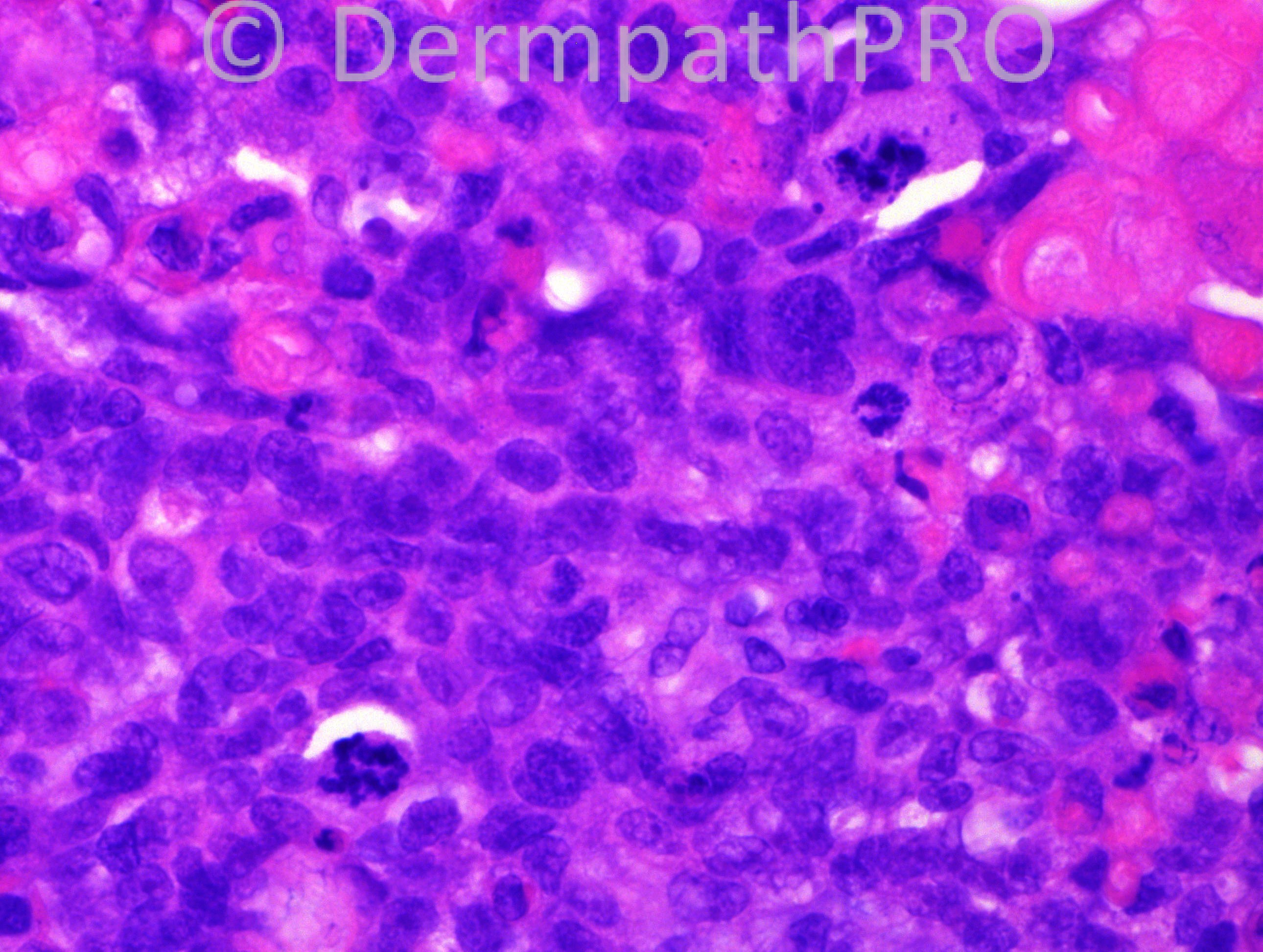
86 years-old male with a scalp lesion.

Case posted by Dr. Hafeez Diwan.

  Report Record

User Feedback


pilomatrix carcinoma

Share this comment


Link to comment
share_externally

? pilomatrix carcinoma (not seen one before)

Share this comment


Link to comment
share_externally

Guest Romualdo

Posted

Pilomatrix carcinoma

Share this comment


Link to comment
share_externally

Sasi Attili

Posted

Looks atypical and agree that there are ghost cells in keeping with a pilomatrical differentiation (though not 100% specific for pilomatrical tumours- Ghost cells may also be seen in various follicular neoplasms and BCC). The lesion looks fairly well circumscribed and I don't see an infiltrating pattern. Proliferating pilomatrixoma and pilomatrix carcinoma would be in my differentials. Feeling a bit incapacitated being unable to see the full silhouette.

Share this comment


Link to comment
share_externally

Guest Mario George

Posted

Things to be discussed are many and include AFX, hidardenocarcinom, and cutaneous metastasis.

Share this comment


Link to comment
share_externally

Guest Dr Engin Sezer

Posted

I'd like to add rhabdoid SCC to differentials.

Share this comment


Link to comment
share_externally

Agree, pilomatrix carcinoma. I had seen one before on my desk, but I´m not so confortable without the full silhouette.

Share this comment


Link to comment
share_externally

Guest Jaime Mejía MD

Posted

What we do in our center is r/o epithelioid angiosarcoma by IHC in all epithelioid nodules of bizarre looks or location in elderly people.

Having said that, Is it a far-fetched possibility thinking of an epithelioid angiosarcoma?

Share this comment


Link to comment
share_externally

Guest Juan Carlos Garcés, Ecuador

Posted

Agree with pilomatrix carcinoma

Share this comment


Link to comment
share_externally

Robledo F. Rocha

Posted

Most of us agree this is a malignant hair follicle tumor with matrical differentiation. Once I found it a well-circunscribed lesion, I will favor malignant proliferating trichilemmal tumor, probably arising into its benign counterpart, hence the lack of infiltrative growth pattern.

Share this comment


Link to comment
share_externally

Guest Jim Davie MD

Posted

Agree with Mark that sebaceous carcinoma should be high in the differential. That was my initial impression based on the photos...some rounded spaces have eosinophilic cuticles that suggest sebaceous duct differentiation, high grade atypia, and messy pleomorphic geographic nature of the ghost cells/necrosis.

Share this comment


Link to comment
share_externally

Dr. Richard Carr

Posted

Image quality aside!! I see ghosted nuclei and pleomorphism. I favour pilomatrical carcinoma.

Share this comment


Link to comment
share_externally

Eman El-Nabarawy

Posted

Thought of sebaceous carcinoma.

Share this comment


Link to comment
share_externally

Iskander H. Chaudhry

Posted

Dear All,

Hafeez is away for 2 weeks and I will be covering for him during this time posting the diagnosis, although he has provided the cases.

Nice to see a good range of differentials here, this was a difficult case.

The diagnosis for this is trichilemmal carcinoma, which is not a well recognized entity!

Share this comment


Link to comment
share_externally

Robledo F. Rocha

Posted

Contrary to that I mentioned above, proliferating trichilemmal tumor does not show predominant matrical differentiation, but predominant differentiation toward the outer root sheath at the isthmus, according to Kazakov et al's Cutaneous Adnexal Tumors.

Share this comment


Link to comment
share_externally

Guest adlen

Posted

proliferating epidermoid cell

Share this comment


Link to comment
share_externally

Guest adlen

Posted

[quote name='Guest' timestamp='1371076876']
proliferating epidermoid cyst
[/quote]

Share this comment


Link to comment
share_externally


×
×
  • Create New...