Jump to content
In this section we have spot diagnoses posted on a daily basis since June 2010, now over 4000! You can review the archived cases and read the suggested diagnoses by users and the final comment by the contributors.
Case are uploaded each week day by 10 am UK time with the correct diagnosis will generally be posted at 8 pm UK time. Why not view the most recent spot diagnosis and proffer a diagnosis?

Case Number : Case 780 - 13th May Posted By: Guest

Please read the clinical history and view the images by clicking on them before you proffer your diagnosis.
Submitted Date :
   (0 reviews)

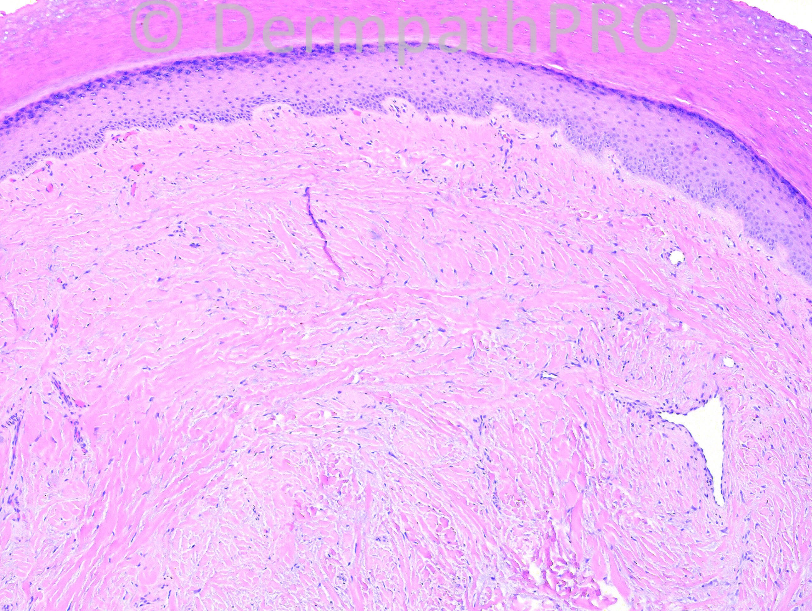
28 years-old hispanic female with lesion on right hand, 3rd finger, previously biopsied and diagnosed as digital fibrokeratoma.

Case posted by Dr. Hafeez Diwan.


  Report Record

User Feedback


Guest Dr Engin Sezer

Posted

Pleomorphic fibroma

Share this comment


Link to comment
share_externally

Guest Bansal_

Posted

Pleomorphic fibroma

Share this comment


Link to comment
share_externally

Guest Maria George

Posted

Agree with my colleague,[color=#1C2837][font=arial, verdana, tahoma, sans-serif][size=4]Pleomorphic fibroma.[/size][/font][/color]

Share this comment


Link to comment
share_externally

Pleomorphic fibroma with some myxoid change.

Share this comment


Link to comment
share_externally

Robledo F. Rocha

Posted

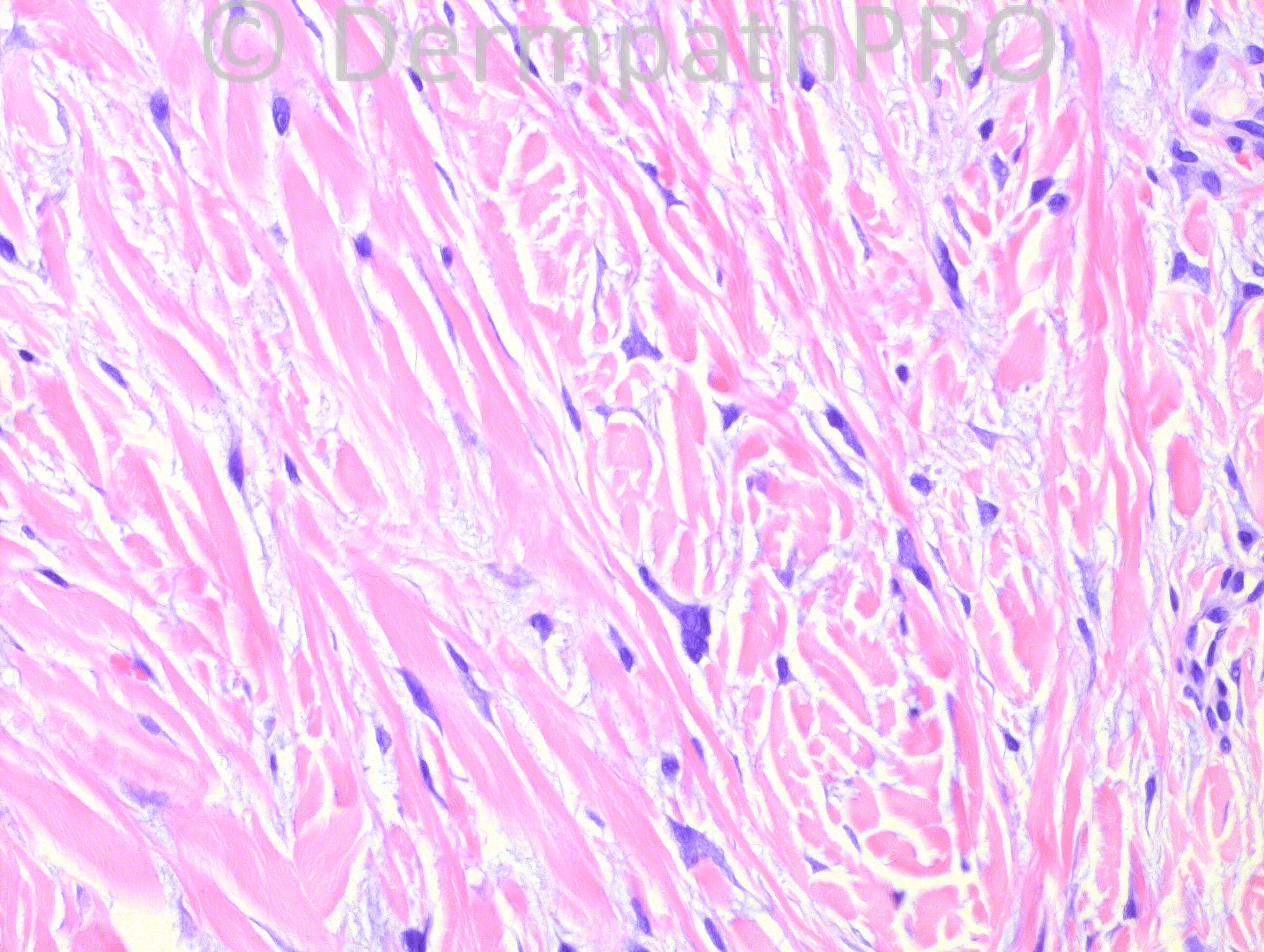
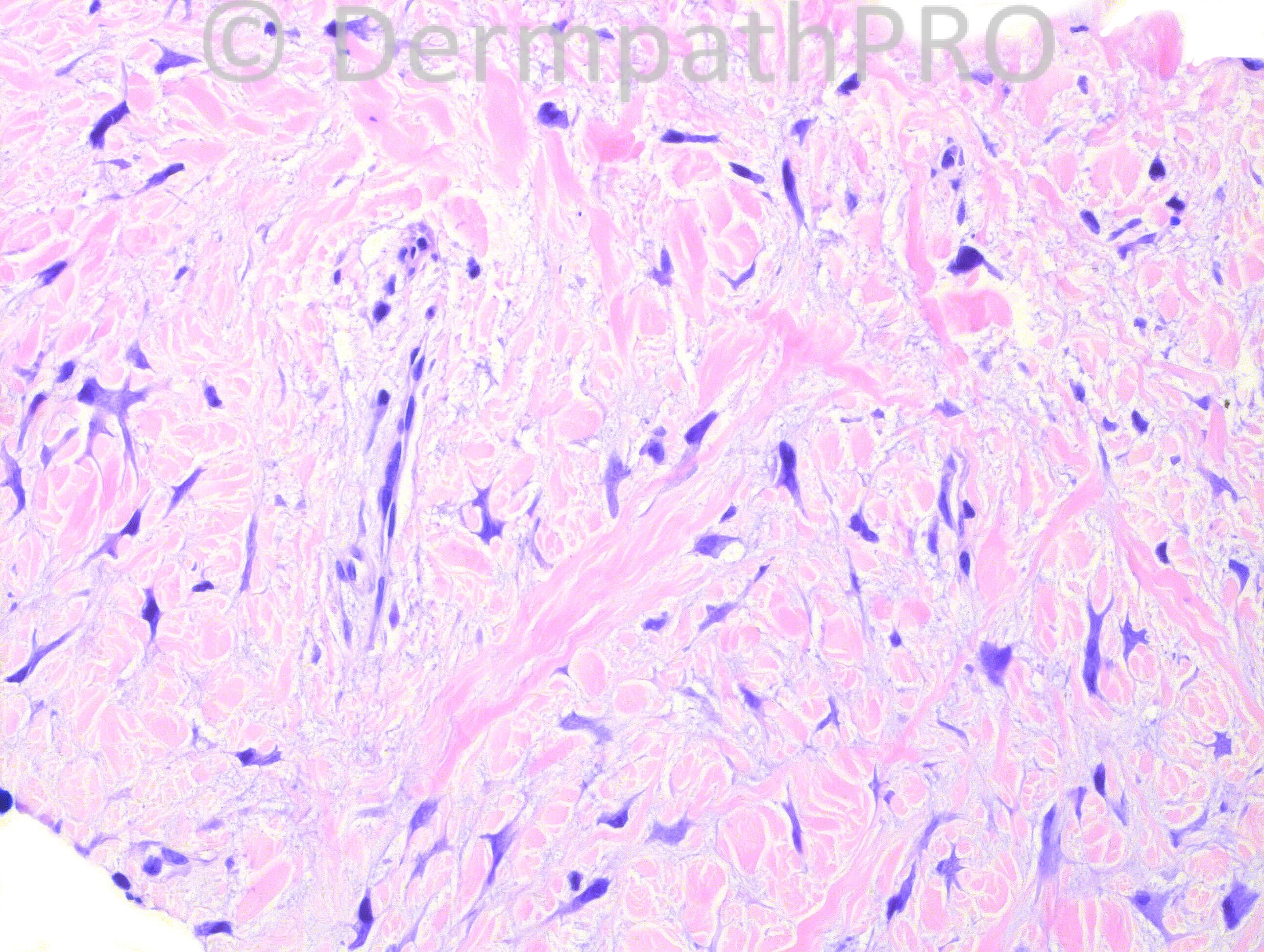
An intact epidermis covering a densely and haphazardly collagenized dermis with a somewhat myxoid change amid which there are scattered pleomorphic cells, some of them showing dendritic processes.
Agree with Dr. Sezer and my other colleagues. It's pleomorphic fibroma of the skin.

Share this comment


Link to comment
share_externally

Guest Juan Carlos Garcés, Ecuador

Posted

Pleomorphic fibroma

Share this comment


Link to comment
share_externally

Dr. Mona Abdel-Halim

Posted

Pleomorphic fibroma

Share this comment


Link to comment
share_externally

Guest Jaime A Mejia MD

Posted

Well, it is not a child, but it made think of giant cell fibroblastoma; however this one does not show the vessel-like structures of the other. Well, pleomorphic fibroma it is...

Share this comment


Link to comment
share_externally

Guest Jim Davie MD

Posted

Pleomorphic fibroma.

Share this comment


Link to comment
share_externally

Guest Romualdo

Posted

I would like to suggest superficial acral fibromyxoma as a possibility, a lesion almost limited to the digits. As pleomorphic fibroma, a tumor seen in almost every location, superficial acral fibromyxoma may also exhibit irregular and multinucleated cells.

Share this comment


Link to comment
share_externally

Iskander H. Chaudhry

Posted

Well done Romualdo, this is a superficial acral fibromyxoma.

Share this comment


Link to comment
share_externally

Robledo F. Rocha

Posted

May I ask why this is not pleomorphic fibroma? I expect a superficial acral fibromyxoma to show increased vascularity, and to display alternating areas of fibrous dermis and extensive myxoid change. Furthermore, superficial acral fibromyxoma has higher cellularity, featuring bland spindle cells and, only in exceptional cases, a few mildly atypical cells.

Share this comment


Link to comment
share_externally

Guest Jim Davie MD

Posted

[size=4]A lesion like this could, in my opinion, be classifiied either way. Classification would likely hinge on what criteria are most important to the viewer. I would (for example) look for increased mast cells in a superficial acral fibromyxoma.

Some pleomorphic fibromas have been reported to have prominent myxoid stroma...just like some superficial acral fibromyxomas less frequently show nonmucinous sclerotic stroma, cellular atypia and/or atypia/multinucleation.

Immunostaining is relatively nonspecific, and will not save the day, but could help push things one way or the other, as pleomorphic fibroma is variable but most frequently actin(+) / CD34(-), with superficial acral fibromyxoma also variable, but usually actin(-) / CD34(+).[/size]

Share this comment


Link to comment
share_externally

Iskander H. Chaudhry

Posted

[size=4][font=arial, helvetica, sans-serif]Thank you both. I tend to agree that this degree of atypia is not see typically in superficial acral fibromyxoma and also such a lesion tends to be more cellular - but I suppose an involuting lesion would be difficult to distinguish from a pleomorphic fibroma. Finally as suggested CD34 would be positive in the former.

I hope Dr Hafeez can address this in more detail on his return or other DermpathPRO editors.[/font][/size]

Share this comment


Link to comment
share_externally

Dr. Richard Carr

Posted

I was leaning towards superficial acral fibromyxoma based on anatomic location (more than histological features!). Not all lesions are that myxoid and some are more cellular (not this case) and the latter I tend to label as cellular digital fibromas (variation of SAF). CD34 is positive in these variations. Provided you recognise them as benign I guess it is a little academic bearing in mind I often take the 5th amendment on soft tissue lesions!

Share this comment


Link to comment
share_externally


×
×
  • Create New...