Jump to content
In this section we have spot diagnoses posted on a daily basis since June 2010, now over 4000! You can review the archived cases and read the suggested diagnoses by users and the final comment by the contributors.
Case are uploaded each week day by 10 am UK time with the correct diagnosis will generally be posted at 8 pm UK time. Why not view the most recent spot diagnosis and proffer a diagnosis?

Case Number : Case 781 - 14 June Posted By: Guest

Please read the clinical history and view the images by clicking on them before you proffer your diagnosis.
Submitted Date :
   (0 reviews)

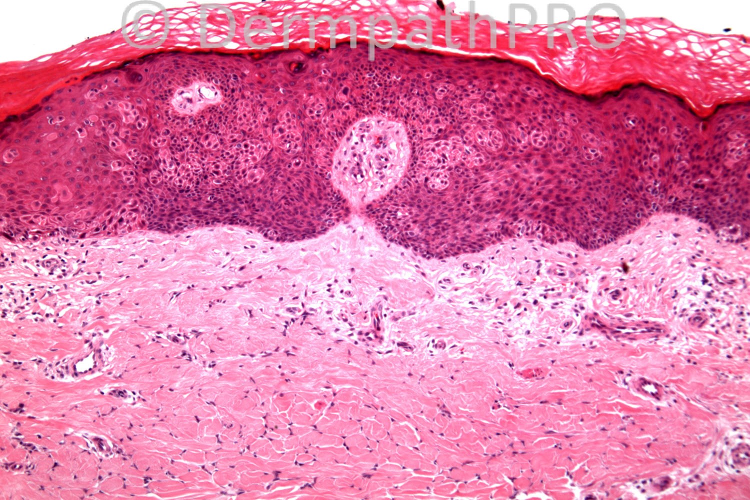
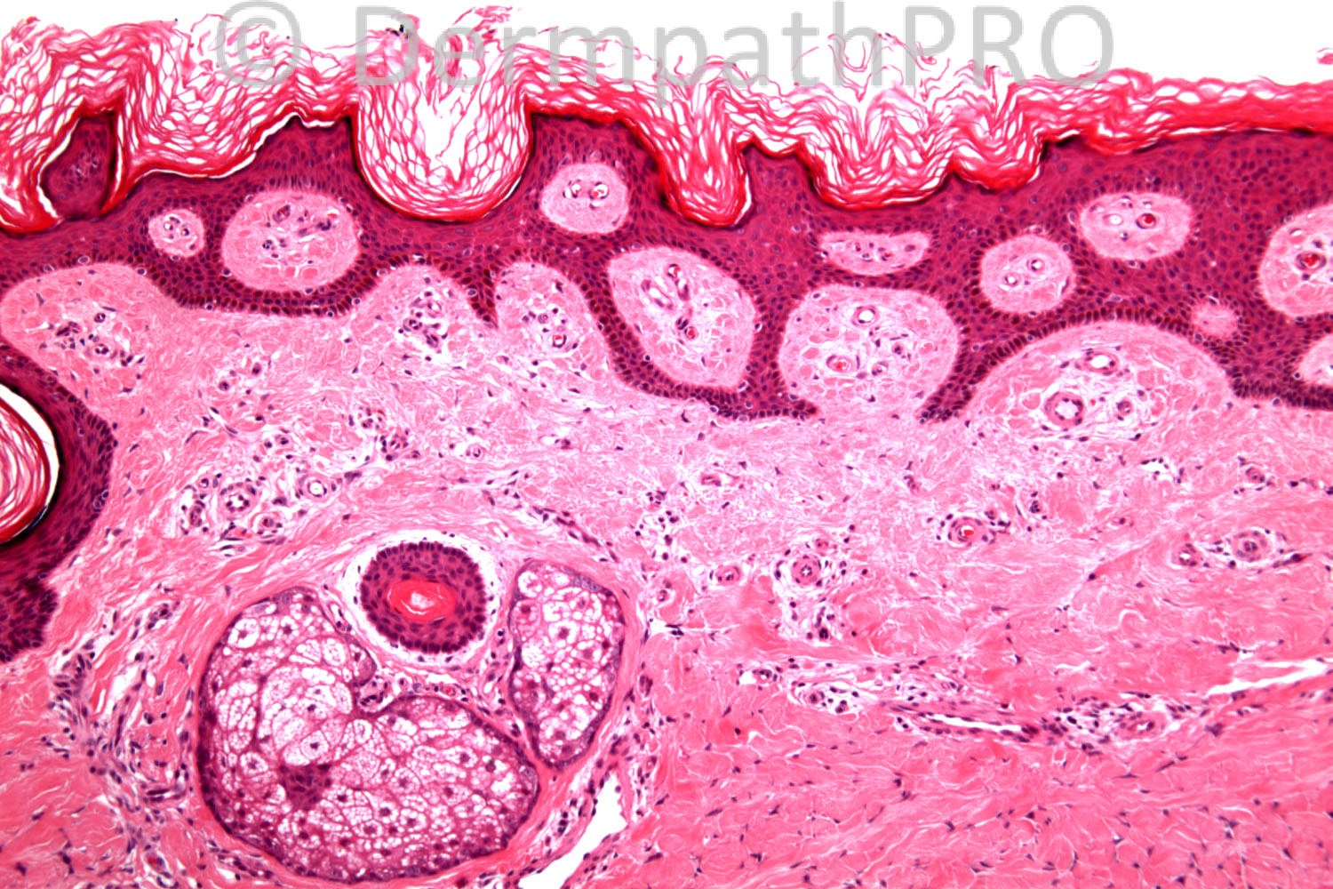
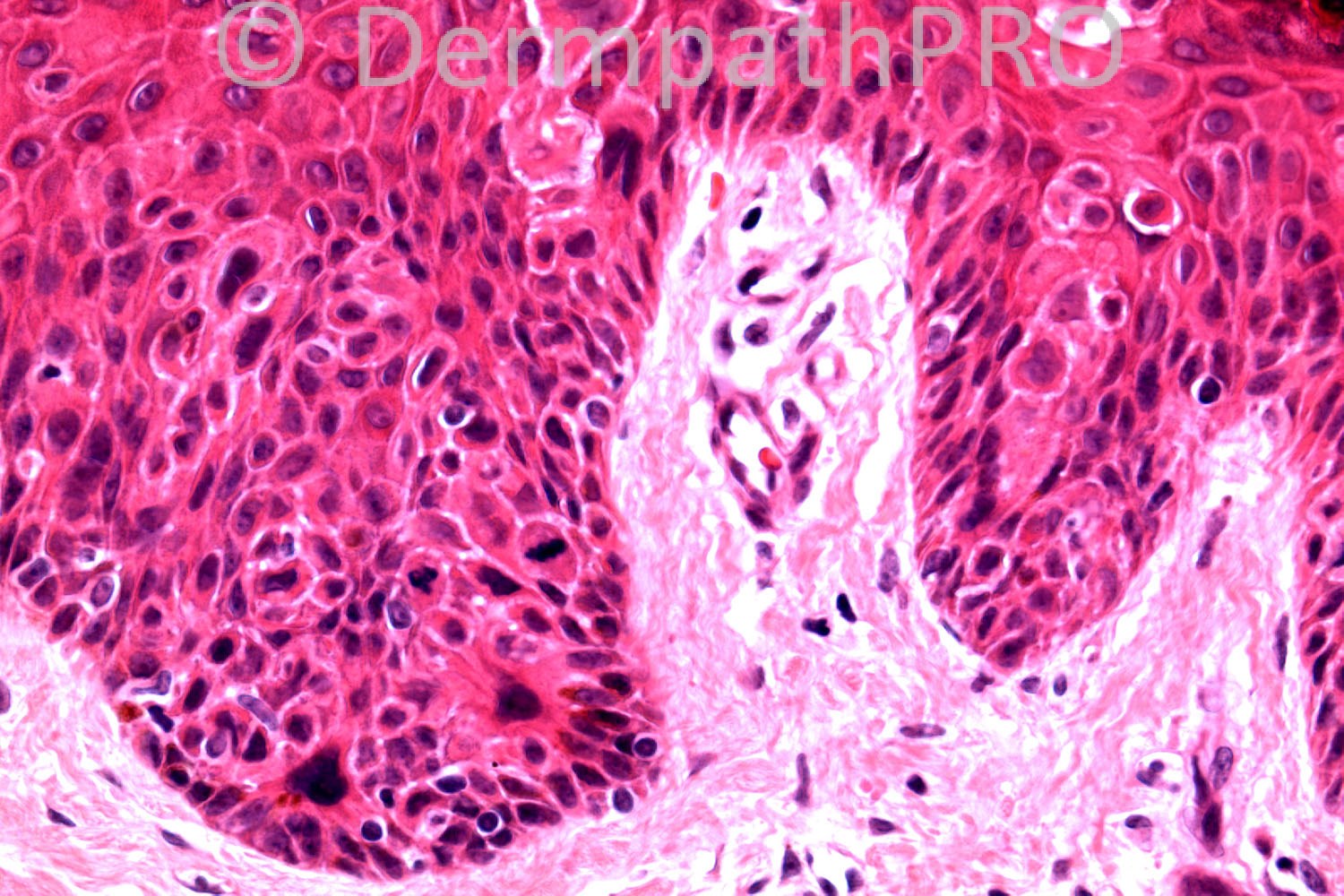
50 years-old male, shin lesion ?DF

Case posted by Dr. Richard Carr.


  Report Record

User Feedback


Guest Dr Engin Sezer

Posted

Epidermal induction by DF.

Share this comment


Link to comment
share_externally

Guest Romualdo

Posted

Bowen's disease induced by dermatofibroma.

Share this comment


Link to comment
share_externally

Guest Maria George

Posted

Is this a combo of Bowen disease and dermatofibroma ,trichodiscoma,or dermatomyfibroma.Or is it a recurrent Bowen disease with scar of previous excision?
Need history.

Share this comment


Link to comment
share_externally

Robledo F. Rocha

Posted

I don't feel this is dermatofibroma because there are sweat gland ducts within the lesion itself, a feature not expected to find in a dermatofibroma. And epidermal hyperplasia extends beyond the limits of the underlying fibrous lesion (upper right of picture #1).
Superficial located fibrocytes of a dermatofibroma are known to act as follicular papilae and induce folliculosebaceous structures from the above epidermis, sometimes with follicular germinative appearance that simulates a true basal cell carcinoma. I don't believe dermatofibroma has the capability of induce Bowen's disease.
Like Dra. Maria George, I favor a recurrent Bowen's disease over the scar of a previous biopsy.

Share this comment


Link to comment
share_externally

Dr. Mona Abdel-Halim

Posted

Definitly there is Bowen's disease. As regards the underlying dermis I agree with Maria's idea

Share this comment


Link to comment
share_externally

Dr. Richard Carr

Posted

I reported this as a dermatofibroma with subtle clonal and pagetoid bowen's. Really this is a very typical sclerosing late DF but I can see the confusion as we have discussed before selected images can be misleading. But certainly scar's would not have preserved sweat glands and the absolutely typical epidermal hyperplaisa in image lower left (from a different cross-section to image top left) and the beautiful collagen entrapment typical of DF lower border and periphery on the left side in the low power. As another point of interest elastic fibres tend to be preserved in DF (and DFSP for that matter) but not in scars. In this old sclerotic stage I cannot see the elastic too well in these particular images. I made a note of the immunos as follows: Focal moderate p16 (a little dissappointing); p53 ?lost but difficult be because subtle lesion; Ki67 not particularly helpful. The Bowen's extended out beyond the DF in two blocks and I assumed was just another chance collision of two common diagnoses. This is the first time I have seen the association.

Share this comment


Link to comment
share_externally

Guest Jim Davie MD

Posted

Too late to jump in on this one (sigh). Agree of course. Classic DF and Bowen's disease as a collision tumor. My observations to support the final DX:

1. The Bowen's disease is asymmetric and only appears in the right side of the epidermis (top left photo), and has border with the benign epidermis just right of photo center. There is basal layer sparing (eyeliner sign) and pagetoid involvement of adjacent epidermis (bottom right photo) near the junction with adjacent normal epidermis at the photo left border.

2. DF: Classic 'basaloid follicular induction' resembling superficial basal cell carcinoma actually seems very rare in the DFs that I see....despite what dermpath texts suggest. The sharply angulated 'delicate' reticulated SK-like epidermal induction with hyperpigmentation, and/or subtle basaloid palisading (as seen in bottom left photo) is often the [i]only [/i]clue of an underlying DF in many unfortunate and suboptimal [i]'rule out DF[/i]' shave biopsies.

[i](Imagine a superficial shave of the area in the bottom left photo...one that doesn't include the telltale sclerosis or fibrocytes at the deep edge of the photo....try confirming that as a DF...ouch! A good reason to encourage clinicians to avoid shaves for a rule-out-DF case). [/i]

The sclerotic area is expansile...it is pushing everything around it... including epidermal contour, encased adnexa and adjacent deep reticular dermis and long rete peg (on far left edge)... away from the center of the sclerotic zone in the top left photo.

Share this comment


Link to comment
share_externally


×
×
  • Create New...