Jump to content
In this section we have spot diagnoses posted on a daily basis since June 2010, now over 4000! You can review the archived cases and read the suggested diagnoses by users and the final comment by the contributors.
Case are uploaded each week day by 10 am UK time with the correct diagnosis will generally be posted at 8 pm UK time. Why not view the most recent spot diagnosis and proffer a diagnosis?

Case Number : Case 783 - 18th June Posted By: Guest

Please read the clinical history and view the images by clicking on them before you proffer your diagnosis.
Submitted Date :
   (0 reviews)

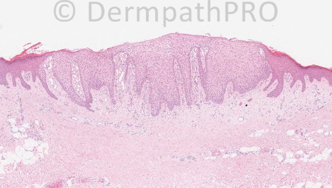
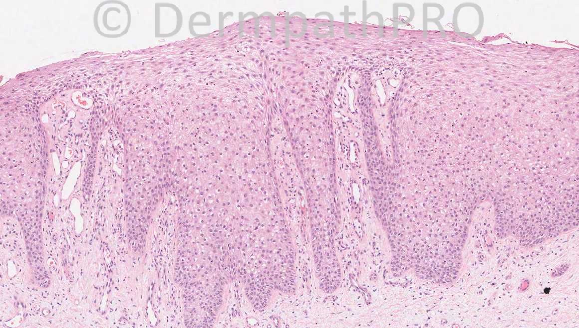
77 years-old female, lesion left shin ?BCC ?SCC

Thank you to Dr. Leena Joseph for providing this case.


  Report Record

User Feedback


Dr. Phillip McKee

Posted

[color=#1C2837][font=arial, verdana, tahoma, sans-serif][size=4]Thanks again for all your kind comments yesterday. I will still be looking at the difficult cases etc, but I have handed over the lead role to Mark Hurt, who I know will be a wonderful replacement. I can see the site growing from strength to strength under his leadership.[/size][/font][/color]

Share this comment


Link to comment
share_externally

Guest Romualdo

Posted

This is a clear cell acanthoma. A happy retirement to you, Dr. McKee!!!

Share this comment


Link to comment
share_externally

Guest Hazem Hamed

Posted

Dear Dr McKee, Thank you for your great effort in teaching us and wish you the best in your retirement.
Agree, this is a clear cell acanthoma.

Share this comment


Link to comment
share_externally

Robledo F. Rocha

Posted

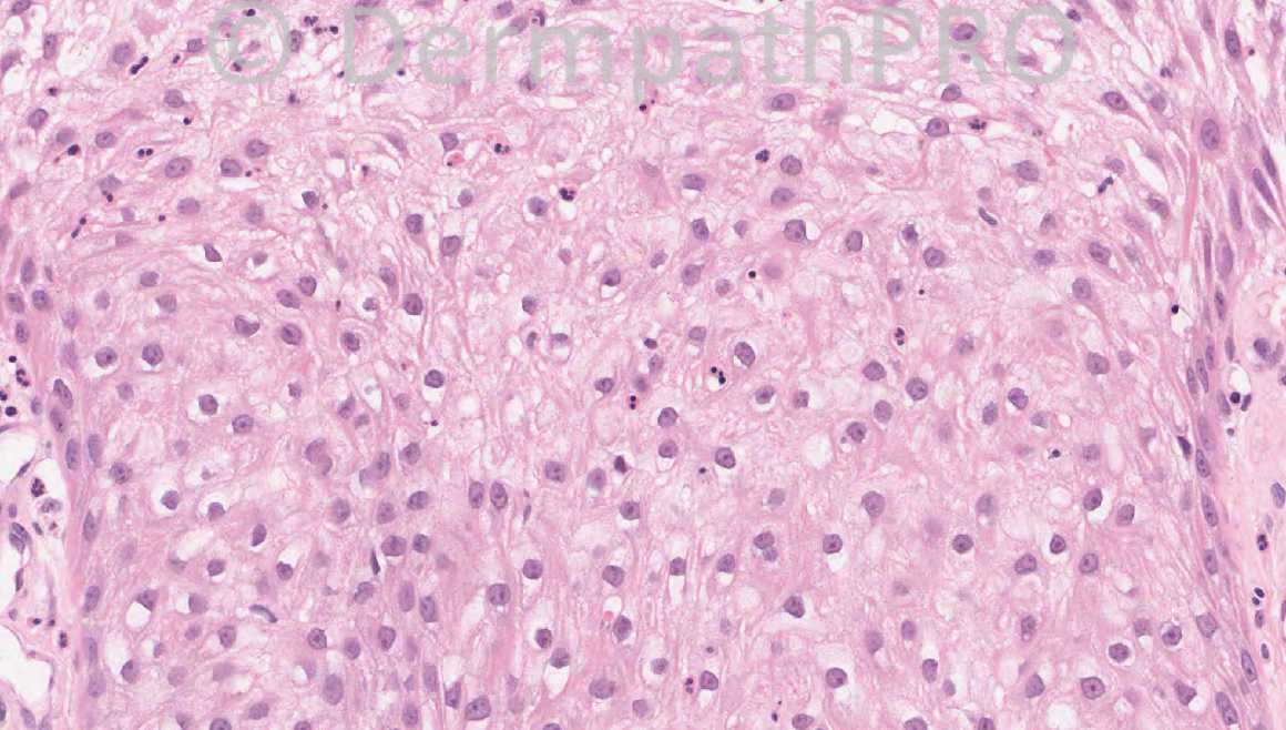
A sharply demarcated psoriasiform hyperplasia of the epidermis showing clear cell change, loss of the granular layer, and some neutrophilic exocytosis. This is a beautiful case of clear cell acanthoma.

Share this comment


Link to comment
share_externally

Guest Rodrigo Restrepo

Posted

Dear Dr Mckee. it’s been a pleasure working with you. I hope you enjoy your retirement and all your future expectations be fulfilled.
My diagnosis for this case is clear cell acanthoma.

Share this comment


Link to comment
share_externally

Guest Dr Engin Sezer

Posted

Agree clear cell acanthoma

Share this comment


Link to comment
share_externally

Guest Maria George

Posted

[color=#1C2837][font=arial, verdana, tahoma, sans-serif][size=4]Agree with [/size][/font][/color][color=#1C2837][font=arial, verdana, tahoma, sans-serif][size=4][right]Romualdo and other coleagues, [/right][/size][/font][/color][color=#1C2837][font=arial, verdana, tahoma, sans-serif][size=4]clear cell acanthoma.[/size][/font][/color]

Share this comment


Link to comment
share_externally

Guest Graham Reilly

Posted

I agree clear cell acanthoma. All the best to Dr Phillip Mckee - thank you for all your input to the site and enjoy your retirement in France.

Share this comment


Link to comment
share_externally

Guest Amira Tawdy

Posted

agree with clear cell acanthoma. Dear Dr Mckee nothing is enough to express our gratitude to you and to

your wonderful site , thank you our dear professor and hoping you best retirement.

Share this comment


Link to comment
share_externally

Guest Bansal_

Posted

Agree, clear cell acanthoma

Share this comment


Link to comment
share_externally

Guest Marcia

Posted

Agree, clear cell acanthoma

Share this comment


Link to comment
share_externally

Dr. Mona Abdel-Halim

Posted

Beautiful clear cell acanthoma...

Share this comment


Link to comment
share_externally

Iskander H. Chaudhry

Posted

I can confirm the correct diagnosis is [color=#1C2837][font=arial, verdana, tahoma, sans-serif][size=4]clear cell acanthoma.[/size][/font][/color]

Share this comment


Link to comment
share_externally

Guest Jim Davie MD

Posted

Nice example of clear cell acanthoma, with classic presentation on leg of a female.

Clonal population of clear cells (sharply demarcated with respect to adjacent epidermis with no pagetoid spread), presence of sparse individual neutrophils in the spinous layer, absence of granular layer with sparse parakeratosis, and "random" rectangular vs. jagged hyperplasia of the epidermis that contrasts with the more regular hyperplasia of psoriasis.

Share this comment


Link to comment
share_externally


×
×
  • Create New...