Jump to content
In this section we have spot diagnoses posted on a daily basis since June 2010, now over 4000! You can review the archived cases and read the suggested diagnoses by users and the final comment by the contributors.
Case are uploaded each week day by 10 am UK time with the correct diagnosis will generally be posted at 8 pm UK time. Why not view the most recent spot diagnosis and proffer a diagnosis?

Case Number : Case 784 - 19th June Posted By: Guest

Please read the clinical history and view the images by clicking on them before you proffer your diagnosis.
Submitted Date :
   (0 reviews)

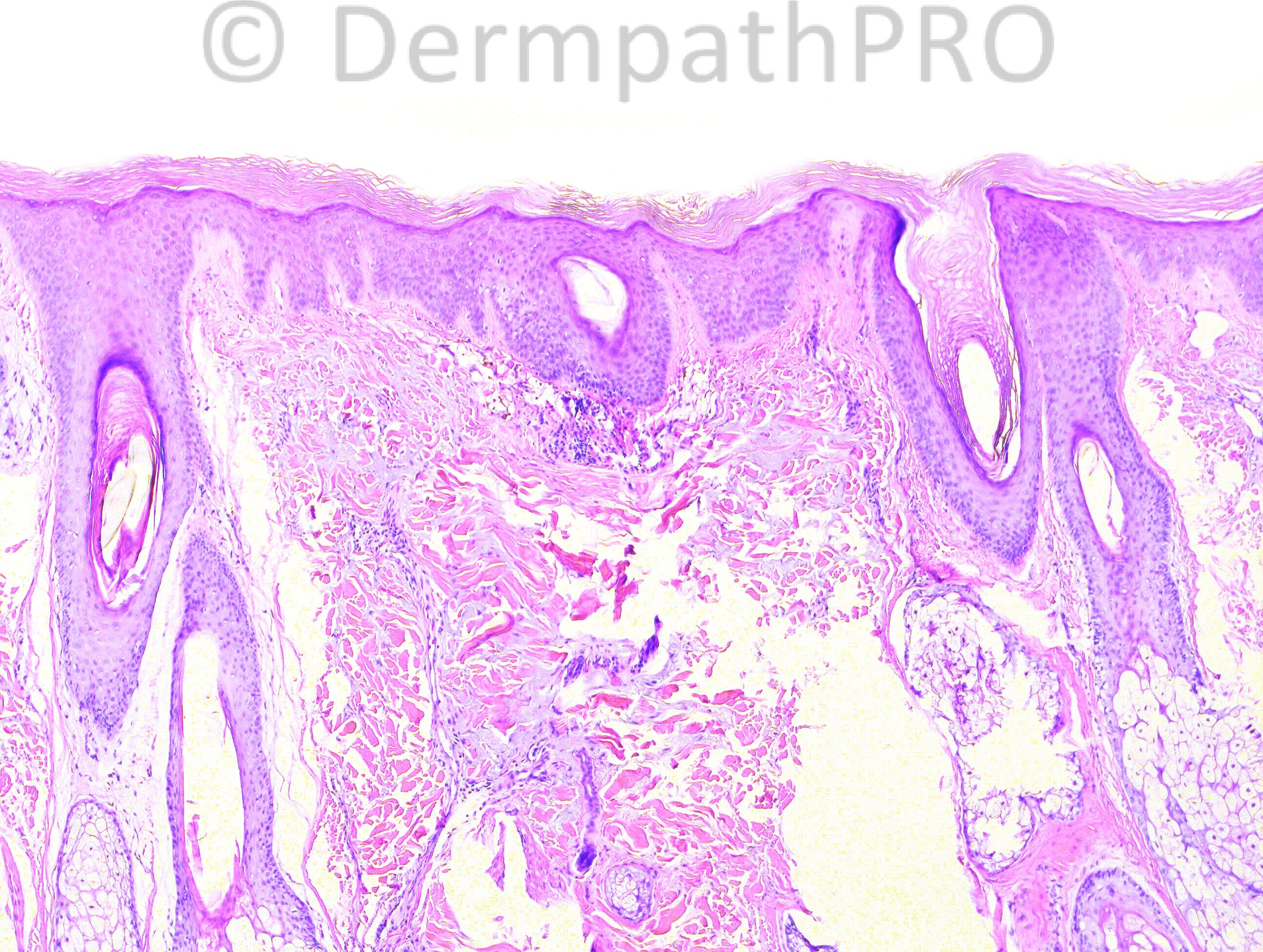
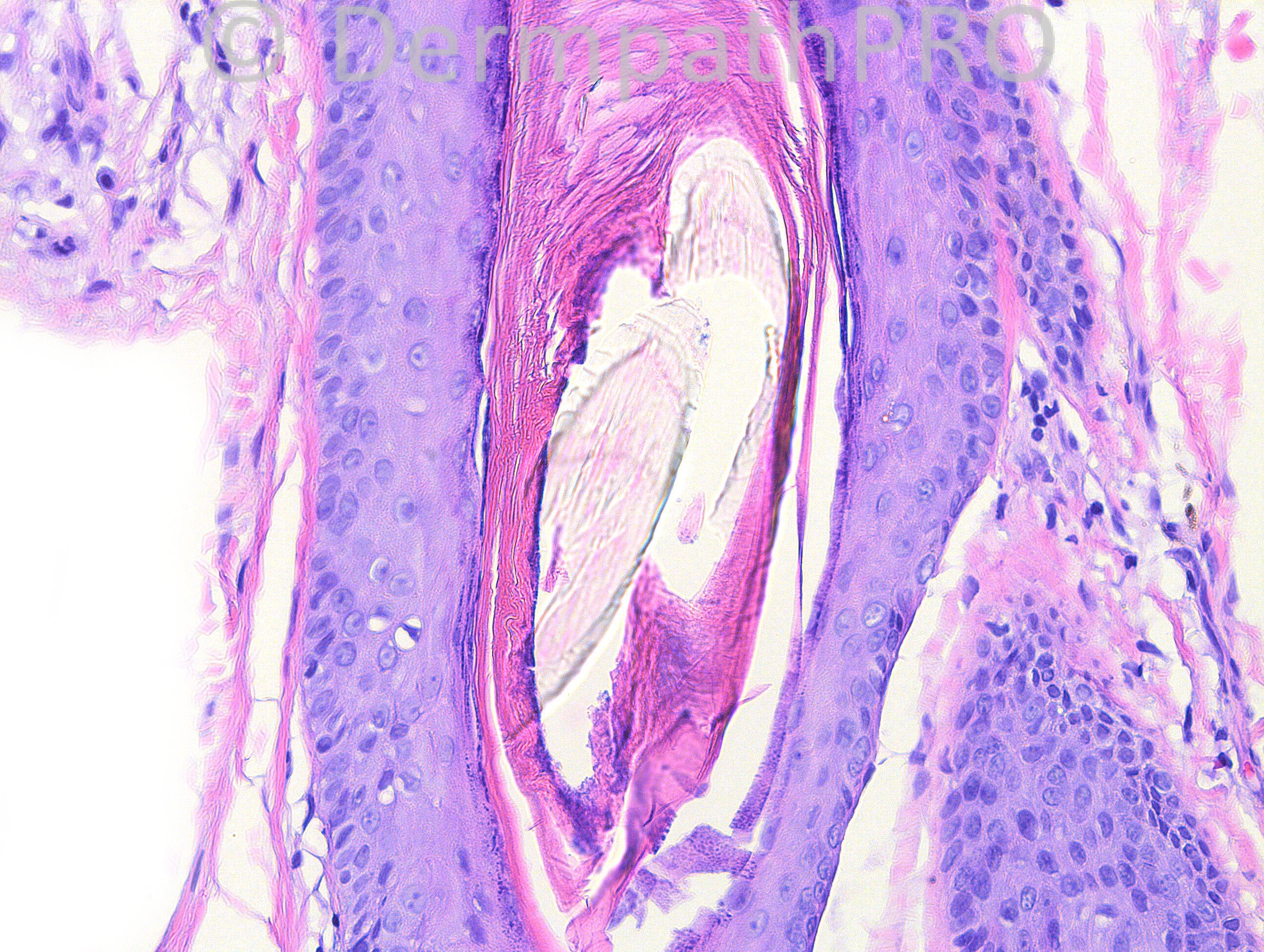
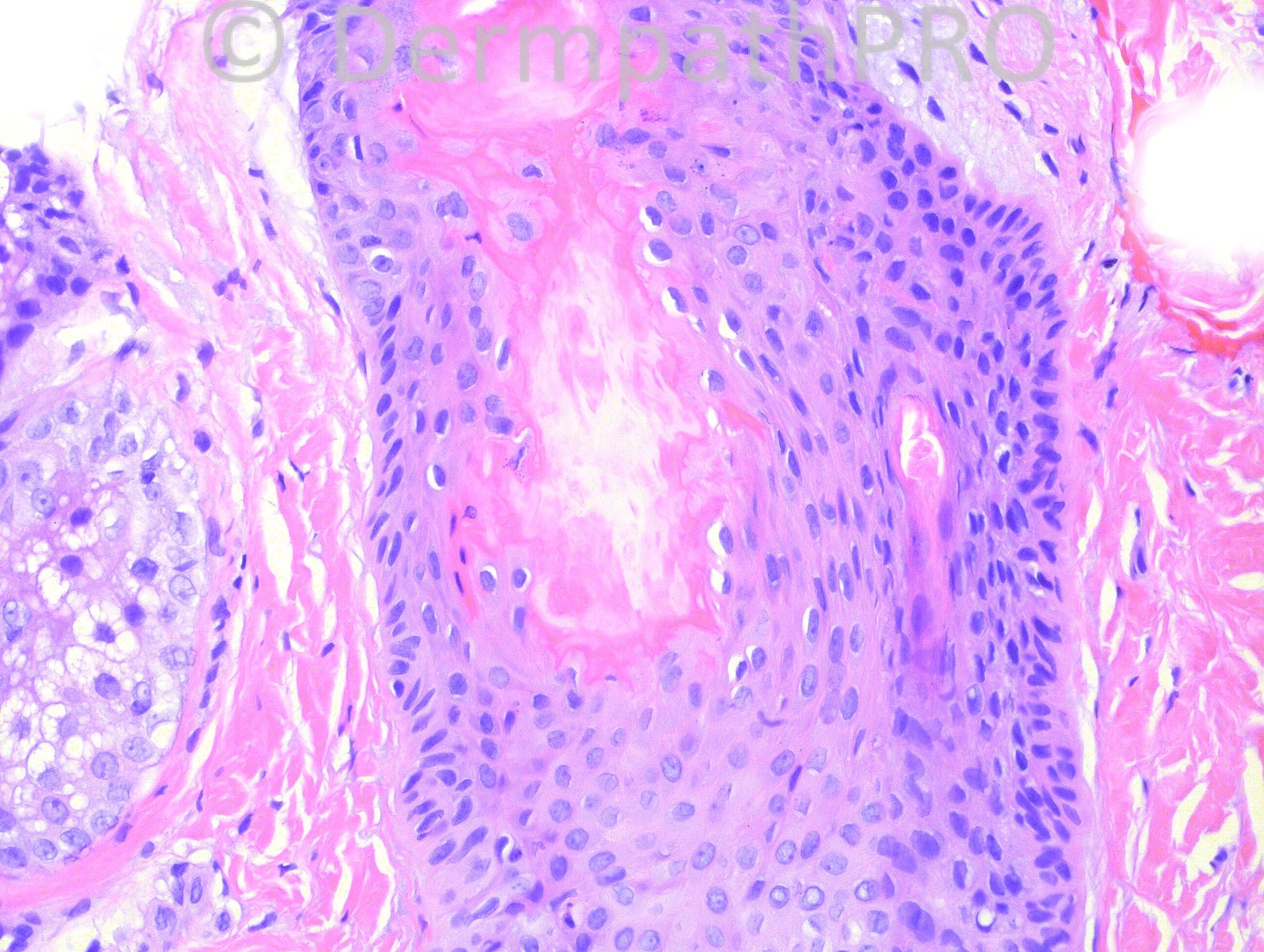
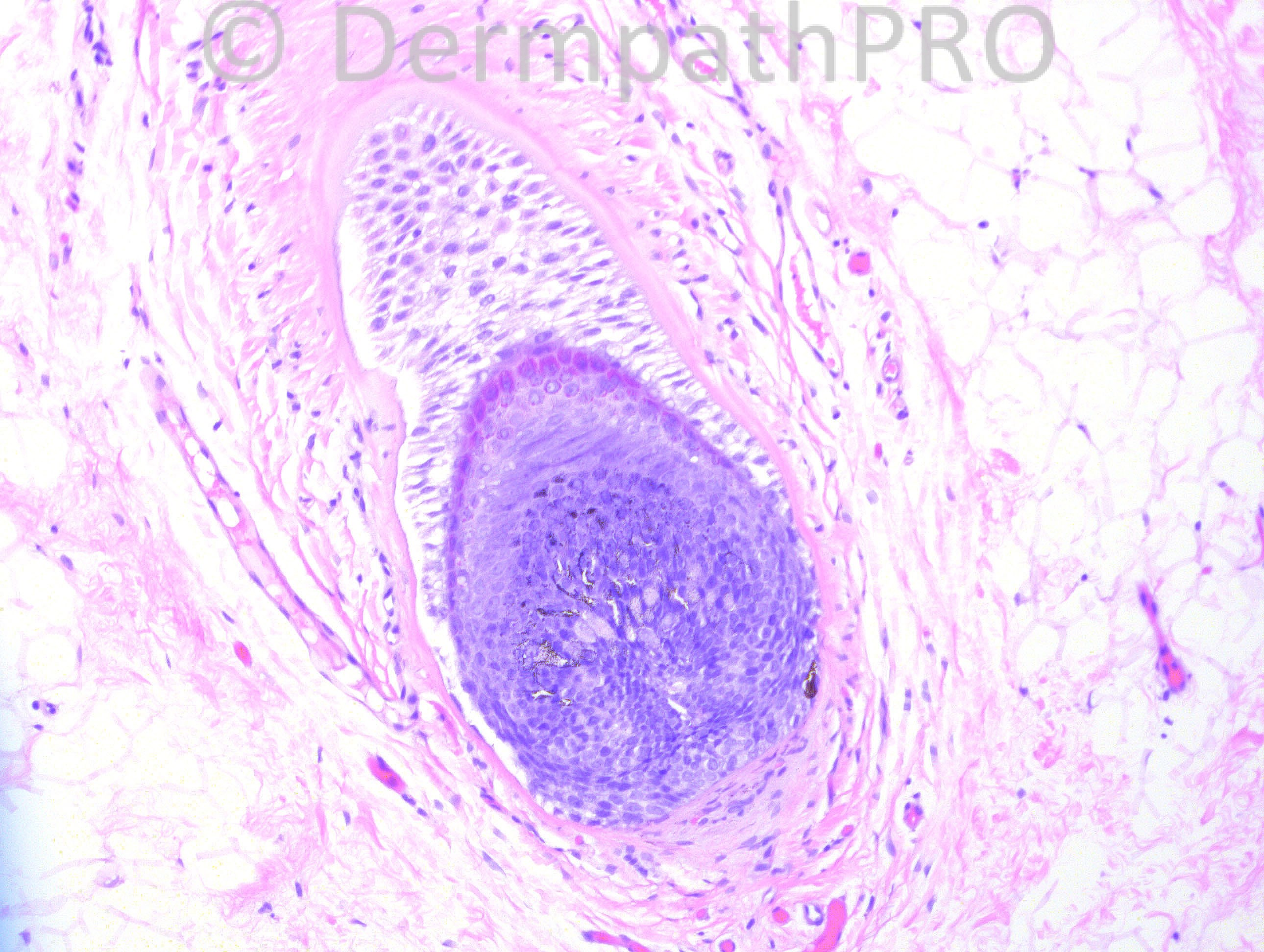
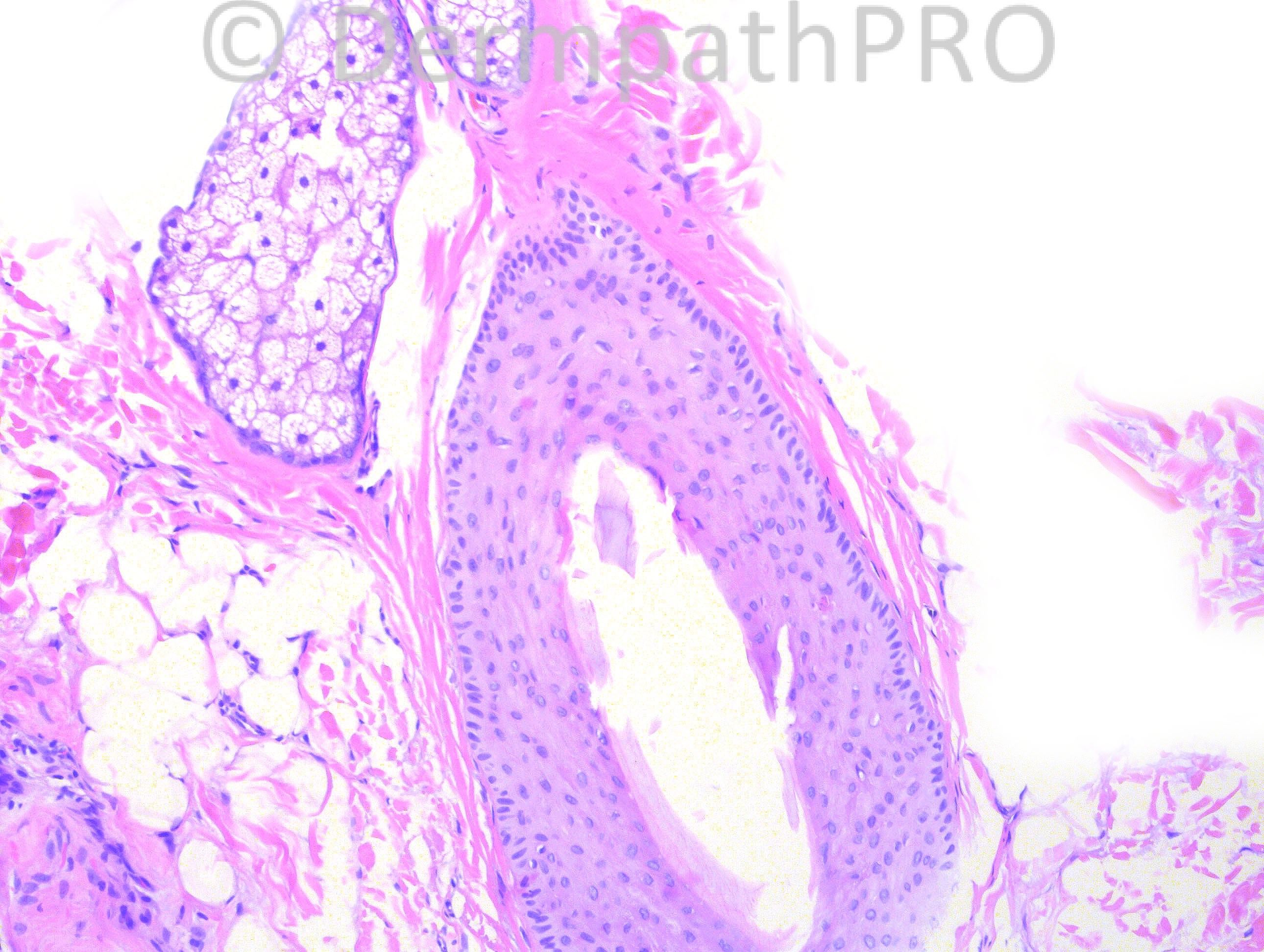
64 years-old veteran with hair loss, believed to be pulling out his hair. This is a vertex biopsy.

Case posted by Dr. Hafeez Diwan.

  Report Record

User Feedback


nick turnbull

Posted

No significant inflamation. Trichomalacia and fractured hair shafts. Damaged inner root sheath. Not sure I can see pigment casts. Maybe there is some haemorrhage around the hair bulb. Dx: Trichotillomania

Share this comment


Link to comment
share_externally

Robledo F. Rocha

Posted

Agree with clinical suspicion of trichotillomania. It's a non-inflammatory and non-scarring alopecia showing signs of follicular damage: irregularly shaped hair follicles, loss of hair shafts, and trichomalacia.
A catagen hair follicle with thickened corrugated basement membrane can also be seen, probably indicating that there is increased number of follicles in this phase of hair growth.

Share this comment


Link to comment
share_externally

Guest Romualdo

Posted

Agree with trichotillomania, although I would be more confident if melanin casts were present.

Share this comment


Link to comment
share_externally

Guest Dr Engin Sezer

Posted

Trichotillomania

Share this comment


Link to comment
share_externally

Guest Maria George

Posted

There are twisted hair shaft or multiple hair shafts in one follicle,Follicular drop out,Follicular plugging by keratin,Perifollicular fibrosis, and premature keratinization and damage of inner root sheath .
If in the skin , I might thought of some exotic conditions like scurvy or Keratosis follicularis spinulosa decalvans, ..etc.
But in scalp , I will say this is central centrfugal alopecia or hot comb alopecia.

Share this comment


Link to comment
share_externally

Sasi Attili

Posted

Agree that it is not quite enough for trichotillomania but can't make it into anything else, given the clinical.

Share this comment


Link to comment
share_externally

Guest Jim Davie MD

Posted

Alopecia diagnosis on these images with limited clinical may be a challenge.
There are numerous hairs present so will presume this is a perilesional biopsy.

[i]Clinical questions: [/i]
1. Do we know the pattern of hair loss (asymmetric patchy or geographic would be expected in trichotillomania). Is it diffuse or dense?
2. Is patient on steroids that may decrease inflammation?

We can't assess CAT follicle ratios or vellus hair ratios.
But if the images are taken for what they are, at least narrowing down the differential may be possible.
I agree with Maria that this leans more towards the central centrifugal scarring alopecia (CCSA) direction:

[b]A. Trichotillomania (disfavored):[/b]
[i]Supportive findings[/i]: Minimal inflammation. Moderate distortion of architecture in the catagen bulb (mid left photo), and mild distortion of the anagen bulb outline in the mid-right photo). Clinical suspicion of trichotillomania.
[i]Dismissive findings[/i]: No pigment casts. No collapsed outer root sheaths (bottom left photo shows artifactual loss of hair shaft, but no collapse). Lack of significant trichomalacia/distortion of the terminal hairs present (ignoring the expected sectioning artefact).

[b]B. CCSA / scarring alopecia (favored): [/b]
[i]Supportive findings[/i]: polytrichia/tufting with possible follicle fusion (top two photos). Hint of premature desquamation of inner root sheath, and especially notable: asymmetric thinning or atrophy of the follicular epithelium (top left photo on left edge center). Follicular plugging. Location on vertex (most common).
[i]Dismissive findings:[/i] No significant perifollicular lamellar fibrosis. No significant perifollicular chronic inflammation. Sebaceous glands appear intact (not obliterated/scarred out).

Share this comment


Link to comment
share_externally

Iskander H. Chaudhry

Posted

The diagnosis for this was [color=#1C2837][font=arial, verdana, tahoma, sans-serif][size=4]trichotillomania.[/size][/font][/color]

Share this comment


Link to comment
share_externally

Guest Rodrigo Restrepo

Posted

Agree, trichotillomania.

Share this comment


Link to comment
share_externally


×
×
  • Create New...