Jump to content
In this section we have spot diagnoses posted on a daily basis since June 2010, now over 4000! You can review the archived cases and read the suggested diagnoses by users and the final comment by the contributors.
Case are uploaded each week day by 10 am UK time with the correct diagnosis will generally be posted at 8 pm UK time. Why not view the most recent spot diagnosis and proffer a diagnosis?

Case Number : Case 785 - 20th June Posted By: Guest

Please read the clinical history and view the images by clicking on them before you proffer your diagnosis.
Submitted Date :
   (0 reviews)

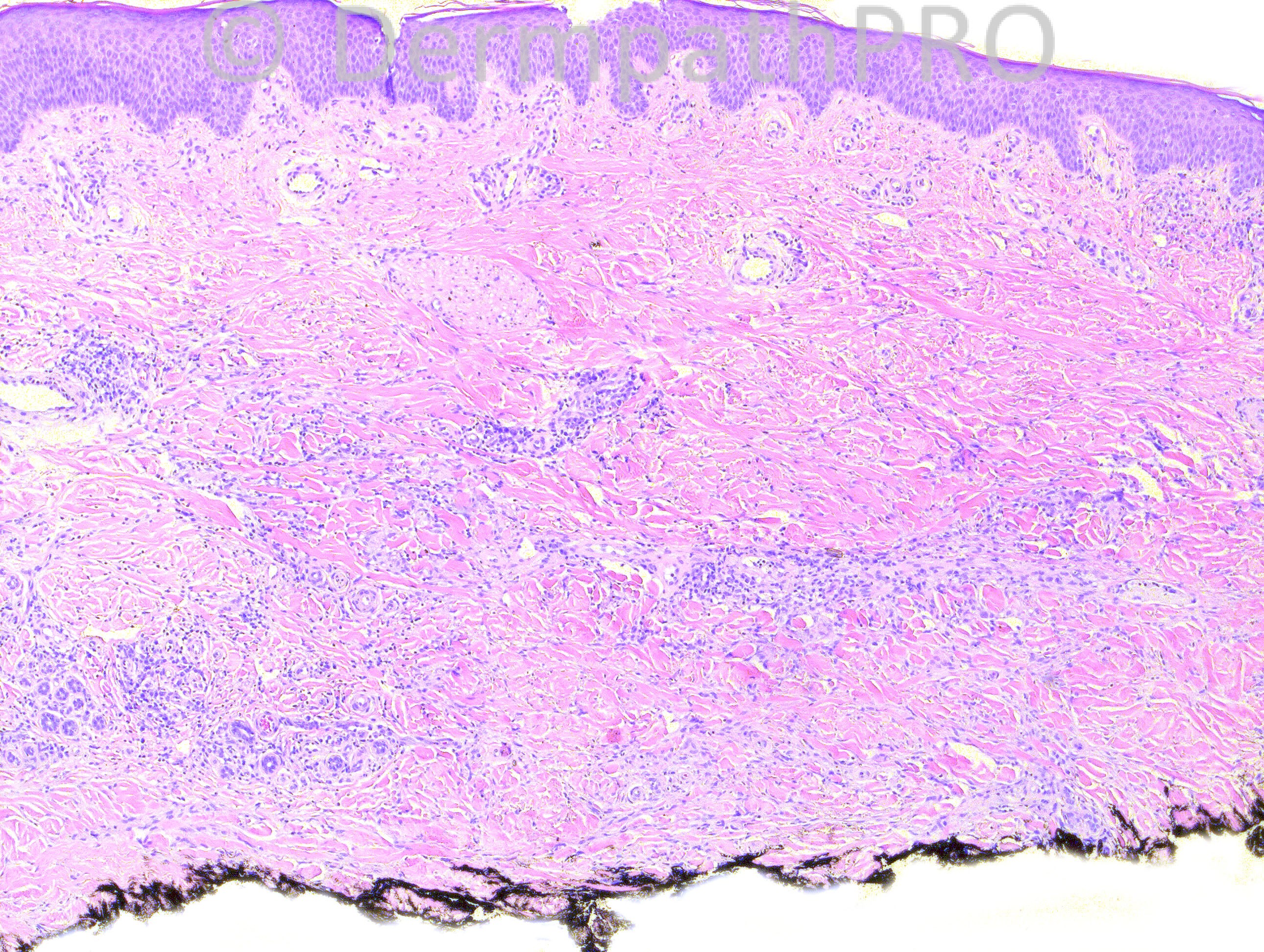
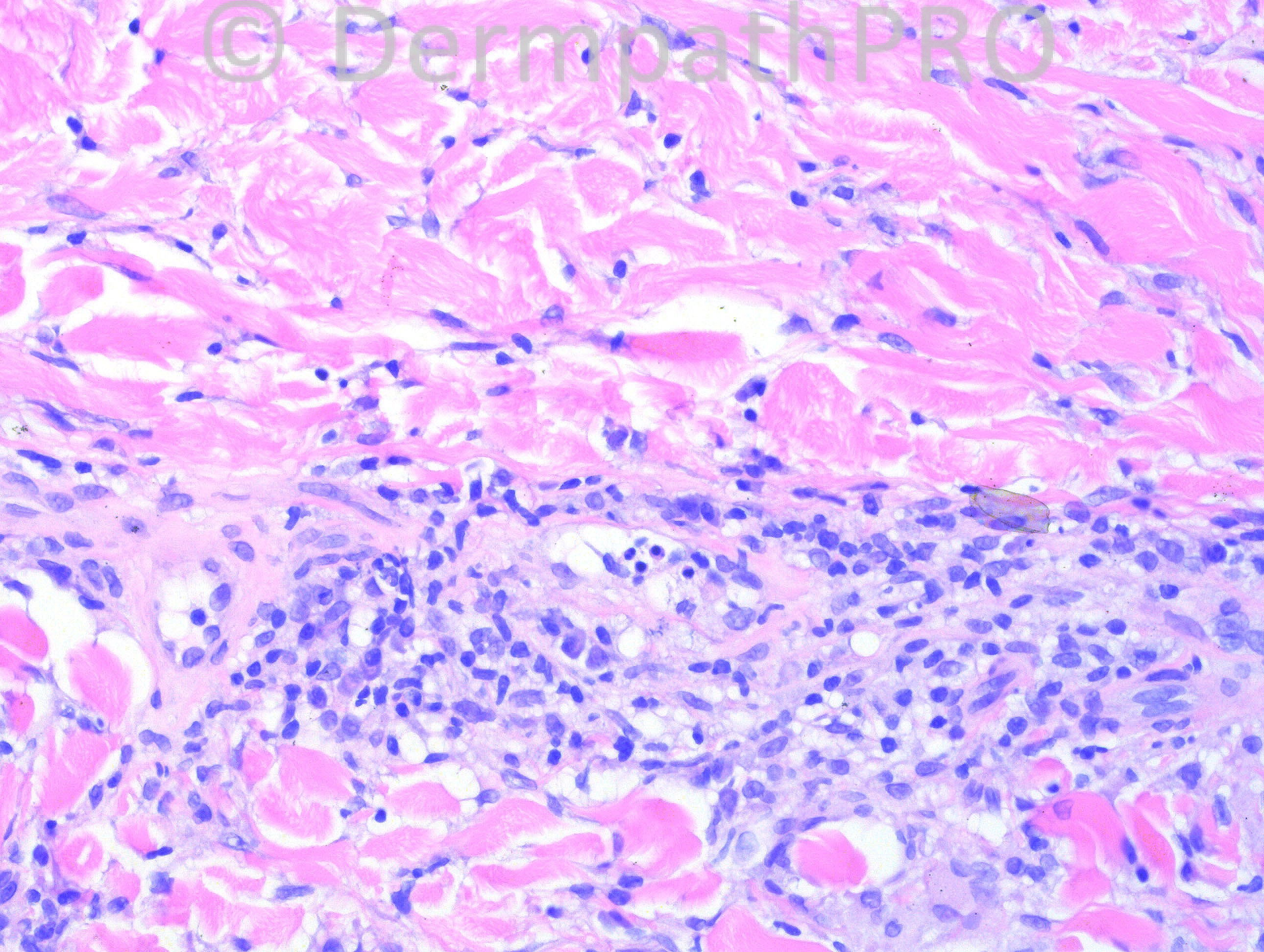
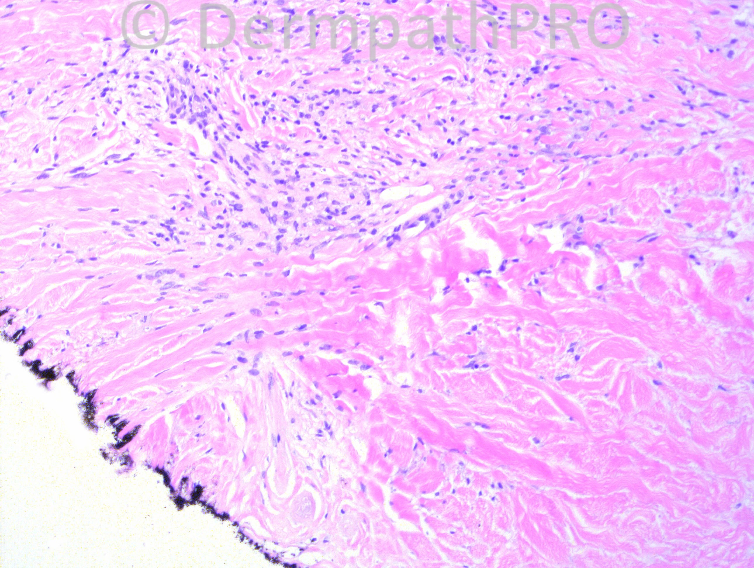
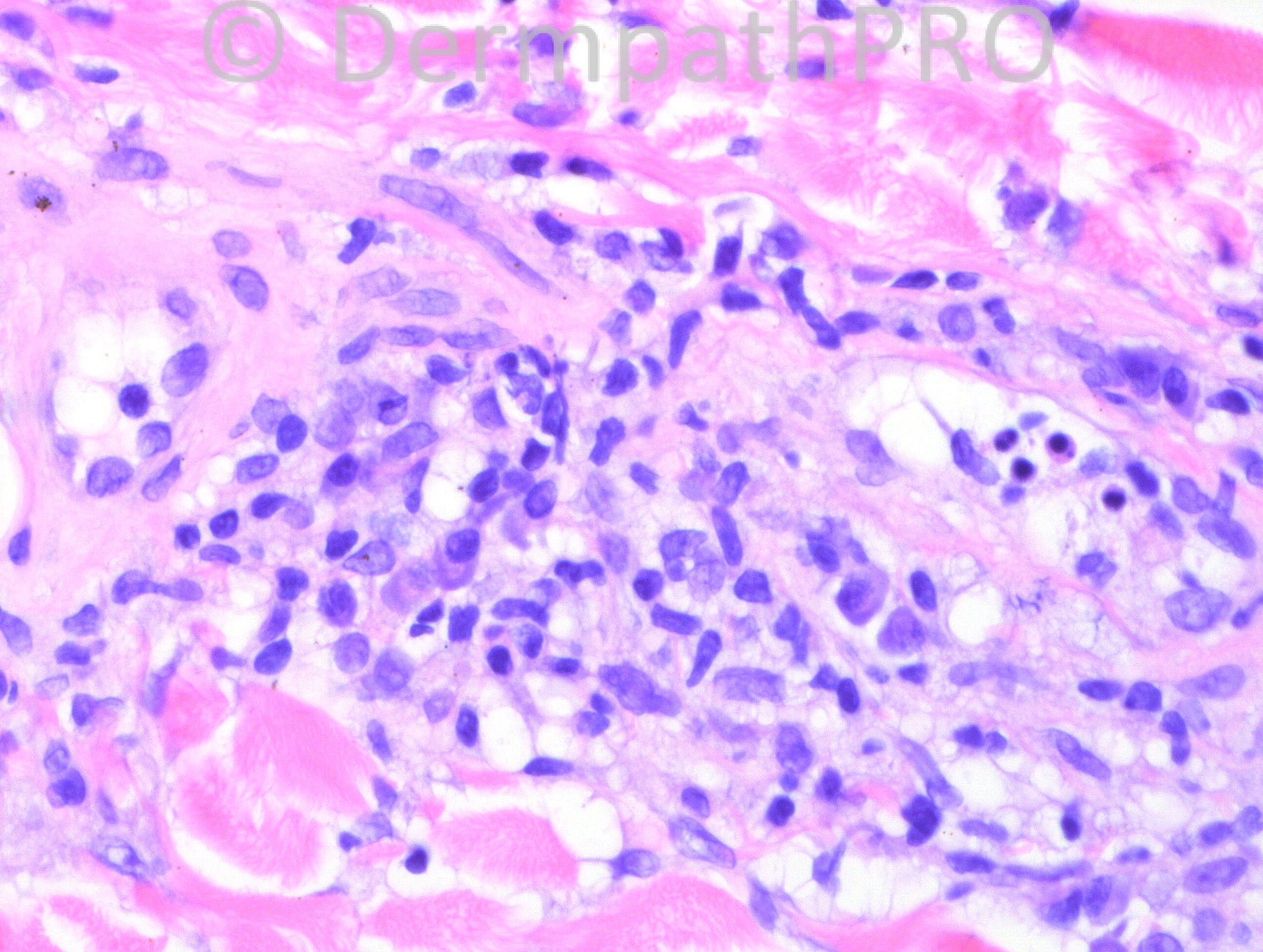
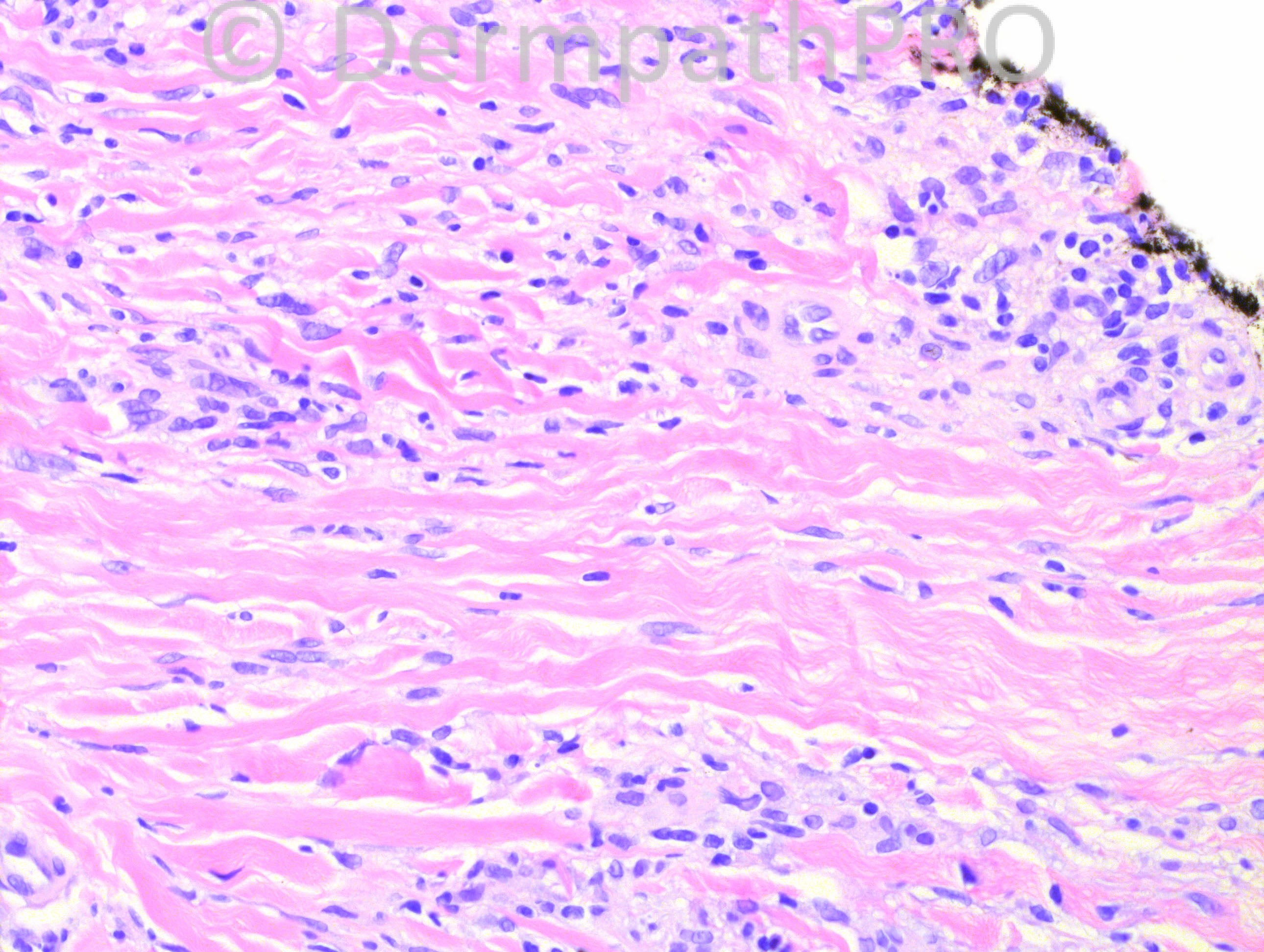
22 years-old female with lesion on shin.

Case posted by Dr. Hafeez Diwan.

  Report Record

User Feedback


Guest Romualdo

Posted

Although I can't see large necrobiotic foci there is a suggestion of a layered pattern with horizontal bands of granulomatous inflammatory infiltrates alternating with hypocellular sclerotic bands. The inflammatory cells are represented by lymphocytes, histiocytes and plasma cells. I think this is an early lesion of necrobiosis lipoidica.

Share this comment


Link to comment
share_externally

Necrobiosis lipoidica

Share this comment


Link to comment
share_externally

Guest Maria George

Posted

Although seeing plasma cells always alert me to non-visble amyloidosis in the skin, I do agree with [color=#1C2837][font=arial, verdana, tahoma, sans-serif][size=4][right]Romualdo and [/right][/size][/font][/color][color=#1C2837][font=arial, verdana, tahoma, sans-serif][size=4][right]Marcela for [/right][/size][/font][/color][color=#1C2837][font=arial, verdana, tahoma, sans-serif][size=4] necrobiosis lipoidica.[/size][/font][/color]

Share this comment


Link to comment
share_externally

I observe a foci of necrobiosis on mid lett photo at the rigth bottom egde, fibrosis and lots of plasma cells, but everything became easier after Romualdo´s sugestion.

Share this comment


Link to comment
share_externally

Robledo F. Rocha

Posted

Pictures show three features that make me favor early necrobiosis lipoidica over interstitial granuloma annulare: infiltrate is more concentrated on lower part of the reticular dermis, frequent plasma cells, and horizontally oriented tiers of histiocytes. Furthermore, shin is the most typical site for necrobiosis lipoidica.
I couldn’t check unequivocally, based only in the available pictures, the most reliable feature to distinguish necrobiosis lipoidica and granuloma annulare, to wit, the intervening dermis, which is altered in the former and normal in the latter.

Share this comment


Link to comment
share_externally

Guest Juan Carlos Garcés, Ecuador

Posted

Necrobiosis lipoidical

Share this comment


Link to comment
share_externally

Dr. Mona Abdel-Halim

Posted

Favor NLD also

Share this comment


Link to comment
share_externally

I can see this being NLD. Anyone think it could me morphea? That also crossed my mind given the plasma cells and sclerosis.

Share this comment


Link to comment
share_externally

Guest Jim Davie MD

Posted

I would favor an old lesion NLD, given the horizontal tiering of the collagen bundles and inflammatory infiltrate, overall dermal thickening, mixed lymphoplasmacytic inflammatory infiltrate, and clinical context of female shin.

Having said that, there is absence of classic necrobiosis, and limited scope of the images.
I will therefore keep the following in the differential:

1. Morphea[list]
[*]Obliteration of fat around the eccrine coils (bound-down adnexa) best seen in the first (top left) photo.
[*]Mixed lyphoplasmacytic cells in inflammatory/early stage.
[*]Thick dermis (no subcutis).
[/list]
2. Dermatofibroma :[list]
[*]Frequently have an associated lymphocytic or lymphoplasmacytic inflammatory reaction (predominantly peripheral).
[*]Mulitnucleated and single cells that wrap around collagen bundles.
[*]Thickened collagen bundles.
[*]Epidermal hyperplasia and/or hyperpigmentation.
[/list]

Share this comment


Link to comment
share_externally

Iskander H. Chaudhry

Posted

Well done, it is necrobiosis lipoidica diabeticorum.

Share this comment


Link to comment
share_externally

Guest Sandro Casavilca

Posted

Well, in my opinion morphea, granuloma anulare and necrobiosis lipoidica were possible diagnosis. Thank you very much for share this case whith us.

Share this comment


Link to comment
share_externally


×
×
  • Create New...