Jump to content
In this section we have spot diagnoses posted on a daily basis since June 2010, now over 4000! You can review the archived cases and read the suggested diagnoses by users and the final comment by the contributors.
Case are uploaded each week day by 10 am UK time with the correct diagnosis will generally be posted at 8 pm UK time. Why not view the most recent spot diagnosis and proffer a diagnosis?

Case Number : Case 786 - 21st June Posted By: Guest

Please read the clinical history and view the images by clicking on them before you proffer your diagnosis.
Submitted Date :
   (0 reviews)

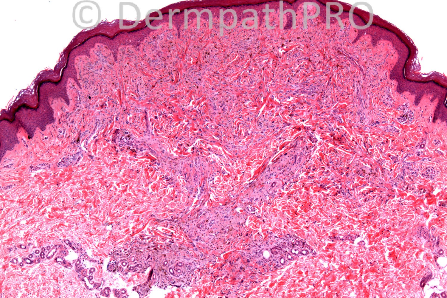
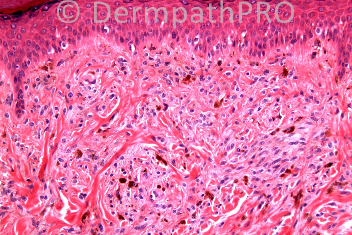
16 years-old female, lesion on foot. ?Spitz naevus

Case posted by Dr. Richard Carr.

  Report Record

User Feedback


Guest Maria George

Posted

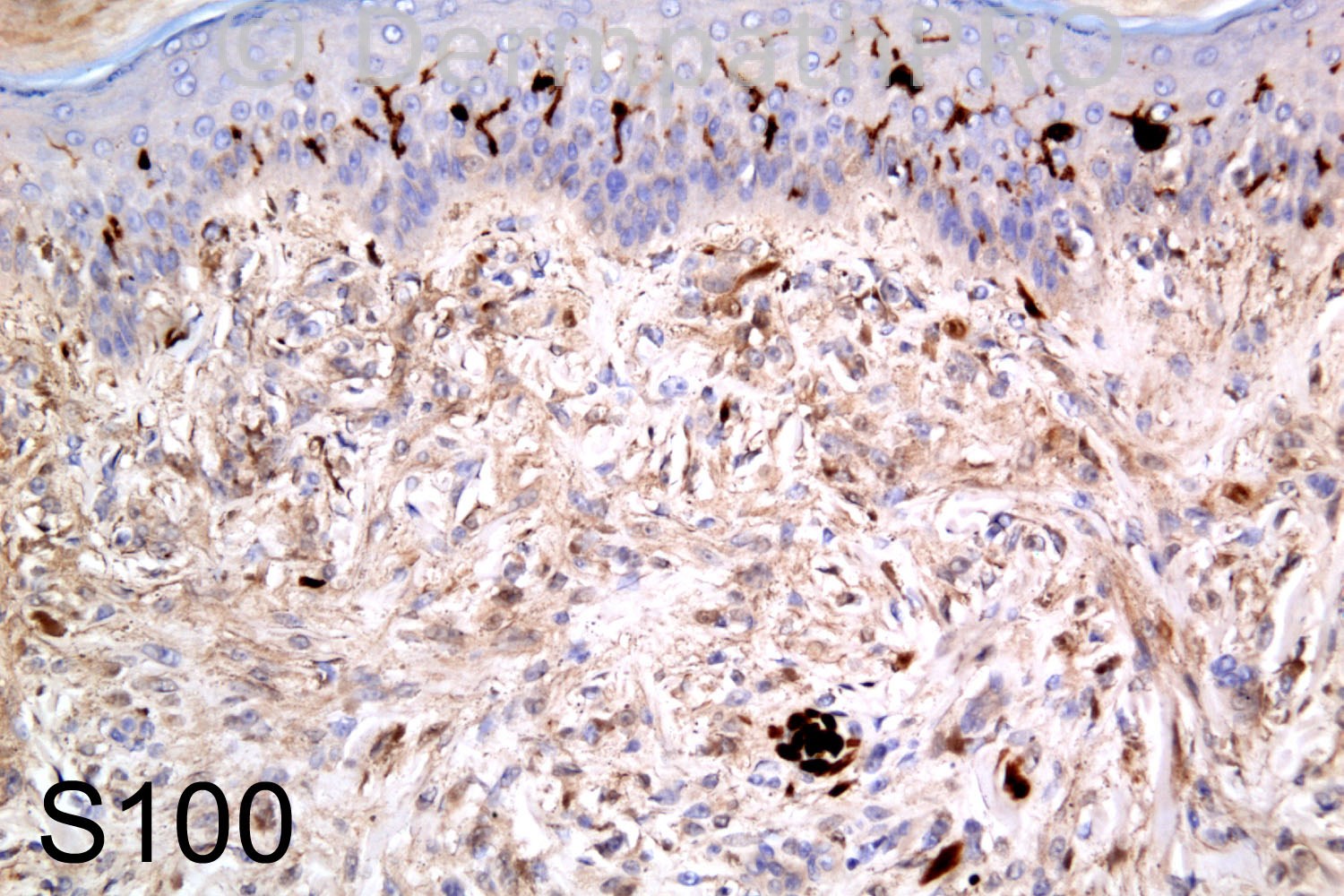
Could it be a nevus which lost s100 staining or cellular neurothekoma which is not staining with s 100.

Share this comment


Link to comment
share_externally

Guest Maria George

Posted

Just to supplment the IHC suggested by Mark, I did find the paragraphs in this online link useful;
[url="http://emedicine.medscape.com/article/2034472-overview#aw2aab6b6"]http://emedicine.medscape.com/article/2034472-overview#aw2aab6b6[/url]

Share this comment


Link to comment
share_externally

Dr. Mona Abdel-Halim

Posted

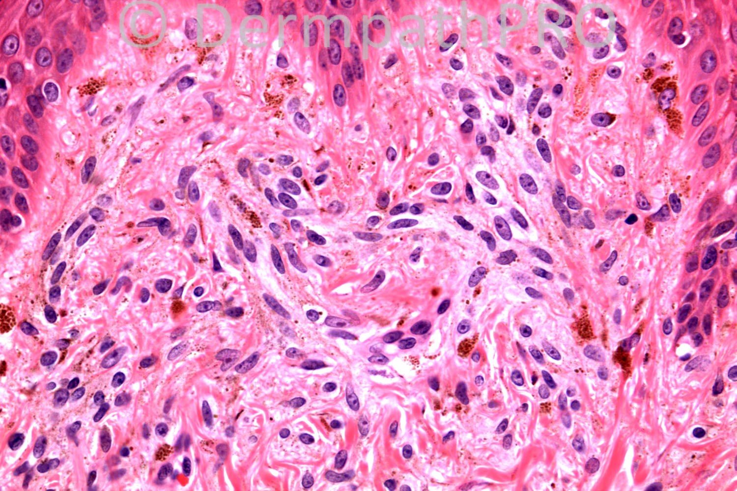
And what about the melanophages/ melanin granules dispersed among the lesion? I am not sure that S100 here is negative. Thought of hypopigmented type blue nevus or less likely desmoplastic nevus??

Share this comment


Link to comment
share_externally

Sclerotic hypopigmented blue nevus vs Desmoplastic nevus

Share this comment


Link to comment
share_externally

Hypopigmented blue nevus. I don´t think that there is sclerosis or a juntional component for a Desmoplastic nevus. Nice case. Waiting for more comments.

Share this comment


Link to comment
share_externally

Robledo F. Rocha

Posted

I think this is a desmoplastic Spitz nevus. I found a wedge-shaped intradermal melanocytic proliferation with epithelioid superficial cells and smaller deep ones, all of them embedded in a sclerotic stroma. Typically melanin pigment is sparse and the site is an extremity of a young patient.
S100 is negative, since normal dendritic melanocytes within the epidermis are strongly highlighted by this immunostain. This might mean a loss of s100 expression by lesional cells.

Share this comment


Link to comment
share_externally

Guest Juan Carlos Garcés, Ecuador

Posted

Hypopigmented blue nevus

Share this comment


Link to comment
share_externally

Guest Jim Davie MD

Posted

[size=4]In my opinion, the numerous apparent melanophages diffusely scattered throughout the lesion in oddly uniform fashion (compare papillary and deep dermis) is the histologic 'elephant in the room' that needs to be explained. I agree it is S100 negative.[/size]

[size=4]Differential for me would therefore include (in descending order of favor):[/size]
[size=4]1. [/size][size=4]Cellular neurothekeoma with melanocytosis ([/size][size=4]anomalously uniform instead of patchy).**[/size]
[size=4]2. Blue nevus (with anomalous negative S100).[/size]

[size=4]An NKI-C3 would be helpful, as cellular NK is positive for this, and characteristically S100 negative. [/size]
[size=4]B[/size][size=4]lue nevi are characteristically triple-positive for melanocyte markers S100, HMB45, and MART1/MelanA. [/size][size=4]It would be useful to have these markers to help resolve melanocytic vs other.[/size]

[font=times new roman,times,serif][size=4]**[link] [/size][size=4][url="http://www.ncbi.nlm.nih.gov/pubmed/18190453"]Cellular neurothekeoma with melanocytosis.[/url] [/size][size=4]J Cutan Pathol. 2008 Feb;35(2):241-5. doi: 10.1111/j.1600-0560.2007.00792.x.[/size][/font]

Share this comment


Link to comment
share_externally

Dr. Richard Carr

Posted

I am itching to help you all so I am going to give you a little help. There is mis-information with respect to immunohistochemistry in the comments above. Most textbooks won't help either - a case of "not in the textbooks". Several of you have made the correct H&E diagnosis.

Share this comment


Link to comment
share_externally

Eman El-Nabarawy

Posted

Cellular neurothekeoma with melanocytosis.

Share this comment


Link to comment
share_externally

Guest Jim Davie MD

Posted

I'd favor Blue nevus with anomalous S100 negative staining, if we are dealing with a situation of anomalous negative [size=4]S100 staining. (S100-negative blue nevi are uncommon [<3%], and are usually hypopigmented).[/size]

Share this comment


Link to comment
share_externally

Guest Romualdo

Posted

I agree with the diagnosis of blue nevus with aberrant S100 negative staining.

Share this comment


Link to comment
share_externally

Dr. Richard Carr

Posted

This is a "relatively" hypopigmented blue naevus (HBN). One definition for HBN is melanin detected in <5% of the lesional cells and I guess this lesion qualifies if we exclude the melanophages. The clinical mis-diagnosis of Spitz (usually non-pigmented lesions) would support the "hypopigmented" classification. I agree if you are not familiar with hypopigmented BN then you will consider an odd naevocellular or desmoplastic naevus (cells are quite plump in this case and in other similar lesions we have seen) but as correctly mentioned by responders above there are nice dendritic cells on close inspection and the dendritic nature can easily be confirmed with MelanA which is diffusely positive in all cases. What is not in the textbooks and is very much our experience is that blue naevi are usually either only focally or weakly S100 positive or very commonly, as in this case, completely S100 negative. Putting this case in has prompted me to finish off our paper on this topic. It is not novel information as some authors have mentioned it in the literature but it seems to be totally overlooked in most textbooks. It has to be said that some papers do state S100 as being positive in HBN but in our experience S100-/weak/focal; diffuse melanA positive (dendritic) pattern is a diagnostic clue to blue naevus. We have been caught out by near completely melanin negative case and made the wrong diagnosis of a benign fibrohistiocytic lesion based on S100 neg before spotting very sparse melanin on H&E and confirming the correct diagnosis with MelanA. A subsequent case we correctly diagnosed only because of this prior experience. Thanks for mentioning cellular neurothekeoma as this can now go in to our discussion and I was not familiar with cellular neurothekeoma with melanocytosis (do you have a reference?) but melanA will confirmed the correct diagnosis in this case. Jim I would be interested in your reference that <3% of blue naevi are S100 negative (because that is not our experience that includes cases referred in from other pathologists/labs). I interpreted the S100 positive dendritic cells in the epidermis to be Langerhan's rather than melanocytes but there may be a combination here. Remember S100 nuclear staining is helpful in heavily pigmented lesions. If you look at the S100 again you can see the subtle melanin pigmentation of the dendritic processes in this case.

Share this comment


Link to comment
share_externally

Guest Jim Davie MD

Posted

[color=#003333][font=arial, helvetica, sans-serif][size=4]Richard, here's my source:[/size][/font][/color]

[color=#003333][font=arial, helvetica, sans-serif][size=4]RE: BLUE NEVUS[/size][/font][/color]
[color=#003333][font=arial,helvetica,sans-serif][url="http://www.thedoctorsdoctor.com/diseases/bluenevus.html#ipox"]http://www.thedoctor...nevus.html#ipox[/url] [/font][/color]
[color=#003333][font=arial,helvetica,sans-serif]This has a table with selected refs on blue nevus--and blue nevus variant-- immunostaining properties. [/font][/color]

[color=#003333][font=arial,helvetica,sans-serif]1. One table entry specifically a[/font][font=arial, helvetica, sans-serif]ddresses [u]S100 negativity in blue nevi,[/u] with following detail including figure of 3% (38/1358) and two references:[/font][/color]
[color=#003333][b][font=arial, helvetica, sans-serif] [[/font][font=arial, helvetica, sans-serif][color=#008080]S-100 NEGATIVE blue nevi and cellular blue nevi[/color]][/font][/b][/color]
[color=#003333][b][color=#008080][font=arial, helvetica, sans-serif] 38 / 1358 cases of blue nevi showed minimal or no pigment [/font][/color][/b][/color]
[color=#003333][b][color=#008080][font=arial, helvetica, sans-serif] S100 and HMB45 was focal/weak or absent in all cases.[/font][/color][/b][/color]
[color=#003333][color=#008080][font=arial, helvetica, sans-serif] Am J Dermatopathol 1999;21:225-228[/font][/color][/color]
[color=#003333][color=#008080][font=arial, helvetica, sans-serif] Applied Immunohistochem 1999;7:255-259 ][/font][/color][/color]

[color=#003333][font=arial,helvetica,sans-serif]2. Amelanotic blue nevi are discussed elsewhere on the same webpage, with one reference abstract mentioning S100 staining, stating that [b]most, but not all, are S100 positive[/b]:[/font][/color]
[color=#003333][font=arial,helvetica,sans-serif][color=#008080] Am J Surg Pathol 2002; 26(11):1493-1500 [/color][/font][/color]
[color=#003333][font=arial,helvetica,sans-serif][color=#008080][b] Amelanotic Cellular Blue Nevus: A Hypopigmented Variant of the Cellular Blue Nevus: Clinicopathologic Analysis of 20 Cases [/b]
Artur Zembowicz, M.D., Ph.D.; Scott R. Granter, M.D.; Phillip H. McKee, M.D.; Martin C. Mihm, M.D.[/color][/font][/color]
[color=#003333][font=arial,helvetica,sans-serif][color=#008080][i]Abstract quote[/i][b]: " ..most, but not all, tumors showed reactivity for S-100 and HMB-45."[/b][/color][/font][/color]

---

[size=4][font=arial, helvetica, sans-serif]RE: Cellular neurothekeoma with melanocytosis. [/font][/size]
[font=arial, helvetica, sans-serif]reference (click hyperlink):[/font]
[size=4][font=arial, helvetica, sans-serif][color=#1C2837]**[link] [/color][color=#1C2837][url="http://www.ncbi.nlm.nih.gov/pubmed/18190453"]Cellular neurothekeoma with melanocytosis.[/url] [/color][color=#1C2837]J Cutan Pathol. 2008 Feb;35(2):241-5. doi: 10.1111/j.1600-0560.2007.00792.x.[/color][/font][/size]

Share this comment


Link to comment
share_externally


×
×
  • Create New...