Jump to content
In this section we have spot diagnoses posted on a daily basis since June 2010, now over 4000! You can review the archived cases and read the suggested diagnoses by users and the final comment by the contributors.
Case are uploaded each week day by 10 am UK time with the correct diagnosis will generally be posted at 8 pm UK time. Why not view the most recent spot diagnosis and proffer a diagnosis?

Case Number : Case 790 - 27th June Posted By: Guest

Please read the clinical history and view the images by clicking on them before you proffer your diagnosis.
Submitted Date :
   (0 reviews)

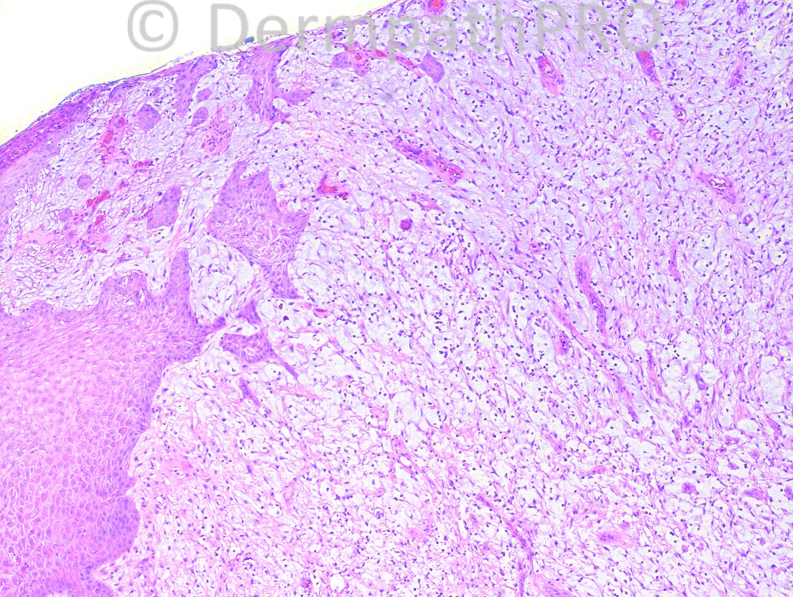
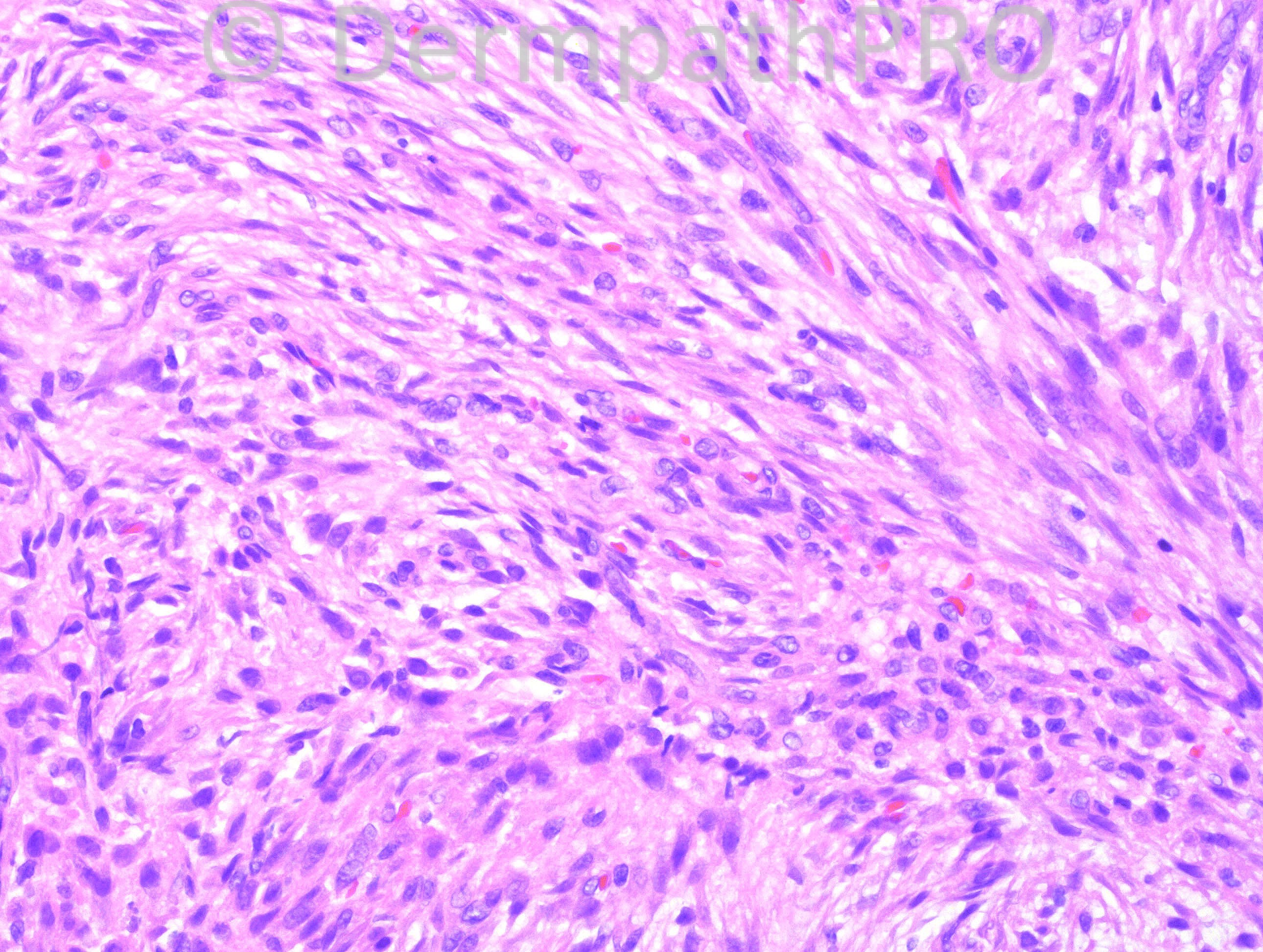
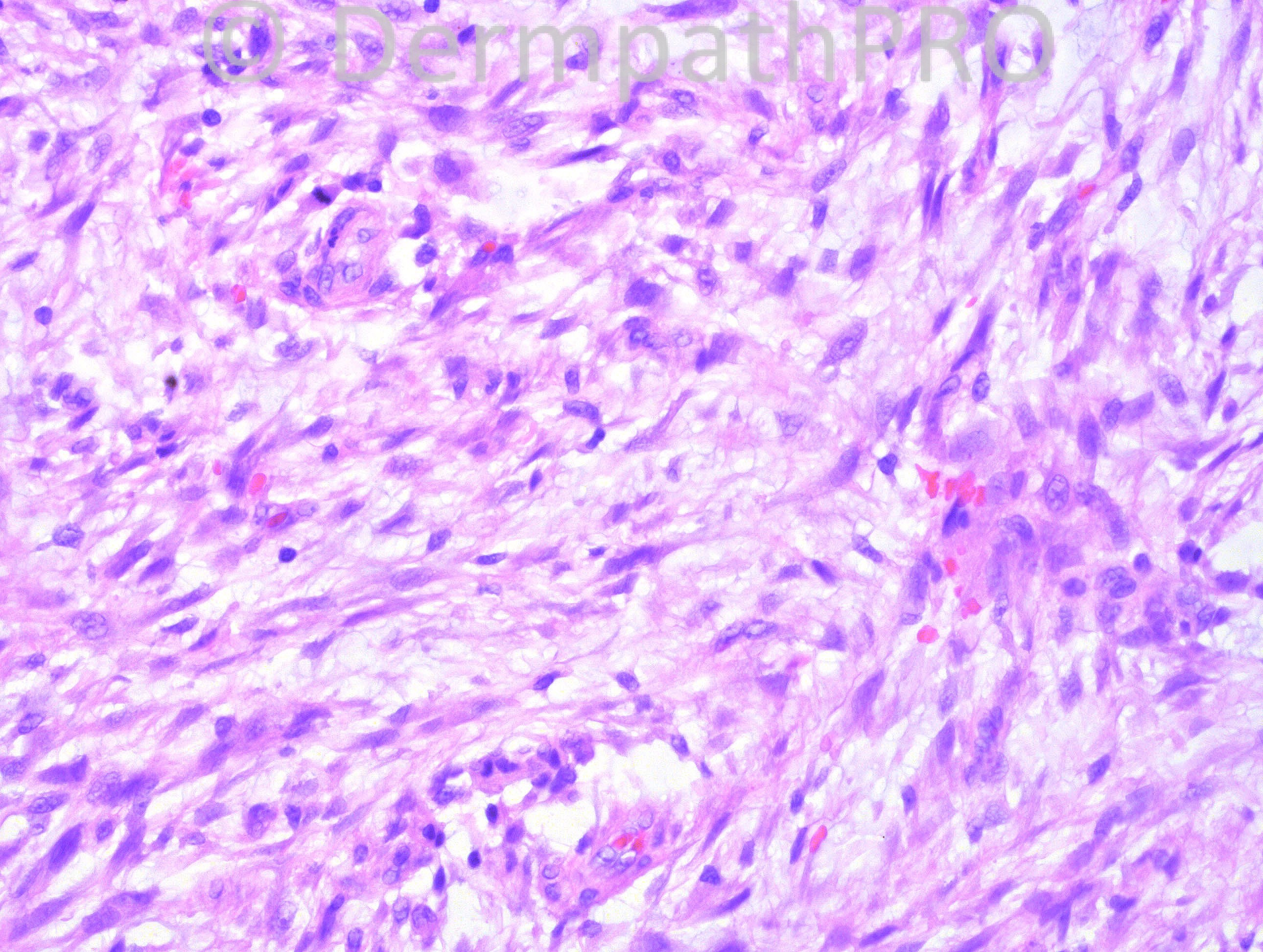
6 years-old female, history of scalp lesion, thought to be abscess, which was drained. The lesion returned and increased in size.

Case posted by Dr Hafeez Diwan.


  Report Record

User Feedback


Guest Dr Engin Sezer

Posted

Spindle cell melanoma (favoring based on conspicious lymphocytes) vs. spindle cell SCC

Share this comment


Link to comment
share_externally

Guest Maria george

Posted

SLAM DUNK named for [url="http://en.wikipedia.org/wiki/Slam_dunk"]http://en.wikipedia.org/wiki/Slam_dunk[/url], and not [url="http://en.wikipedia.org/wiki/Slam_Dunk_(manga)"]http://en.wikipedia.org/wiki/Slam_Dunk_(manga)[/url]

is a good mnemonic for D.D of malignant spindle cell neoplasms.


S - SCC
L - Leiomyosarcoma
A - angiosarcoma
M - melanoma
D - DFSP
U - undifferentiated pleomorphic sarcoma (MFH) and AFX
N - Nodular Fasciitis
K - Kaposi's sarcoma

However, in skin SCC , AFX, and desmoplastic melanoma are the commonest .Hence staining for cytokeratin, CD10, and s100 are indicated for the diseases mentioned respectively.But my gut feeling is for SCC because of epidermal hyperplasia and the age 60 not 6 and the location.

Share this comment


Link to comment
share_externally

Guest Romualdo

Posted

Nodular fasciitis. If there is a lytic bone defect under the scalp lesion the variant of nodular fasciitis known as cranial fasciitis will be the correct diagnosis.

Share this comment


Link to comment
share_externally

Guest Maria George

Posted

I am sorry,I did not recognize that there is no features of malignancy, and given the age and the extravasated RBC, this is a nodular fascitis.

Share this comment


Link to comment
share_externally

Robledo F. Rocha

Posted

[font="Palatino Linotype, serif"][size="4"]Agree with the diagnosis of nodular fasciitis. I appreciate this tissue culture-like pattern of bland spindle cells in a fibromyxoid background. Scattered lymphocytes and microhemorrhages can also be found. Furthermore, head is the most common site in children.[/size][/font]
[font="Palatino Linotype, serif"][size="4"]I don't think this is cranial fasciitis. It occurs almost exclusively in infants during the first months of life and osseous metaplasia is lacking.[/size][/font]
[font="Palatino Linotype, serif"][size="4"]On the other hand, I'm concerned about the report of recurrence with size increasing, once nodular fasciitis rarely reccurs, even after incomplete resection.[/size][/font]

Share this comment


Link to comment
share_externally

Agree with nodular fasciitis. I observe a culture-like patern of spildle-cell proliferation, lymphocitic infiltration, eritrocyte extravasation and mitotic figures in a myxoid background. Care must be taken with the other hypothesis like SCC, Melanoma, AFX, Angiosarcoma and Leiomyosarcoma. The patient is too young for these.

Share this comment


Link to comment
share_externally

Dr. Mona Abdel-Halim

Posted

On this site, I would call it cranial fasciitis ... Nodular fasciitis on the scalp !!!

Share this comment


Link to comment
share_externally

Dr. Hafeez Diwan

Posted

Thanks for the acronym! I called this nodular fasciitis.

Share this comment


Link to comment
share_externally


×
×
  • Create New...