Jump to content
In this section we have spot diagnoses posted on a daily basis since June 2010, now over 4000! You can review the archived cases and read the suggested diagnoses by users and the final comment by the contributors.
Case are uploaded each week day by 10 am UK time with the correct diagnosis will generally be posted at 8 pm UK time. Why not view the most recent spot diagnosis and proffer a diagnosis?

Case Number : Case 791 - 28th June Posted By: Guest

Please read the clinical history and view the images by clicking on them before you proffer your diagnosis.
Submitted Date :
   (0 reviews)

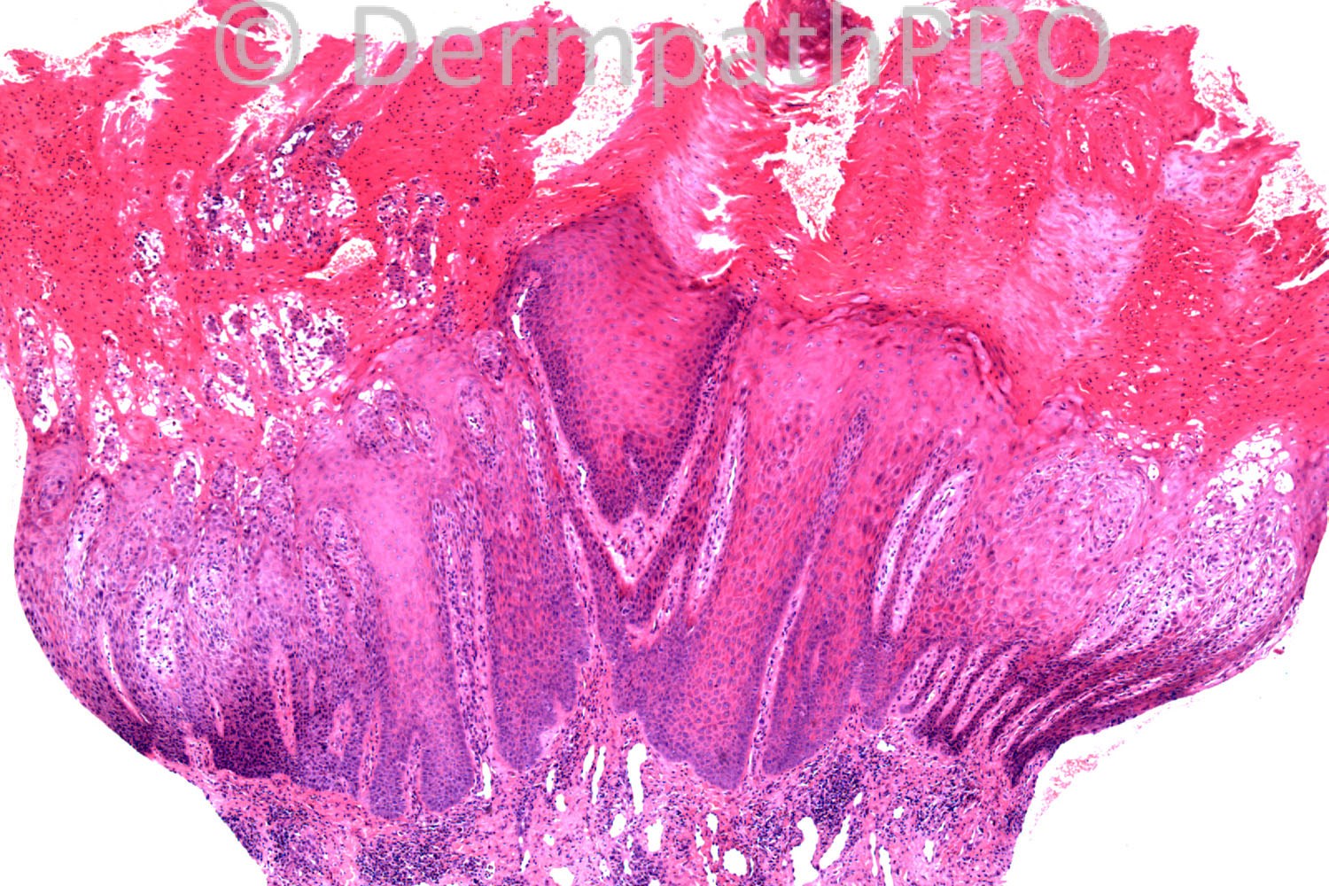
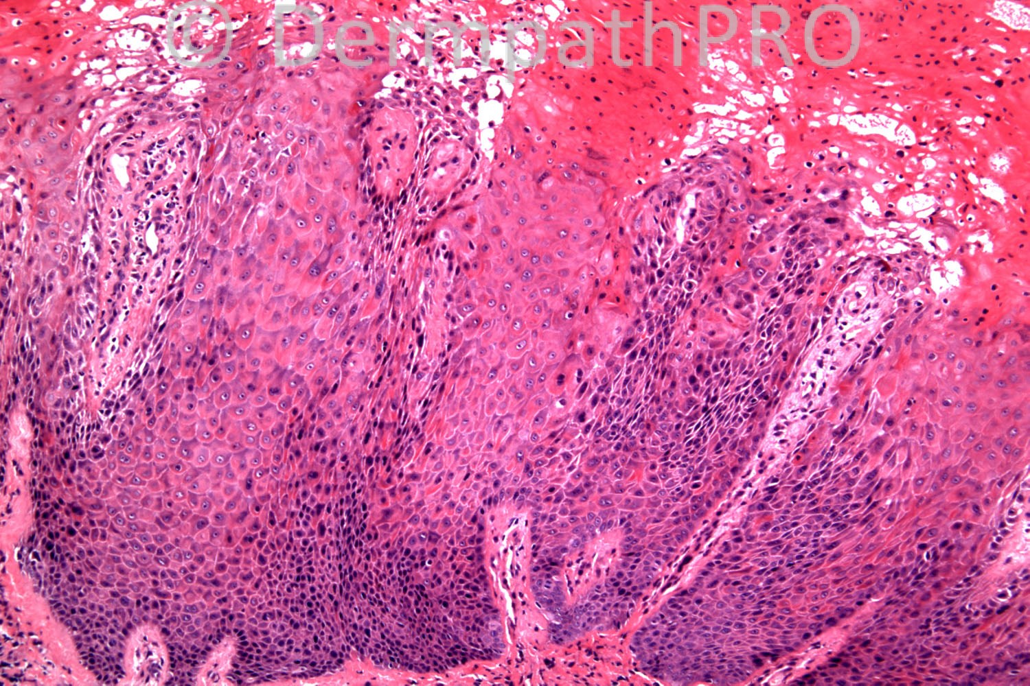
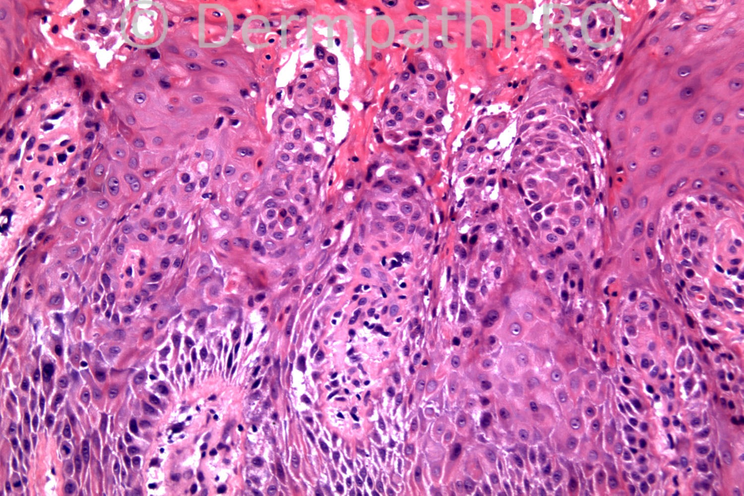
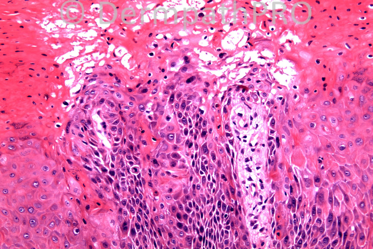
90 years-old female, 6/12 hx of multiple warty plaques natal cleft.?V warts, ?dysplastic/malignant.

Case posted by Dr Richard Carr.


  Report Record

User Feedback


Guest Maria George

Posted

[b] The cells which looks malignant are not taking Ki67.I guess Acantholytic dermatosis localized to genitalia and crural areas or [/b]

[color=#444444][font=arial, sans-serif][size=1][right]Papular [/right][/size][/font][/color][color=#444444][font=arial, sans-serif][size=1][b][right]acantholytic[/right][/b][/size][/font][/color][color=#444444][font=arial, sans-serif][size=1][right] dyskeratosis[b].[/b][/right][/size][/font][/color]

Share this comment


Link to comment
share_externally

Dr. Richard Carr

Posted

This case is very difficult! In addition to the Ki67 shown, p53 was wild type and p16 was negative. As another helpful clue do pay attention to the stratum corneum.

Share this comment


Link to comment
share_externally

Robledo F. Rocha

Posted

Pictures seem to display features of acantholytic dyskeratosis pattern with secondary changes due to persistent injury caused by trauma or friction. Maybe this case represents Darier’s disease whose papules became hypertrophic and therefore encouraged the patient to see the doctor.

Share this comment


Link to comment
share_externally

Guest Dr Engin Sezer

Posted

Psoriasiform keratoses. However, the one that I published was solitary.

Share this comment


Link to comment
share_externally

Guest Jim Davie MD

Posted

[size=4]I favor [b]verruca [/b]vs.[b] low-risk-HPV condyloma[/b] (given negative P16). [/size][size=4] [/size]This looks superficially like an HPV process...moderate pleomorphism, low grade atypia, and disturbed maturation of keratinocytes, binucleation and hyperchromatic smudging in the corneum, clear-cell degenerative ballooning, and delicate papillomatosis with 'church spire' parakeratotic columns within the hyperkeratosis, that hint at an abused verruca, as Mark suggested.

However, there is a peculiar smudgy hyperchromatic multinucleation that seems more at home in zoster.
[size=4]The differential for me would therefore include a [b]verrucous HSV or verrucous herpes zoster[/b] (especially if this is a 'tough' case; s[/size]acral/buttock dermatomes are bulls-eye concentric, so a patch instead of linear distribution would be consistent, if zoster).

[i][ The "suprabasal mitoses" I believe are actually tangential sections of basal or suprabasal keratinocytes, as evidenced by their uniformly low cytoplasmic volume, and proven by the Ki67 which only stains the basal layer (except for some foci that also show tangential basal layer sectioning). ][/i]

Share this comment


Link to comment
share_externally

Guest Romualdo

Posted

Natal cleft? What does it mean?

Share this comment


Link to comment
share_externally

Guest Jim Davie MD

Posted

[i]Natal cleft: [/i][color=#333333][font=verdana, sans-serif][size=3]Also called gluteal cleft, or anal cleft. [/size][/font][/color][color=#333333][font=verdana, sans-serif][size=3]It's the groove or crack between the buttocks that runs from below the sacrum to the perineum; or the groove in which the anus is situated.[/size][/font][/color]

Share this comment


Link to comment
share_externally

Dr. Richard Carr

Posted

Next clue - with the eye of faith I admit - closely opposed collumns of parakeratosis. I took the vacuolation and nuclear pyknosis to be maceration.

Share this comment


Link to comment
share_externally

Guest Maria George

Posted

Porokeratoses.

Share this comment


Link to comment
share_externally

Guest Maria George

Posted

if not could it be an epidermal nevus.

Share this comment


Link to comment
share_externally

Guest Romualdo

Posted

Collumns of parakeratosis, papillomatosis, confluence of rete ridges at the base of the lesion: verruca vulgaris.

Share this comment


Link to comment
share_externally

Dr. Mona Abdel-Halim

Posted

First impression before reaching KI67 was Bowen's disease then I excluded it based on Ki67. Second thought was wart (verruca) but with the clue of Dr Carr that vacuolization and pyknosis r secondary to maceration, I had to exclude this possibility. I am left now with verrucous porokeratosis of the gluteal cleft (porokeratosis ptychotropica)...

Share this comment


Link to comment
share_externally

Guest Romualdo

Posted

Well done Mona and Maria!! You should be correct!!!

Share this comment


Link to comment
share_externally

Robledo F. Rocha

Posted

[quote name='Guest' timestamp='1372432866']
Natal cleft? What does it mean?
[/quote]
Sulco interglúteo.

Share this comment


Link to comment
share_externally

Robledo F. Rocha

Posted

I gonna change my mind and close ranks with Mona and others that are thinking of porokeratosis ptychotropica. I looked for this disease and learned that it typically ocurrs in intergluteal cleft area and features psoriasiform epidermal hyperplasia topped by multiple coalescent cornoid lamellae with underlying hypogranulosis and dyskeratotic keratinocytes.

Share this comment


Link to comment
share_externally

Guest Romualdo

Posted

My English is very poor. I should have written "You must be correct", not "You should be correct"!

Share this comment


Link to comment
share_externally

Dr. Richard Carr

Posted

Well done!! I did say it was difficult and I have to admit to not making the diagnosis at the first attempt. I had reported it as an odd psoriasiform process, probably reactive but then it turned out this patient was one we had reported perianal hypertrophic porokeratosis ten years previously! Based on this I reported this biopsy as compatible with verrucous porokeratosis.

[url="http://www.ncbi.nlm.nih.gov/pubmed/?term=carr+charles-holmes+porokeratosis#"]Clin Exp Dermatol.[/url] 2003 Nov;28(6):676-7.
[b] Hypertrophic perianal porokeratosis in association with superficial actinic porokeratosis of the leg.[/b]
[url="http://www.ncbi.nlm.nih.gov/pubmed?term=Thomas%20C%5BAuthor%5D&cauthor=true&cauthor_uid=14616847"]Thomas C[/url], [url="http://www.ncbi.nlm.nih.gov/pubmed?term=Ogboli%20MI%5BAuthor%5D&cauthor=true&cauthor_uid=14616847"]Ogboli MI[/url], [url="http://www.ncbi.nlm.nih.gov/pubmed?term=Carr%20RA%5BAuthor%5D&cauthor=true&cauthor_uid=14616847"]Carr RA[/url], [url="http://www.ncbi.nlm.nih.gov/pubmed?term=Charles-Holmes%20R%5BAuthor%5D&cauthor=true&cauthor_uid=14616847"]Charles-Holmes R[/url].

Share this comment


Link to comment
share_externally


×
×
  • Create New...