Jump to content
In this section we have spot diagnoses posted on a daily basis since June 2010, now over 4000! You can review the archived cases and read the suggested diagnoses by users and the final comment by the contributors.
Case are uploaded each week day by 10 am UK time with the correct diagnosis will generally be posted at 8 pm UK time. Why not view the most recent spot diagnosis and proffer a diagnosis?

Case Number : Case 707 - 1 Mar Posted By: Guest

Please read the clinical history and view the images by clicking on them before you proffer your diagnosis.
Submitted Date :
   (0 reviews)

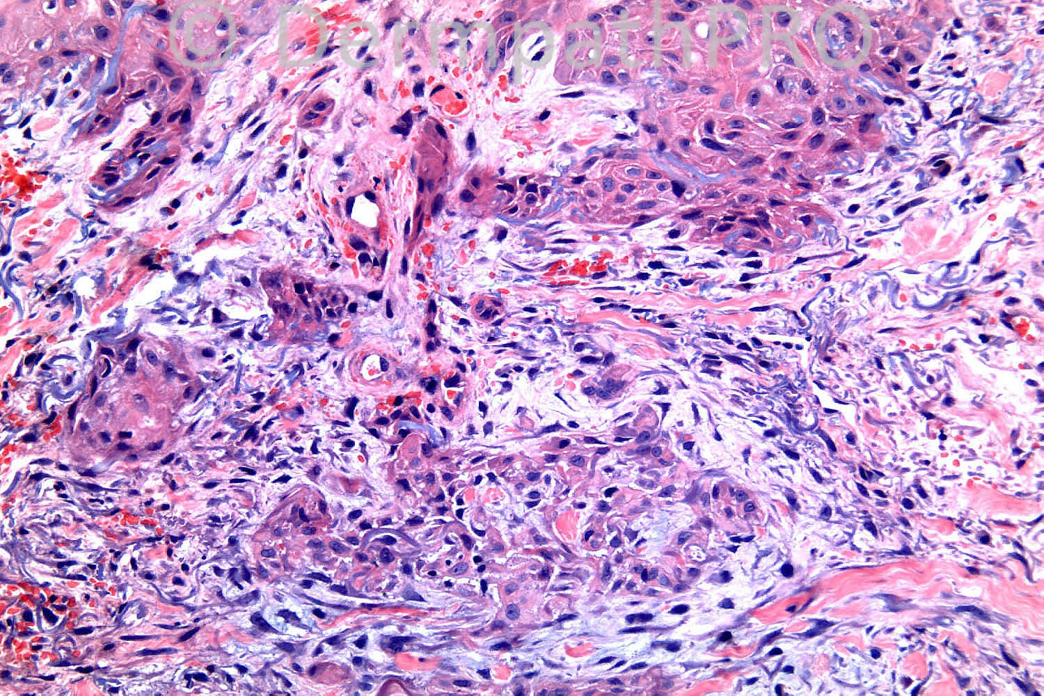
85 years-old male. Scaly patch lower leg ?Squamous cell carcinoma.

Case posted by Dr. Richard Carr


  Report Record

User Feedback


Eman El-Nabarawy

Posted

Eccrine squamous syringometaplasia?

Share this comment


Link to comment
share_externally

Guest Bansal_

Posted

Appears reactive. ? Syringosquamous metaplasia. Needs clinicopathological correlation.

Share this comment


Link to comment
share_externally

Robledo F. Rocha

Posted

Another difficult case, mainly because I think there are two associated conditions. And a striking eccrine squamous syringometaplasia, secondary to these associated conditions, comes just to divert attention.
Blue dermal deposits may represent mucinosis.
Proliferation of small blood vessels and extravasated erythrocytes bring to mind stasis dermatitis.

Share this comment


Link to comment
share_externally

Dr. Mona Abdel-Halim

Posted

Syringosquamous metaplasia...

Share this comment


Link to comment
share_externally

Guest Dr Engin Sezer

Posted

Porocarcinoma

Share this comment


Link to comment
share_externally

Guest MarieMD

Posted

Pseudoep hyperplasias and syringosquamous metaplasia--reactive changes assoc. with stasis dermatitis.

Share this comment


Link to comment
share_externally

Syringosquamus /pseudoepitheliomatous metaplasia

Share this comment


Link to comment
share_externally

Dr. Richard Carr

Posted

This is an exceedingly difficult case. I was very disturbed by the follicular
pattern of proliferation with engulfment of elastic (a feature typical of KA
and some poorly differentiated follicular SCC). I have generally only seen
the latter pattern in invasive tumours and not hyperplasia and on that basis
I raised the possibility of carcinoma. Clinical correlation was essential.
Reviewed at Skin MDM. Biopsy was 2 weeks from onset of the lesion.
Patient had previous episodes of trauma to lower legs but could not remember
anything specific. Lesion was a flatish brown patch in clinic reivew with
no clinically worrying features for malignancy. We decidided this was probably
a resovling KA-like proliferation / superficial lesion with prominent squamous
syringometaplasia and acroangiodermatitis. We adopted a watch and wait policy.
I would certainly be very cautious in such cases and don't be afraid to say
don't know!!
Regards to all and enjoy your weekends.

Share this comment


Link to comment
share_externally

Dr. Mona Abdel-Halim

Posted

Very nice case :-) thanks a lot Dr Carr !!!!

Share this comment


Link to comment
share_externally


×
×
  • Create New...