Jump to content
In this section we have spot diagnoses posted on a daily basis since June 2010, now over 4000! You can review the archived cases and read the suggested diagnoses by users and the final comment by the contributors.
Case are uploaded each week day by 10 am UK time with the correct diagnosis will generally be posted at 8 pm UK time. Why not view the most recent spot diagnosis and proffer a diagnosis?

Case Number : Case 711 - 7 Mar Posted By: Guest

Please read the clinical history and view the images by clicking on them before you proffer your diagnosis.
Submitted Date :
   (0 reviews)

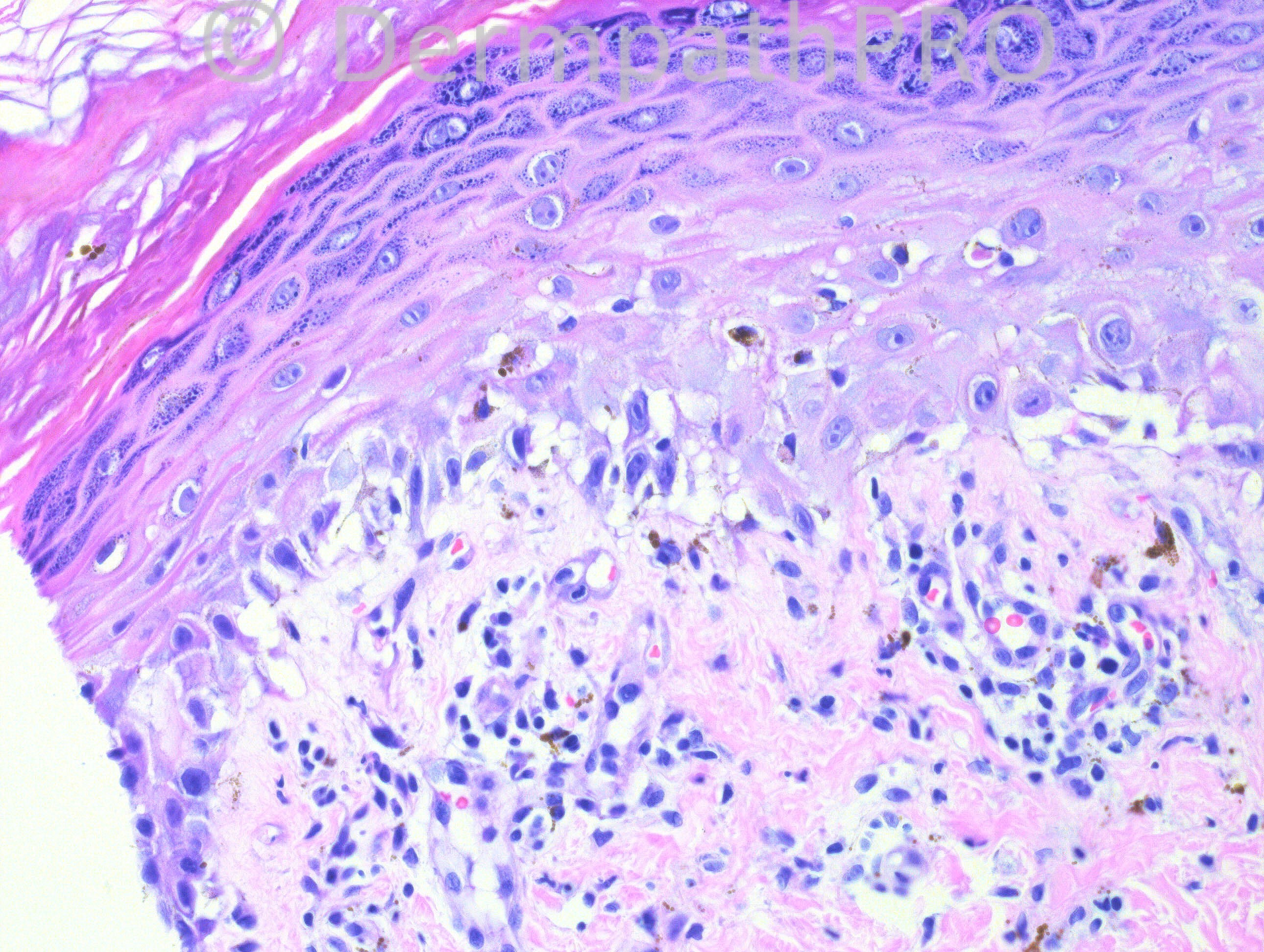
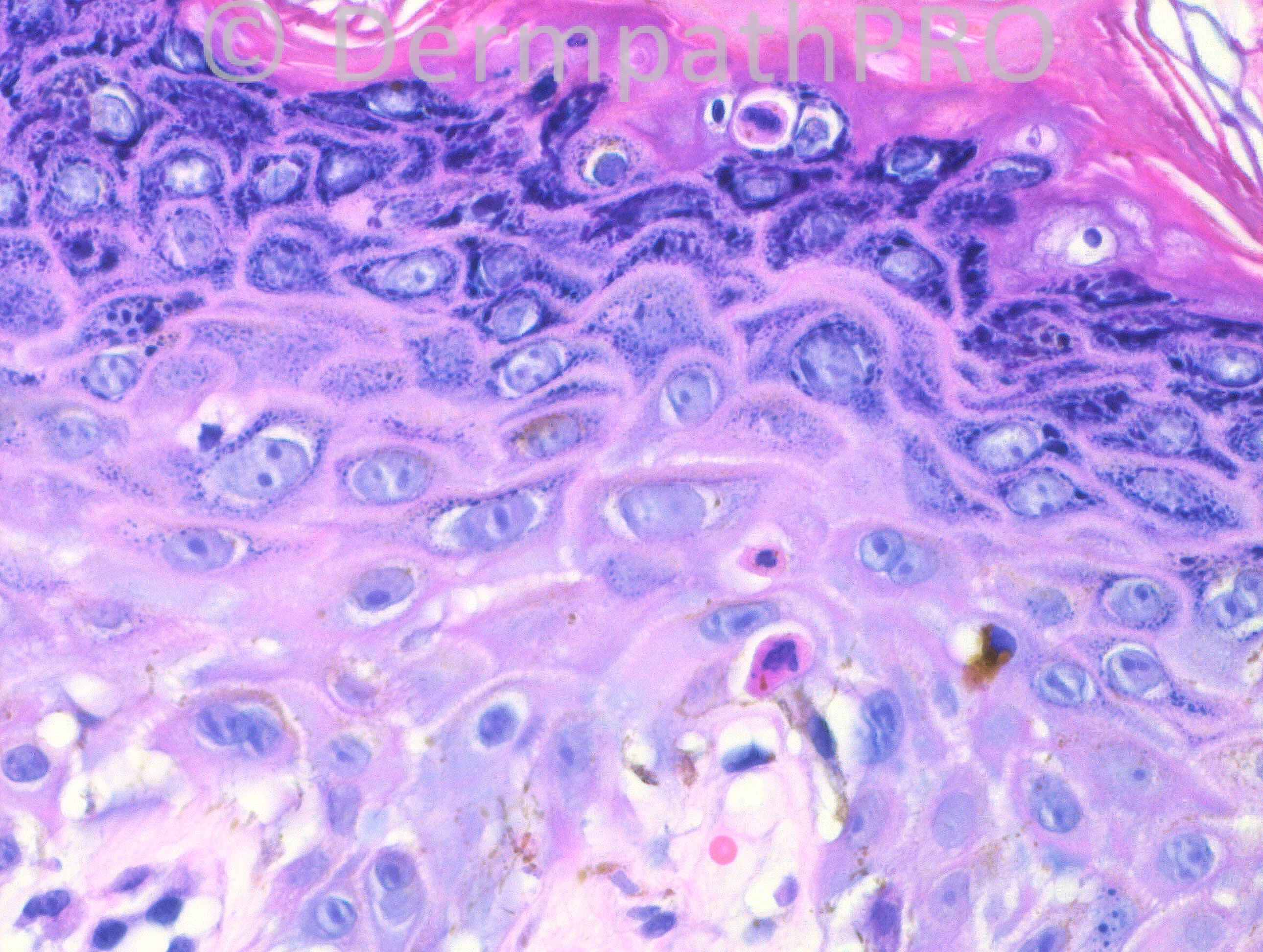
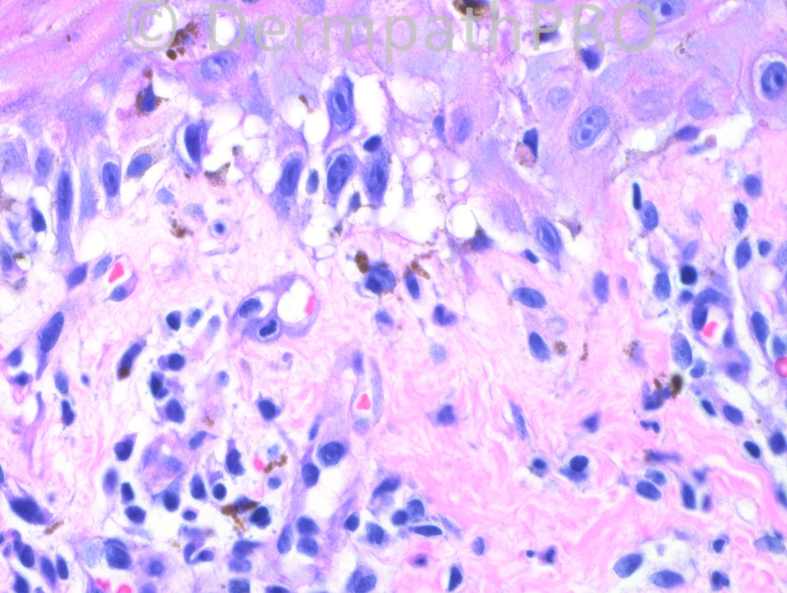
10 months-old female with diffuse hypopigmented macules, patches and papules on face, trunk and extremities. This biopsy is from the right upper back.

Case posted by Dr. Hafeez Diwan.


  Report Record

User Feedback


Eman El-Nabarawy

Posted

PLC leaving postinflammatory hypopigmentation. An infantile case of Pitryriasis lichenoids et varioliformis acuta. Br J Dermatol. 2007 Jul;157(1):194-6.

Share this comment


Link to comment
share_externally

nick turnbull

Posted

Pityriasis lichenoides

Share this comment


Link to comment
share_externally

Robledo F. Rocha

Posted

Interface dermatitis with sparse superficial perivascular lymphocytic infiltrate that extends minimally into the epidermis. There’s also focal basal vacuolization, scattered dyskeratotic keratinocytes, and prominent hypergranulosis with overlying confluent hyperkeratosis.
I think it is pityriasis lichenoides chronica evolving towards postinflammatory hypopigmentation, despite of the fact that the face is not spared.

Share this comment


Link to comment
share_externally

Sasi Attili

Posted

I haven't really seen PLC at such a young age and moreover presenting with hypopigmented macules and papules + on the face, would be quite unusual! Based on the history and interface change I wondered about hypopigmented MF, but do not see atypical lymphocytes. How about a drug reaction or a GVHD like reaction (if the history fits)? 10 months is too old for neonatal lupus. Viral exanthems can sometimes also present as unusual vacuolar dermatoses. Thanks for this extremely interesting case.

Share this comment


Link to comment
share_externally

Dr. Mona Abdel-Halim

Posted

Pathologically fits with PLC but strange enough to be on the face which is usually spared and to be in this age group,,, but life has taught me that diseases do not follow text books !!!!!!!!!!!!!!!

Share this comment


Link to comment
share_externally

Eman El-Nabarawy

Posted

I'd like to add to my DD Gianotti-Crosti syndrome.

Share this comment


Link to comment
share_externally

Guest Jim Davie MD

Posted

[size=4][font=arial, helvetica, sans-serif]Interesting case. The sandwich sign in the stratum corneum implies sudden onset at this biopsy site. In absence of additional clinical information, I favor the general direction of a viral etiology / Gianotti-Crosti that would explain face involvement, the mixed spongiotic and interface inflammatory pattern with necrotic keratinocytes and mononuclear exocytosis, and potential for hypopigmentation/hyperpigmentation. Viral etiology would be common in the age group, but expression as hypopigmented patches would be, however, unusual. [/font][/size]

Share this comment


Link to comment
share_externally

Dr. Mona Abdel-Halim

Posted

I think Gianotti Crosti strikingly spares the trunk, but I agree in this age group that this may just be residual viral infection that have healed with post inflammatory hypopigmentation.

Share this comment


Link to comment
share_externally

Robledo F. Rocha

Posted

I don’t think that Gianotti-Crosti syndrome can explain all clinical and microscopic features of this case. Although this viral exanthem is a more frequent disease in this age group, as a rule it spares the trunk and does not manifest as hypopigmented patches. Microscopically, papillary dermal edema and spongiosis would be present, but both are lacking.
Albeit rare, pityriasis lichenoides do occur during infancy, but PLEVA is the most common pattern in younger children according to two papers (see below). Face is spared in most cases, but facial involvement is much more common in infants than in adults. As Mona remembered us, diseases don’t read medicine textbooks!
Ersoy-Evans S, Greco F, Mancini AJ, et al. Pityriasis lichenoides in childhood. A retrospective review of 124 patients. J Am Acad Dermatol 2007;56:205-210.
Wahie S, Hiscutt E, Natarajan S, et al. Pityriasis lichenoides: the differences between children and adults. Br J Dermatol 2007;157:941-945.

Share this comment


Link to comment
share_externally

Guest Leonardo Mello

Posted

keratosis lichenoides chronica? Lichen planus?

Share this comment


Link to comment
share_externally

Guest Marcia

Posted

I'd like to add erythema multiforme.

Share this comment


Link to comment
share_externally

Dr. Hafeez Diwan

Posted

This turned out to be neonatal lupus.

Share this comment


Link to comment
share_externally

Sasi Attili

Posted

[quote name='Dr. Hafeez Diwan' timestamp='1362755986']
This turned out to be neonatal lupus.
[/quote] Wow! Interesting but confusing. Is this not quite a late presentation to be 'neotatal' LE?

Share this comment


Link to comment
share_externally

Robledo F. Rocha

Posted

Many thanks to Dr. Diwan for posting this challenging case.

Share this comment


Link to comment
share_externally


×
×
  • Create New...